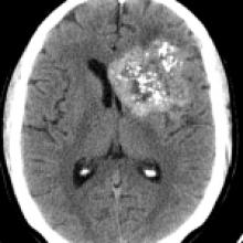
Giant Cavernous Angioma
- Giant cavernomas are benign vascular malformations.
- When small, diagnosis is usually straightforward. Larger lesions result in complex-looking lesions and can be more difficult to diagnose.
- Clinical Presentation: Nonspecific. Headache is a common complaint.
- Key Imaging Features: Small lesions: "Popcorn" appearance. Larger lesions: Complex-appearing, with a heterogeneous appearance on T1WI and T2WI, and with foci of susceptibility within. A complete hemosiderin rim can be seen. Alternatively, in a more acute setting, a fluid-fluid level is seen, suggestive of recent hemorrhage. Heterogeneous enhancement is seen following contrast administration. Presence of an associated developmental venous anomaly, best seen on susceptibility-weighted imaging or contrast-enhanced MPRAGE sequence, helps in making the diagnosis.
- DDx: Primary neuronal mixed neural-glial tumor
- Rx: Conservative if small. Larger lesions may require surgical excision.