Advertisement

Main menu

  • Home
  • Content
    • Current Issue
    • Accepted Manuscripts
    • Article Preview
    • Past Issue Archive
    • AJNR Case Collection
    • Case of the Week Archive
    • Classic Case Archive
    • Case of the Month Archive
  • Special Collections
    • Spinal CSF Leak Articles (Jan 2020-June 2024)
    • 2024 AJNR Journal Awards
    • Most Impactful AJNR Articles
  • Multimedia
    • AJNR Podcast
    • AJNR Scantastics
    • Video Articles
  • For Authors
    • Submit a Manuscript
    • Author Policies
    • Fast publishing of Accepted Manuscripts
    • Graphical Abstract Preparation
    • Manuscript Submission Guidelines
    • Imaging Protocol Submission
    • Submit a Case for the Case Collection
  • About Us
    • About AJNR
    • Editorial Board
  • More
    • Become a Reviewer/Academy of Reviewers
    • Subscribers
    • Permissions
    • Alerts
    • Feedback
    • Advertisers
    • ASNR Home
  • Other Publications
    • ajnr

User menu

  • Alerts
  • Log in

Search

  • Advanced search
American Journal of Neuroradiology

American Journal of Neuroradiology

ASHNR American Society of Functional Neuroradiology ASHNR American Society of Pediatric Neuroradiology ASSR
  • Alerts
  • Log in

Advanced Search

  • Home
  • Content
    • Current Issue
    • Accepted Manuscripts
    • Article Preview
    • Past Issue Archive
    • AJNR Case Collection
    • Case of the Week Archive
    • Classic Case Archive
    • Case of the Month Archive
  • Special Collections
    • Spinal CSF Leak Articles (Jan 2020-June 2024)
    • 2024 AJNR Journal Awards
    • Most Impactful AJNR Articles
  • Multimedia
    • AJNR Podcast
    • AJNR Scantastics
    • Video Articles
  • For Authors
    • Submit a Manuscript
    • Author Policies
    • Fast publishing of Accepted Manuscripts
    • Graphical Abstract Preparation
    • Manuscript Submission Guidelines
    • Imaging Protocol Submission
    • Submit a Case for the Case Collection
  • About Us
    • About AJNR
    • Editorial Board
  • More
    • Become a Reviewer/Academy of Reviewers
    • Subscribers
    • Permissions
    • Alerts
    • Feedback
    • Advertisers
    • ASNR Home
  • Follow AJNR on Twitter
  • Visit AJNR on Facebook
  • Follow AJNR on Instagram
  • Join AJNR on LinkedIn
  • RSS Feeds

Welcome to the new AJNR, Updated Hall of Fame, and more. Read the full announcements.


AJNR is seeking candidates for the position of Associate Section Editor, AJNR Case Collection. Read the full announcement.


Metastasic Ganglioneuroblastoma in the Orbit

  • Background: Ganglioneuroblastoma is a tumor derived from primordial neural crest cells that form in the sympathetic nervous system. It has inmature and mature cell types and is more frequently found in children.
    • Common primary neoplastic sites are adrenal medulla, extra-adrenal retroperitoneum, posterior mediastinum; less common are brain, nasal fossa, neck, ganglion, and pelvis. Bone is the most common site of metastasis.
    • Clinical behavior is extremely variable, with some tumors regressing or maturing, while other progress despite aggressive treatment. Neuroblastic tumors may secrete catecholamines such as vanillylmandelic acid (VMA) and homovanillic acid (HVA).
    • Prognostic parameters include age, stage, histology, tumor differentiation, mitosis-karyorrhexis index, chromosome 11q, and MYCN oncogene status.3 Children older than 1 year with stage 4 tumors have a 3-year event-free survival rate of 15%. Elevated serum levels of neuron specific enolase (>100 ng/mL) and ferritin (>142 ng/mL) indicate a worse prognosis. Expression of TRK-A, a nerve growth factor, correlates with a favorable outcome.1
  • Relevant Clinical Information: Proptosis, periorbital ecchymoses, frontal prominence, pain, weight loss, malaise, irritability, Hutchinson syndrome, Horner syndrome, paraneoplasic syndromes
  • Key Diagnostic Features:
    • CT scan depicts soft tissue mass with calcifications and variable attenuation values due to areas of necrosis and hemorrhage. Vascular encasement and compression or invasion of adjacent structures may be seen too. Metastasis to the bone may present as sclerotic or lytic lesions and suture widening.
    • On MR, lesions are commonly heterogeneous with variable enhancement. Marrow disease appears hypointense on T1WI and hyperintense on T2WI. These findings may differentiate LCH from monostotic fibrous dysplasia (MFD) which presents low signal on T1WI and T2WI (in cases where the fibrous component is predominant).
  • DDx: Sclerotic lesions as in this case may be found in fibrous dysplasia, hemangioma, osteosarcoma, osteoblastoma, and lymphoma. The existence of a primary posterior mediastinal, retroperitoneal or adrenal mass points to the diagnosois.
  • Treatment Options: Steroids, surgery, chemotherapy, and radiation therapy
October 29, 2015
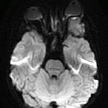
A 6-year-old girl with a history of thoracic mass resection in 2011, now with left proptosis
View Case
  • Read more about 10292015
  • Comments
Advertisement
Subscribe to RSS - Metastasic Ganglioneuroblastoma in the Orbit

Indexed Content

  • Current Issue
  • Accepted Manuscripts
  • Article Preview
  • Past Issues
  • Editorials
  • Editors Choice
  • Fellow Journal Club
  • Letters to the Editor

Cases

  • Case Collection
  • Archive - Case of the Week
  • Archive - Case of the Month
  • Archive - Classic Case

Special Collections

  • Special Collections

Resources

  • News and Updates
  • Turn around Times
  • Submit a Manuscript
  • Author Policies
  • Manuscript Submission Guidelines
  • Evidence-Based Medicine Level Guide
  • Publishing Checklists
  • Graphical Abstract Preparation
  • Imaging Protocol Submission
  • Submit a Case
  • Become a Reviewer/Academy of Reviewers
  • Get Peer Review Credit from Publons

Multimedia

  • AJNR Podcast
  • AJNR SCANtastic
  • Video Articles

About Us

  • About AJNR
  • Editorial Board
  • Not an AJNR Subscriber? Join Now
  • Alerts
  • Feedback
  • Advertise with us
  • Librarian Resources
  • Permissions
  • Terms and Conditions

American Society of Neuroradiology

  • Not an ASNR Member? Join Now

© 2025 by the American Society of Neuroradiology All rights, including for text and data mining, AI training, and similar technologies, are reserved.
Print ISSN: 0195-6108 Online ISSN: 1936-959X

Powered by HighWire