
Article Figures & Data
Figures
- fig 1.
Coronal MR images from a control subject demonstrate the boundaries of the temporopolar, entorhinal, and perirhinal cortices (white outlines). Six rostrocaudal levels are shown (A, most rostral; F, most caudal).
A, The temporopolar cortex is the area between the lateral edge of the superior temporal sulcus and the medial bank of the inferior temporal sulcus.
B, The appearance of the collateral sulcus marks the beginning of the perirhinal cortex.
C, The boundaries of the perirhinal cortex at the level of the limen insula. The collateral sulcus is shallow (depth, < 1 cm), and therefore, the lateral border of the perirhinal cortex is located at the midpoint of the occipitotemporal gyrus (16).
D, The boundaries of the entorhinal and perirhinal cortices at the level of the amygdala. The collateral sulcus is shallow, and therefore, the border between the entorhinal and perirhinal cortices is located at the fundus of the collateral sulcus (arrowhead). The lateral border of the perirhinal cortex is located at the midpoint of the occipitotemporal gyrus.
E, The boundaries of the entorhinal and perirhinal cortices at the level of the hippocampus. The borders of the entorhinal and perirhinal cortices are identical to those in D.
F, The caudal limit of the perirhinal cortex is located two sections behind the end of the uncus. A indicates amygdala; CS, collateral sulcus; EC, entorhinal cortex; HC, hippocampus; LI, limen insula; PRh, perirhinal cortex; TP, temporopolar cortex. Scale bar, 10 mm.
- fig 2.
Scatterplots show the intraobserver variability of repeated measurements in different cortical areas of 10 control subjects. A, Temporopolar cortex; B, entorhinal cortex; and C, perirhinal cortex. The limits of agreement between the first and the second measurements are expressed as the mean difference in volume: [volume in the first measurement minus volume in the second measurement (mm3)] ± 2 SD. Inserts in the lower left corner show the association between the first (y-axis) and second (x-axis) measurements. Mean indicates mean difference in volume; +2 SD, mean difference in volume plus 2 SD; -2 SD, mean difference in volume minus 2 SD
- fig 3.
Percentage of TLE patients with damage to the hippocampus (A), entorhinal cortex (B), temporopolar cortex (C), or perirhinal cortex (D). Contra indicates the side contralateral to the seizure focus; Dam+, a volume reduction of at least 2 SD from the control mean; Dam-, a volume reduction of less than 2 SD from the control mean; EC, entorhinal cortex; Focus, side of the seizure focus; HC, hippocampus; n, number of patients; PRh, perirhinal cortex; TP, temporopolar cortex
- fig 4.
Scatterplots show the correlation between the volumes of the left (A–C) or right (D–F) hippocampus and the volumes of the entorhinal, temporopolar, and total perirhinal (ie, combined perirhinal and temporopolar volumes) cortices on that side in patients with TLE. Closed circles refer to patients with right TLE and open circles to patients with left TLE. Mean indicates mean volume in control subjects; -1SD, mean volume in control subjects minus 1 SD; -2 SD, mean volume in control subjects minus 2 SD; n, number of patients; r, Pearson's correlation coefficient
- fig 5.
Coronal MR images of a 32-year-old man with cryptogenic right TLE. The duration of epilepsy at the time of imaging was 29 years, and the seizure frequency was 36 complex partial seizures per year. The patient subsequently underwent surgery and is currently seizure-free (Engel's class IA).
A, Ipsilaterally, the volume of the entorhinal cortex is 81% of that (mean volume) in control subjects and 78% of that on the contralateral side.
B, MR image taken from a more caudal level shows the volume reduction in the ipsilateral hippocampus (volume 41% of that in control subjects and 50% of that on the contralateral side). The volume of right perirhinal cortex is 2597 mm3; left, 2924 mm3 (both within normal range). A indicates amygdala; EC, entorhinal cortex; HC, hippocampus; L, left; PRh, perirhinal cortex; R, right. Scale bar, 10 mm.


Tables
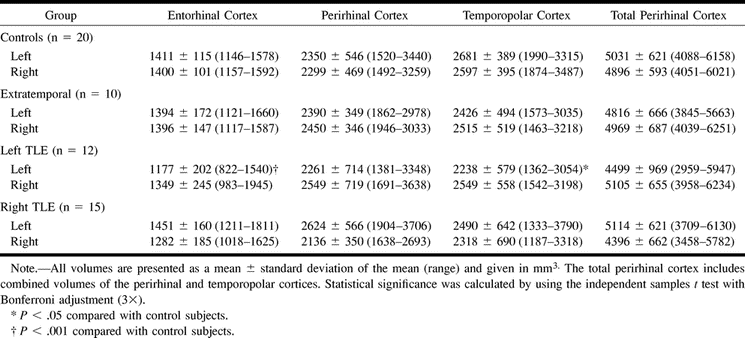
 TABLE 1:
Normalized volumes of the left and right entorhinal, perirhinal, temporopolar, and total perirhinal cortices in control subjects and patient groups
- TABLE 2:
Asymmetry ratios of various medial temporal lobe areas in control subjects and patient groups
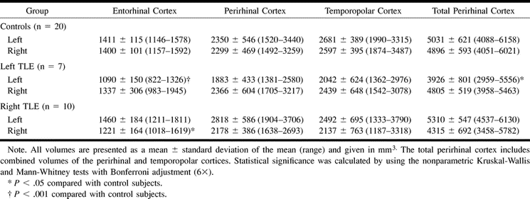
- TABLE 3:
Normalized volumes of the left and right entorhinal, perirhinal, temporopolar, and total perirhinal cortices in control subjects and TLE patients with a reduction of at least 2 SD from the mean of control subjects in the ipsilateral hippocampal volume

In this issue


Jump to section
Related Articles
Cited By...
- Distinct changes to hippocampal and medial entorhinal circuits emerge across the progression of cognitive deficits in epilepsy
- Basal temporal sulcal morphology in healthy controls and patients with temporal lobe epilepsy
- Recurrent Circuits in Layer II of Medial Entorhinal Cortex in a Model of Temporal Lobe Epilepsy
- Hyperexcitability, interneurons, and loss of GABAergic synapses in entorhinal cortex in a model of temporal lobe epilepsy.
- Progression in temporal lobe epilepsy: Differential atrophy in mesial temporal structures
- Structural abnormalities remote from the seizure focus: A study using T2 relaxometry at 3 T
- Neuroprotective Properties of Topiramate in the Lithium-Pilocarpine Model of Epilepsy
- Medial temporal lobe atrophy in patients with refractory temporal lobe epilepsy
- Why study mesial temporal atrophy in patients with intractable temporal lobe epilepsy?
- Reduced Inhibition and Increased Output of Layer II Neurons in the Medial Entorhinal Cortex in a Model of Temporal Lobe Epilepsy
- Long term outcome of temporal lobe epilepsy surgery: analyses of 140 consecutive patients