Advertisement

Main menu

  • Home
  • Content
    • Current Issue
    • Accepted Manuscripts
    • Article Preview
    • Past Issue Archive
    • Video Articles
    • AJNR Case Collection
    • Case of the Week Archive
    • Case of the Month Archive
    • Classic Case Archive
  • Special Collections
    • AJNR Awards
    • Low-Field MRI
    • Alzheimer Disease
    • ASNR Foundation Special Collection
    • Photon-Counting CT
    • View All
  • Multimedia
    • AJNR Podcasts
    • AJNR SCANtastic
    • Trainee Corner
    • MRI Safety Corner
    • Imaging Protocols
  • For Authors
    • Submit a Manuscript
    • Submit a Video Article
    • Submit an eLetter to the Editor/Response
    • Manuscript Submission Guidelines
    • Statistical Tips
    • Fast Publishing of Accepted Manuscripts
    • Graphical Abstract Preparation
    • Imaging Protocol Submission
    • Author Policies
  • About Us
    • About AJNR
    • Editorial Board
    • Editorial Board Alumni
  • More
    • Become a Reviewer/Academy of Reviewers
    • Subscribers
    • Permissions
    • Alerts
    • Feedback
    • Advertisers
    • ASNR Home

User menu

  • Alerts
  • Log in

Search

  • Advanced search
American Journal of Neuroradiology

American Journal of Neuroradiology

ASHNR American Society of Functional Neuroradiology ASHNR American Society of Pediatric Neuroradiology ASSR
  • Alerts
  • Log in

Advanced Search

  • Home
  • Content
    • Current Issue
    • Accepted Manuscripts
    • Article Preview
    • Past Issue Archive
    • Video Articles
    • AJNR Case Collection
    • Case of the Week Archive
    • Case of the Month Archive
    • Classic Case Archive
  • Special Collections
    • AJNR Awards
    • Low-Field MRI
    • Alzheimer Disease
    • ASNR Foundation Special Collection
    • Photon-Counting CT
    • View All
  • Multimedia
    • AJNR Podcasts
    • AJNR SCANtastic
    • Trainee Corner
    • MRI Safety Corner
    • Imaging Protocols
  • For Authors
    • Submit a Manuscript
    • Submit a Video Article
    • Submit an eLetter to the Editor/Response
    • Manuscript Submission Guidelines
    • Statistical Tips
    • Fast Publishing of Accepted Manuscripts
    • Graphical Abstract Preparation
    • Imaging Protocol Submission
    • Author Policies
  • About Us
    • About AJNR
    • Editorial Board
    • Editorial Board Alumni
  • More
    • Become a Reviewer/Academy of Reviewers
    • Subscribers
    • Permissions
    • Alerts
    • Feedback
    • Advertisers
    • ASNR Home
  • Follow AJNR on Twitter
  • Visit AJNR on Facebook
  • Follow AJNR on Instagram
  • Join AJNR on LinkedIn
  • RSS Feeds

AJNR Awards, New Junior Editors, and more. Read the latest AJNR updates

OtherBrain

Unusual Small Choroid Plexus Cyst Obstructing the Foramen of Monroe: Case Report

Majdi M. Radaideh, Norman E. Leeds, Ashok J. Kumar, Janet M. Bruner and Raymond Sawaya
American Journal of Neuroradiology May 2002, 23 (5) 841-843;
Majdi M. Radaideh
  • Find this author on Google Scholar
  • Find this author on PubMed
  • Search for this author on this site
Norman E. Leeds
  • Find this author on Google Scholar
  • Find this author on PubMed
  • Search for this author on this site
Ashok J. Kumar
  • Find this author on Google Scholar
  • Find this author on PubMed
  • Search for this author on this site
Janet M. Bruner
  • Find this author on Google Scholar
  • Find this author on PubMed
  • Search for this author on this site
Raymond Sawaya
  • Find this author on Google Scholar
  • Find this author on PubMed
  • Search for this author on this site
  • Article
  • Figures & Data
  • Info & Metrics
  • Responses
  • References
  • PDF
Loading

Abstract

Summary: This is a case report of unusual case of choroid plexus cyst at the right foramen of Monro in the anterior third ventricle that caused unilateral obstructive hydrocephalus. The value of small-FOV thin-section MR imaging in the diagnosis of small lesions of the foramen of Monroe is demonstrated. The immunohistochemical findings in choroid epithelial cysts in comparison with those of other types of cysts at this location are discussed.

Choroid plexus cyst in the anterior third ventricle is a relatively common finding at autopsy, but the cyst is usually asymptomatic. To our knowledge, only seven symptomatic choroid cysts were reported between 1956 and 1984, and none were located at the foramen of Monroe (1).

Case Report

A 53-year-old woman presented with recurrent headaches since 1998 as well as multiple falls without loss of consciousness. CT and MR imaging of the brain at an outside institution revealed only asymmetry of the lateral ventricles, with enlargement of the right lateral ventricle. The patient came to our institution for further workup. Repeat T1- (5000/12/5 [TR/TE/NEX]) and T2-weighted (5000/128/5) MR imaging was performed with a modified protocol that included a small FOV (16 × 16 cm) and thin-section (3-mm) MR imaging at the level of foramen of Monroe. The images revealed a small cystic lesion at the right foramen with a signal intensity similar to that of CSF that produces unilateral right-sided hydrocephalus (Figs 1–3).

Fig 1.
  • Download figure
  • Open in new tab
  • Download powerpoint
Fig 1.

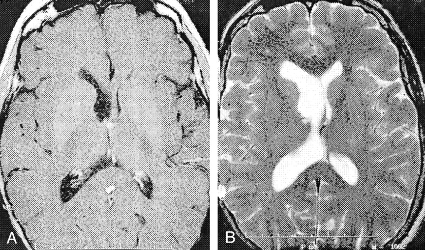
Axial MR images demonstrate unilateral right-sided hydrocephalus with deviation of the septum pellucidum to the left.

A, T1-weighted contrast-enhanced image.

B, T2-weighted image.

Fig 2.
  • Download figure
  • Open in new tab
  • Download powerpoint
Fig 2.

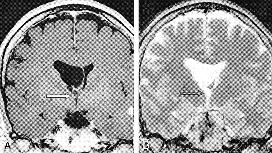
Coronal MR images demonstrate a 5-mm cystic lesion (arrow) with CSF-like signal intensity.

A, T1-weighted contrast-enhanced image.

B, T2-weighted image. The signal intensity of the lesion is slightly higher than that of CSF and is likely to be related to the to the higher protein content of the cyst compared with that of the CSF.

Fig 3.
  • Download figure
  • Open in new tab
  • Download powerpoint
Fig 3.

Sagittal MR images demonstrate a 5-mm nonenhancing cystic lesion (arrow) at the right foramen of Monro.

A, T1-weighted contrast-enhanced image.

B, T2-weighted image.

Surgery

The patient underwent resection of anterior third ventricular cyst via a right transcallosal approach with the use of a Surgiscope DeeMed, Grenoble, France and intraoperative sonography (Figs 4, 5).

Fig 4.
  • Download figure
  • Open in new tab
  • Download powerpoint
Fig 4.

Intraoperative image reveals a cystic lesion (straight arrow) at the right Foramen of Monro. The curved yellow arrow indicates the normal choroid plexus exiting the lateral ventricle through the foramen of Monro; curved white arrow, the septal vein.

Fig 5.
  • Download figure
  • Open in new tab
  • Download powerpoint
Fig 5.

The lesion is completely resected, and the foramen of Monro is open.

Pathology

The surgical specimen consisted of a smooth thin-walled cyst that measured 5 mm and contained CSF-like fluid. Histopathologic study revealed a fibrous vascular stroma covered by cuboidal epithelium. The appearance was that of choroid plexus neuroepithelial cells. Findings with immunohistochemical staining were confirmatory, with a positive reaction for transthyretin and negative reactions for epithelial membrane antigen (EMA) and mucin (mucicarmine). Other considerations eliminated as diagnostic possibilities included colloid cyst (negative mucin result) and arachnoid cyst (negative EMA result).

Follow-up

The patient had an uncomplicated postoperative course, with symptomatic relief.

Discussion

Choroid plexus cysts can occur throughout the ventricular system, but they are most frequently present in the glomus of the lateral ventricles. They are usually more common in children than in adults (2). These cysts arise from the primitive neuroepithelium. They contain CSF, and their wall is composed of connective tissue interspersed with epithelial cells. The typical choroid plexus cyst has low signal intensity on T1-weighted MR images and high signal intensity on T2-weighted MR images. In some cases, T2-weighted images may show that the cysts have higher signal intensity than that of CSF (Figs 2B, 3B). This finding is attributed to higher protein content of the cyst compared with that of the CSF (2).

The differential diagnosis of a cyst in the anterior third ventricle includes a colloid cyst, ependymal cyst, arachnoid cyst, Rathke cyst, cystosercosis, and histocytosis X (3). Immunohistochemical studies are useful in differentiating between these different types of cysts. The epithelium of choroid and ependymal cysts shows immunoreactivity for markers of normal neuroepithelial structures of the CNS. Choroid plexus epithelium has positive results with transthyretin, and ependymal epithelium has positive result with glial fibrillary acidic protein. On the contrary, the epithelial wall of colloid cysts and Rathke cleft cysts express epithelial markers, such as mucin (mucicarmine) (Fig 6). These markers support the theory of a neuroepithelial origin of a choroid plexus cyst or ependymal cyst and n endodermal origin of a colloid cyst or Rathke cleft cyst (4, 5).

Fig 6.
  • Download figure
  • Open in new tab
  • Download powerpoint
Fig 6.

Flowchart shows the immunohistochemical findings in different anterior third ventricle cysts. +ve indicates a positive finding.

Conclusion

Conventional MR images of small lesions in the foramen of Monroe may not show the lesion but only their secondary effect, namely, hydrocephalus. If a focused high-resolution (thin-section small-FOV) MR imaging technique is used, diagnosis is possible, and the findings aid in surgical planning.

Footnotes

  • Presented at the ASNR meeting in Boston, MA, 2001.

References

  1. ↵
    S. Hatashita, S. Takagi, T. Sakakibara. Choroid plexus cyst of the lateral ventricle in an elderly man, case report. J Neurosurg 1984;60:435–437
    PubMed
  2. ↵
    Kennedy KA, Carey JC. Choroid plexus cysts: significance and current management practices. Semin US CT MR 1993;14:23–30
  3. ↵
    Carmel PW. Tumours of the third ventricle. Acta Neurochir (Wien) 1985;75:136–14
    CrossRefPubMed
  4. ↵
    Coca S, Martinez A, Vaquero J, et al. Immunohistochemical study of intracranial cysts. Histol Histopathol 1993;8:651–654
    PubMed
  5. ↵
    Lach B, Scheithauer BW, Gregor A, Wick MR. Colloid cyst of the third ventricle: a comparative immunohistochemical study of neuraxis cysts and choroid plexus epithelium. J Neurosurg 1993;78:101–111
    PubMed
  • Received November 21, 2001.
  • Accepted after revision November 12, 2001.
  • Copyright © American Society of Neuroradiology
View Abstract
PreviousNext
Back to top

In this issue

American Journal of Neuroradiology: 23 (5)
American Journal of Neuroradiology
Vol. 23, Issue 5
1 May 2002
  • Table of Contents
  • Index by author
Advertisement
Print
Download PDF
Email Article

Thank you for your interest in spreading the word on American Journal of Neuroradiology.

NOTE: We only request your email address so that the person you are recommending the page to knows that you wanted them to see it, and that it is not junk mail. We do not capture any email address.

Enter multiple addresses on separate lines or separate them with commas.
Unusual Small Choroid Plexus Cyst Obstructing the Foramen of Monroe: Case Report
(Your Name) has sent you a message from American Journal of Neuroradiology
(Your Name) thought you would like to see the American Journal of Neuroradiology web site.
CAPTCHA
This question is for testing whether or not you are a human visitor and to prevent automated spam submissions.
Cite this article
Majdi M. Radaideh, Norman E. Leeds, Ashok J. Kumar, Janet M. Bruner, Raymond Sawaya
Unusual Small Choroid Plexus Cyst Obstructing the Foramen of Monroe: Case Report
American Journal of Neuroradiology May 2002, 23 (5) 841-843;

Citation Manager Formats

  • BibTeX
  • Bookends
  • EasyBib
  • EndNote (tagged)
  • EndNote 8 (xml)
  • Medlars
  • Mendeley
  • Papers
  • RefWorks Tagged
  • Ref Manager
  • RIS
  • Zotero
0 Responses
Respond to this article
Share
Bookmark this article
Unusual Small Choroid Plexus Cyst Obstructing the Foramen of Monroe: Case Report
Majdi M. Radaideh, Norman E. Leeds, Ashok J. Kumar, Janet M. Bruner, Raymond Sawaya
American Journal of Neuroradiology May 2002, 23 (5) 841-843;
del.icio.us logo Twitter logo Facebook logo Mendeley logo
  • Tweet Widget
  • Facebook Like
  • Google Plus One

Jump to section

  • Article
    • Abstract
    • Case Report
    • Discussion
    • Conclusion
    • Footnotes
    • References
  • Figures & Data
  • Info & Metrics
  • Responses
  • References
  • PDF

Related Articles

  • No related articles found.
  • PubMed
  • Google Scholar

Cited By...

  • No citing articles found.
  • Crossref
  • Google Scholar

This article has not yet been cited by articles in journals that are participating in Crossref Cited-by Linking.

More in this TOC Section

  • Usefulness of Quantitative Susceptibility Mapping for the Diagnosis of Parkinson Disease
  • White Matter Alterations in the Brains of Patients with Active, Remitted, and Cured Cushing Syndrome: A DTI Study
  • Qualitative and Quantitative Analysis of MR Imaging Findings in Patients with Middle Cerebral Artery Stroke Implanted with Mesenchymal Stem Cells
Show more Brain

Similar Articles

Advertisement

Indexed Content

  • Current Issue
  • Accepted Manuscripts
  • Article Preview
  • Past Issues
  • Editorials
  • Editor's Choice
  • Fellows' Journal Club
  • Letters to the Editor
  • Video Articles

Cases

  • Case Collection
  • Archive - Case of the Week
  • Archive - Case of the Month
  • Archive - Classic Case

More from AJNR

  • Trainee Corner
  • Imaging Protocols
  • MRI Safety Corner
  • Book Reviews

Multimedia

  • AJNR Podcasts
  • AJNR Scantastics

Resources

  • Turnaround Time
  • Submit a Manuscript
  • Submit a Video Article
  • Submit an eLetter to the Editor/Response
  • Manuscript Submission Guidelines
  • Statistical Tips
  • Fast Publishing of Accepted Manuscripts
  • Graphical Abstract Preparation
  • Imaging Protocol Submission
  • Evidence-Based Medicine Level Guide
  • Publishing Checklists
  • Author Policies
  • Become a Reviewer/Academy of Reviewers
  • News and Updates

About Us

  • About AJNR
  • Editorial Board
  • Editorial Board Alumni
  • Alerts
  • Permissions
  • Not an AJNR Subscriber? Join Now
  • Advertise with Us
  • Librarian Resources
  • Feedback
  • Terms and Conditions
  • AJNR Editorial Board Alumni

American Society of Neuroradiology

  • Not an ASNR Member? Join Now

© 2025 by the American Society of Neuroradiology All rights, including for text and data mining, AI training, and similar technologies, are reserved.
Print ISSN: 0195-6108 Online ISSN: 1936-959X

Powered by HighWire