Advertisement

Main menu

  • Home
  • Content
    • Current Issue
    • Accepted Manuscripts
    • Article Preview
    • Past Issue Archive
    • AJNR Case Collection
    • Case of the Week Archive
    • Classic Case Archive
    • Case of the Month Archive
  • Special Collections
    • Spinal CSF Leak Articles (Jan 2020-June 2024)
    • 2024 AJNR Journal Awards
    • Most Impactful AJNR Articles
  • Multimedia
    • AJNR Podcast
    • AJNR Scantastics
    • Video Articles
  • For Authors
    • Submit a Manuscript
    • Author Policies
    • Fast publishing of Accepted Manuscripts
    • Graphical Abstract Preparation
    • Manuscript Submission Guidelines
    • Imaging Protocol Submission
    • Submit a Case for the Case Collection
  • About Us
    • About AJNR
    • Editorial Board
  • More
    • Become a Reviewer/Academy of Reviewers
    • Subscribers
    • Permissions
    • Alerts
    • Feedback
    • Advertisers
    • ASNR Home
  • Other Publications
    • ajnr

User menu

  • Alerts
  • Log in

Search

  • Advanced search
American Journal of Neuroradiology

American Journal of Neuroradiology

ASHNR American Society of Functional Neuroradiology ASHNR American Society of Pediatric Neuroradiology ASSR
  • Alerts
  • Log in

Advanced Search

  • Home
  • Content
    • Current Issue
    • Accepted Manuscripts
    • Article Preview
    • Past Issue Archive
    • AJNR Case Collection
    • Case of the Week Archive
    • Classic Case Archive
    • Case of the Month Archive
  • Special Collections
    • Spinal CSF Leak Articles (Jan 2020-June 2024)
    • 2024 AJNR Journal Awards
    • Most Impactful AJNR Articles
  • Multimedia
    • AJNR Podcast
    • AJNR Scantastics
    • Video Articles
  • For Authors
    • Submit a Manuscript
    • Author Policies
    • Fast publishing of Accepted Manuscripts
    • Graphical Abstract Preparation
    • Manuscript Submission Guidelines
    • Imaging Protocol Submission
    • Submit a Case for the Case Collection
  • About Us
    • About AJNR
    • Editorial Board
  • More
    • Become a Reviewer/Academy of Reviewers
    • Subscribers
    • Permissions
    • Alerts
    • Feedback
    • Advertisers
    • ASNR Home
  • Follow AJNR on Twitter
  • Visit AJNR on Facebook
  • Follow AJNR on Instagram
  • Join AJNR on LinkedIn
  • RSS Feeds

Welcome to the new AJNR, Updated Hall of Fame, and more. Read the full announcements.


AJNR is seeking candidates for the position of Associate Section Editor, AJNR Case Collection. Read the full announcement.


OtherBRAIN

Virtual Arterial Endoscopy as a Diagnostic Aid in a Patient with Basilar Artery Fenestration and Thromboembolic Pontine Infarct

Axel Scherer, Mario Siebler and Albrecht Aulich
American Journal of Neuroradiology August 2002, 23 (7) 1237-1239;
Axel Scherer
  • Find this author on Google Scholar
  • Find this author on PubMed
  • Search for this author on this site
Mario Siebler
  • Find this author on Google Scholar
  • Find this author on PubMed
  • Search for this author on this site
Albrecht Aulich
  • Find this author on Google Scholar
  • Find this author on PubMed
  • Search for this author on this site
  • Article
  • Figures & Data
  • Info & Metrics
  • Responses
  • References
  • PDF
Loading

Abstract

Summary: Focal signal intensity loss of the basilar artery on MR angiograms obtained in a 69-year-old man was considered to be caused by an embolus, and thrombolytic therapy was initiated. On the follow-up MR angiograms, the same oval signal intensity loss of the basilar artery was observed. On the basis of a virtual endoscopic look into the basilar artery, the diagnosis of a rare vascular anomaly—a fenestration of the basilar artery—was confirmed and the presence of a thrombus at the site of the signal intensity loss was excluded.

Basilar artery fenestrations are the most frequently observed fenestrations of the cerebral arteries, with vertebral and middle cerebral artery fenestrations being the next most frequent. Although their clinical significance is controversial, a basilar artery fenestration might be misinterpreted as an arterial dissection or thrombosis, especially in patients with stroke, thereby leading to a wrong decision regarding further diagnostic approaches or therapies. Virtual endoscopy may facilitate the qualitative analysis and enhance the diagnostic capabilities in the presence of basilar artery fenestrations.

Case Report

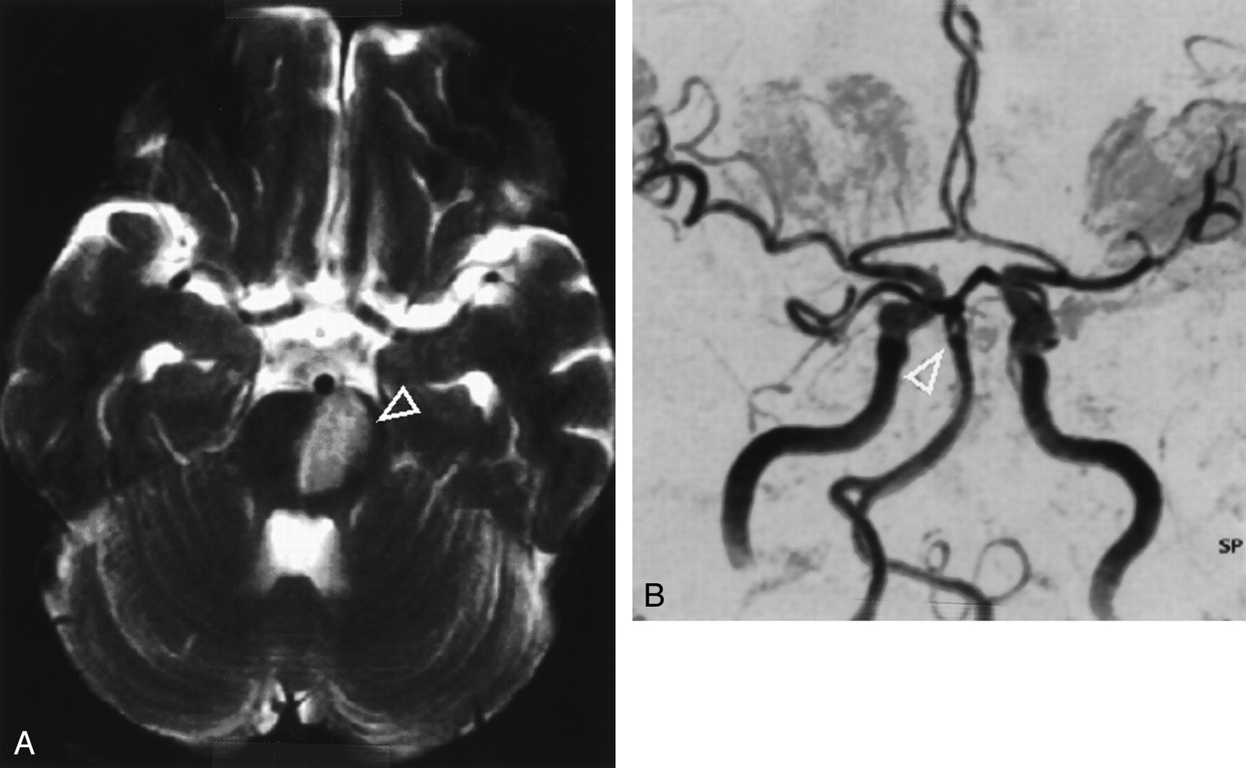
A 69-year-old man suddenly developed several episodes of dysarthria, dysphagia, and right-sided paresis 3 days before admission. At admission, he had severe dysarthria and paresis of the right side but without disturbances of gaze. The vascular risk profile included hypertension, diabetes, and hypercholesterinemia. Diffusion- and T2-weighted MR imaging performed the same day revealed high signal intensity in the left pontine area, compatible with an ischemic lesion (Fig 1A). MR angiography, using time-of-flight technique, displayed a focal signal intensity loss within the basilar artery (Fig 1B). According to these findings, diagnosis of an acute thromboembolic brain stem infarction was made that was treated with anticoagulation (heparin and thrombocyte aggregation inhibitors) (1). On the follow-up MR angiograms, the same oval signal intensity loss of the basilar artery was observed. On the basis of a virtual endoscopic look into the basilar artery (Fig 2), reconstructed from the MR angiographic data sets, the diagnosis of a small slitlike fenestration was confirmed and a thrombus was deemed unlikely. The precise illustration of the intraluminal symmetric appearance of a tiny membranous pathologic finding of the fly-through technique was especially helpful in indicating this vascular anomaly. According to the literature, two types of fenestrations of cerebral arteries exist: true duplications and arterial slits (2). Because of the incomplete central fusion shown by the virtual endoscopy, we speculated that this was not a true duplication-type fenestration but an incomplete abortive slitlike fenestration.

Fig 1.
  • Download figure
  • Open in new tab
  • Download powerpoint
Fig 1.

Images from the case of a 69-year-old man with acute pons infarction.

A, Axial T2-weighted MR image reveals hyperintense signals in the left pontine area (arrowhead). Note the normal flow void in the middle cerebral and basilar arteries.

B, Maximum intensity projection of the 3D time-of-flight MR angiogram shows a circumscript, small signal intensity loss of the basilar artery at its terminal segment (arrowhead).

Fig 2.
  • Download figure
  • Open in new tab
  • Download powerpoint
Fig 2.

Virtual endoscopy (point of view is marked by green arrowhead in small black window) of the basilar artery shows an eight-shaped lumen due to incomplete fenestration (white arrowheads). Complete virtual endoscopic fly is available at www.ajnr.org.

Discussion

Basilar artery fenestrations are the most frequently observed fenestrations of the cerebral arteries, with vertebral and middle cerebral artery fenestrations being the next most frequent. The angiographically determined incidence of basilar artery fenestrations is reported to be 0.3–0.6% (2). The clinical significance is controversial, but a basilar artery fenestration might be misinterpreted as an arterial dissection or thrombosis, especially in patients with stroke, thereby leading to a wrong decision regarding further diagnostic approaches or therapies. Fenestrations of the cerebral arteries can be multiple, and associated with cerebral aneurysm, cerebral arteriovenous malformation, or persistent trigeminal artery (3). None of these additional findings were observed in our patient. It is unlikely that the fenestration had played a role as an embolic source in our case. To date, we know of only one case report that speculates about an association between brain stem infarction and vertebral artery fenestration (4).

Virtual endoscopy combines perspective volume rendering and interactive visualization to a new approach featuring the motion of postprocessing procedures in MR imaging (5). Comparing maximum intensity projection and virtual endoscopy, we found relevant additional information regarding virtual endoscopy, such as depth information, intraluminal perspective, lighting, and color. The ability to obtain an internal view would facilitate the qualitative analyses and would thus enhance diagnostic capabilities and could improve the pretherapeutic visualization of intracranial vascular malformations. Despite these improvements, this technique is time-consuming and pitfalls exist (eg, suitable threshold values). It also does not provide more data than do available single sections, but it does allow a practicable evaluation of the complex morphology of intracranial vessels. Therefore, in our opinion, virtual endoscopy has to be applied in select cases and must be evaluated combined with the conventional data sets.

Conclusion

Basilar artery fenestrations are rare findings that should not be misinterpreted as thrombosis, even if a brain stem infarction is noted. Virtual arterial endoscopic measures may be helpful in these cases, but online reconstruction requires a high power of computation.

Acknowledgments

We gratefully acknowledge the excellent assistance of U. Junghans, MD, J. Berg, MD, and L.-W. Poll, MD.

Footnotes

  • Supported by the Bundesministerium für Bildung und Forschung (Kompetenznetzwerk Schlaganfall).

References

  1. ↵
    Junghans U, Seitz RJ, Aulich A, Freund HJ, Siebler M. Bleeding risk of GPIIb/IIIa platelet receptor antagonist tirofiban in progressive ischemic stroke. Cerebrovasc Dis 2001;12:308–312
    PubMed
  2. ↵
    Uchino A, Kato A, Abe M, Kudo S. Association of cerebral arteriovenous malformation with cerebral arterial fenestration. Eur Radiol 2001;11:493–496
    CrossRefPubMed
  3. ↵
    Uchino A, Kato A, Takase Y, Kudo S. Basilar artery fenestrations detected by MR angiography. Radiat Med 2001;19:71–74
    PubMed
  4. ↵
    Matusumoto Y, Sugino T. Infantile brainstem infarction due to vertebral artery fenestration: a case report. No Shinkei Geka 1995;23:1045–1049
    PubMed
  5. ↵
    Fellner F, Blank M, Fellner C, Bohm-Jurkovic H, Bautz W, Kalender WA . Virtual cisternoscopy of intracranial vessels: a novel visualization technique using virtual reality. Magn Reson Imaging 1998;16:1013–1022
    CrossRefPubMed
  • Received January 28, 2002.
  • Accepted after revision March 17, 2002.
  • Copyright © American Society of Neuroradiology
View Abstract
PreviousNext
Back to top

In this issue

American Journal of Neuroradiology: 23 (7)
American Journal of Neuroradiology
Vol. 23, Issue 7
1 Aug 2002
  • Table of Contents
  • Index by author
Advertisement
Print
Download PDF
Email Article

Thank you for your interest in spreading the word on American Journal of Neuroradiology.

NOTE: We only request your email address so that the person you are recommending the page to knows that you wanted them to see it, and that it is not junk mail. We do not capture any email address.

Enter multiple addresses on separate lines or separate them with commas.
Virtual Arterial Endoscopy as a Diagnostic Aid in a Patient with Basilar Artery Fenestration and Thromboembolic Pontine Infarct
(Your Name) has sent you a message from American Journal of Neuroradiology
(Your Name) thought you would like to see the American Journal of Neuroradiology web site.
CAPTCHA
This question is for testing whether or not you are a human visitor and to prevent automated spam submissions.
Cite this article
Axel Scherer, Mario Siebler, Albrecht Aulich
Virtual Arterial Endoscopy as a Diagnostic Aid in a Patient with Basilar Artery Fenestration and Thromboembolic Pontine Infarct
American Journal of Neuroradiology Aug 2002, 23 (7) 1237-1239;

Citation Manager Formats

  • BibTeX
  • Bookends
  • EasyBib
  • EndNote (tagged)
  • EndNote 8 (xml)
  • Medlars
  • Mendeley
  • Papers
  • RefWorks Tagged
  • Ref Manager
  • RIS
  • Zotero
0 Responses
Respond to this article
Share
Bookmark this article
Virtual Arterial Endoscopy as a Diagnostic Aid in a Patient with Basilar Artery Fenestration and Thromboembolic Pontine Infarct
Axel Scherer, Mario Siebler, Albrecht Aulich
American Journal of Neuroradiology Aug 2002, 23 (7) 1237-1239;
del.icio.us logo Twitter logo Facebook logo Mendeley logo
  • Tweet Widget
  • Facebook Like
  • Google Plus One

Jump to section

  • Article
    • Abstract
    • Case Report
    • Discussion
    • Conclusion
    • Acknowledgments
    • Footnotes
    • References
  • Figures & Data
  • Info & Metrics
  • Responses
  • References
  • PDF

Related Articles

  • No related articles found.
  • PubMed
  • Google Scholar

Cited By...

  • Endovascular structures of the basilar artery: forms of the basilar nonfusion spectrum
  • CTA Evaluation of Basilar Septations: An Entity Better Characterized as Aberrant Basilar Fenestrations
  • Crossref
  • Google Scholar

This article has not yet been cited by articles in journals that are participating in Crossref Cited-by Linking.

More in this TOC Section

  • Fast Contrast-Enhanced 4D MRA and 4D Flow MRI Using Constrained Reconstruction (HYPRFlow): Potential Applications for Brain Arteriovenous Malformations
  • Quiet PROPELLER MRI Techniques Match the Quality of Conventional PROPELLER Brain Imaging Techniques
  • Predictors of Reperfusion in Patients with Acute Ischemic Stroke
Show more Brain

Similar Articles

Advertisement

Indexed Content

  • Current Issue
  • Accepted Manuscripts
  • Article Preview
  • Past Issues
  • Editorials
  • Editors Choice
  • Fellow Journal Club
  • Letters to the Editor

Cases

  • Case Collection
  • Archive - Case of the Week
  • Archive - Case of the Month
  • Archive - Classic Case

Special Collections

  • Special Collections

Resources

  • News and Updates
  • Turn around Times
  • Submit a Manuscript
  • Author Policies
  • Manuscript Submission Guidelines
  • Evidence-Based Medicine Level Guide
  • Publishing Checklists
  • Graphical Abstract Preparation
  • Imaging Protocol Submission
  • Submit a Case
  • Become a Reviewer/Academy of Reviewers
  • Get Peer Review Credit from Publons

Multimedia

  • AJNR Podcast
  • AJNR SCANtastic
  • Video Articles

About Us

  • About AJNR
  • Editorial Board
  • Not an AJNR Subscriber? Join Now
  • Alerts
  • Feedback
  • Advertise with us
  • Librarian Resources
  • Permissions
  • Terms and Conditions

American Society of Neuroradiology

  • Not an ASNR Member? Join Now

© 2025 by the American Society of Neuroradiology All rights, including for text and data mining, AI training, and similar technologies, are reserved.
Print ISSN: 0195-6108 Online ISSN: 1936-959X

Powered by HighWire