
Abstract
Summary: Nine embolization experiments of a silicone aneurysm model were conducted by using helical and complex-shaped coils. Coils were introduced up to the point when the adjunct of a supplementary coil caused protrusion into the parent vessel. Packing ratios (volume of coils-aneurysm volume) ranged between 31% and 38%. Optimal packing was achieved with complex-shaped coils used in a concentric fashion. The complex-shaped coils allowed a better aneurysm filling than did helical coils.
Detachable coils are now widely used for the treatment of cerebral aneurysms. When the aneurysm is not tightly occluded, however, the coils have a propensity to gather together, being pushed and displaced toward the dome by the arterial pulsatile flow. To avoid this problem, it has been proposed that “dense packing” should be done. Recently, complex-shaped platinum coils (Complex Fill; Cordis, Miami Lakes, FL) have been introduced on the market. Our clinical experience gave us the subjective impression that the use of these complex-shaped platinum coils allows a better filling of the aneurysm sac than can be achieved by using helical platinum coils. The purpose of our experimental study was to determine whether the use of these complex-shaped platinum coils may improve the packing of cerebral aneurysms.
Description of the Technique
A silicone sidewall aneurysm model (Flowtek, Inc., San Diego, CA) was used for these experiments (dome, 7 mm; neck, 3.5 mm; parent vessel, 4.3 mm). To measure the volume of the aneurysm precisely, the model was imaged with three-dimensional (3D) rotational angiography. The aneurysm volume was determined by using dedicated software (Integris 3D-RA; Philips, Best, the Netherlands); the volume calculation was repeated five times, and we regarded the average as the definitive aneurysm volume. The aneurysm model was then connected to a circulatory system and to a pump that provided a pulsatile flow. Pressure values delivered by the pump were set to match physiologic conditions. Normal saline solution was used as circulating fluid. Under fluoroscopic guidance (angiography [Integris Allura; Philips]) a microcatheter (Prowler Plus; Cordis) was navigated to the aneurysm dome for coil delivery. Dense packing of the aneurysm was then attempted with the detachable platinum coils (Trufill Detachable Coil System; Cordis). Digital subtraction angiography was performed after each coil was introduced. The point of maximal packing density (PD) was defined as that point at which the introduction of additional coil caused slight protrusion into the parent artery (ie, the point at which more coils could not be safely placed by using the standard technique of coil embolization). We performed nine embolization experiments with three different scenarios of dense packing in our aneurysm model: three experiments were conducted with the use of complex-shaped coils only (scenario 1); three with helical-shaped coils only (scenario 2); and, finally, three with a mix of complex and helical-shaped coils (scenario 3). As we routinely do in the clinical setting, we introduced first the larger and longer coils to create a basket in which smaller coils were subsequently positioned to obtain the best aneurysm filling. The calculation of the total volume of the coils introduced into each aneurysm was based on the diameter and length of the coils that were introduced in the aneurysm. The volumetric ratios of maximal PD (total volume of introduced coils-volume of the aneurysm) regarding the actual aneurysm volume were then calculated. Volumetric ratios from the embolization experiments are shown in the Table. The nine coiling experiments resulted in maximal PD ranging between 31% and 38%. The results of the three different packing scenarios were compared for statistical significance by a nonparametric test (Kruskal-Wallis). Key points for optimal packing were the use of complex-shaped coils to create a basket and then the use of smaller complex-shaped coils inside the previously deposited coils (scenario 1), which enabled the concentric filling of the aneurysmal sac (Fig 1). The three experiments of scenario 1 did not provide statistically significantly (P .05) greater PD than did those of the three experiments of scenario 2, even if the results showed a trend toward greater PD with complex-shaped coils.

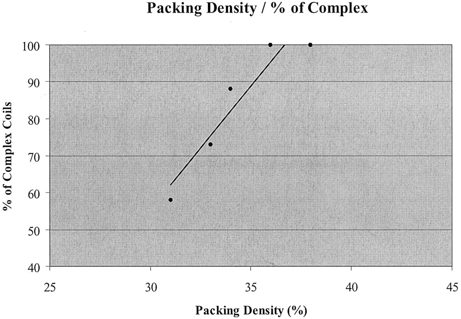
Packing density and percentage of complex-shaped coils. The linear curve of tendency shows that the use of complex-shaped coils tends to improve the filling of the aneurysm.
Results of the nine experiments
Complex (scenario 1) versus Helical coils (scenerio 2)
Discussion
The goal of endovascular treatment of cerebral aneurysms is to obtain a complete, stable exclusion of the sac from arterial circulation with preservation of the parent vessel. Ideally, aneurysm thrombosis followed by endothelialization across the aneurysm orifice should be obtained. Coils have now been used extensively to treat ruptured and unruptured aneurysms. Until now, the mechanism of aneurysm occlusion with platinum or tungsten coils has been mechanical, and increasing clinical experience has shown that there is a positive relationship between packing density and long-term occlusion rate (1). The presence of a residual neck in the aneurysm inflow zone precludes complete aneurysm occlusion in most of the cases. When an aneurysm is embolized incompletely with coils, there is a potential risk of aneurysm recanalization. Residual arterial flow in the aneurysm prevents endothelial cell proliferation across the neck of the aneurysm and anatomic isolation of the aneurysm from the blood circulation. Many undergoing investigations are focusing on the development of biologically “active” coils, which should promote the organization of the initial thrombus that forms during or just after coil deposition and the use of liquid polymer (2–5). The few clinical reports concerning early and late histopathologic findings after endovascular therapy with coils in human have shown that there is a lack of permanent thrombus organization (6–8). Although an aneurysm packed with coils may appear attenuated radiographically, experimental studies have indicated that a significant part of the sac becomes acutely occluded with thrombus after coiling, but this clot has no permanency in many cases, exposing the sac to the possibility of recanalization and coil compaction (9).
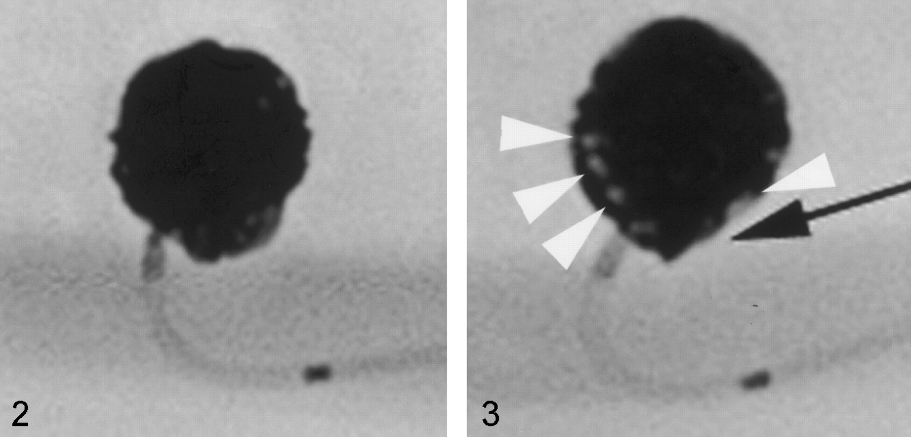
Complex-shaped or 3D coils have been introduced recently on the market to treat wide-necked aneurysms or aneurysms having an unfavorable neck-fundus ratio, which are known to be difficult to embolize with helical coils without the use of balloon remodeling or other supplemental methods (10–12). Until now, the potential usefulness of complex-shaped coils to gain a better packing of the aneurysm sac has not been emphasized. They have a propensity to form a 3D cage after deployment, and their conformability is superior to that of helical coils with subsequently less compartmentalization, allowing more homogeneous aneurysm filling (Figs 2 and 3); however, our experimental work had some limitations. Although most aneurysms are located at arterial bifurcations and our model was a sidewall aneurysm, we felt it was satisfactory for the basic purpose of this study. The conditions, however, did not reflect those present in clinical situations. For instance, the circulating saline fluid was not as viscous and did not clot as blood. In vivo endosaccular thrombosis, even if temporary, occurs with detachable coils and means that fewer coils are required to achieve maximal PD. Volumetric ratios of in vivo aneurysm packing would therefore be expected to be lower, so the volumetric ratios here were perhaps not entirely realistic. On the other hand, the smallest available diameter for the Cordis complex-shaped coils is 4 mm; it could be argued that the use of 3- or 2-mm-diameter complex-shaped coils would have allowed us to obtain a higher PD.

Aneurysm model (nonsubtracted lateral view) at the end of the first experiment (scenario 1) showing that both the sac and the neck are densely packed (packing attenuation, 38%).

Aneurysm model (nonsubtracted lateral view) at the end of the fifth experiment (scenario 2) showing that both the sac (interstices inside the coil mesh, white arrowheads) and the neck (black arrow) are less packed than on Fig 1 (packing attenuation, 32%).
Conclusion
Our experimental results confirmed our sense that the use of complex-shaped platinum coils shows a trend, even if not statistically significant, that allows better filling of aneurysm sac than helical platinum coils. The best packing was obtained by forming a basket with a complex-shaped coil that was subsequently filled concentrically with smaller complex-shaped coils in an “onionlike” fashion. Nevertheless, because the highest packing density obtained was 38%, much space is still left within the sac after coiling a sidewall aneurysm as completely as possible.
Addendum
We later performed seven additional experiments with complex-shaped coils only (scenario 1) and seven other experiments with helical coils only (scenario 2). The resulting 10 coil experiments of the scenario 1 resulted in a mean packing density of 37%, whereas the 10 coil experiments of the scenario 2 resulted in a mean packing density of 34.5% (see chart). The results of the two different packing scenarios (scenario 1 and scenario 2) were compared for statistical significance by a non-parametric Mann-Whitney U test. Scenario 1 provided statistically significant (P =.015) greater packing density than that scenario 2. These new results confirm the trend toward greater packing density that can be achieved with complex-shaped coils.
References
- Received January 24, 2003.
- Accepted after revision March 16, 2003.
- Copyright © American Society of Neuroradiology
In this issue


Jump to section
Related Articles
Cited By...
- In vitro measurement of the permeability of endovascular coils deployed in cerebral aneurysms
- Optimal first coil selection to avoid aneurysmal recanalization in endovascular intracranial aneurysmal coiling
- Three-dimensional printing of anatomically accurate, patient specific intracranial aneurysm models
- Aneurysm permeability following coil embolization: packing density and coil distribution
- Initial experience with Penumbra Coil 400 versus standard coils in embolization of cerebral aneurysms: a retrospective review
- Complex shaped detachable platinum coil system for the treatment of cerebral aneurysms: The Codman Trufill DCS and Trufill DCS Orbit Detachable Coil System COMPLEX Registry final results
- Analysis and quantification of endovascular coil distribution inside saccular aneurysms using histological images
- The impact of coil shape design on angiographic occlusion, packing density and coil mass uniformity in aneurysm embolization: an in vitro study
- Cerecyte versus Platinum Coils in the Treatment of Intracranial Aneurysms: Packing Attenuation and Clinical and Angiographic Midterm Results
- Brain Aneurysms and Arteriovenous Malformations: Advancements and Emerging Treatments in Endovascular Embolization