
Article Figures & Data
Figures
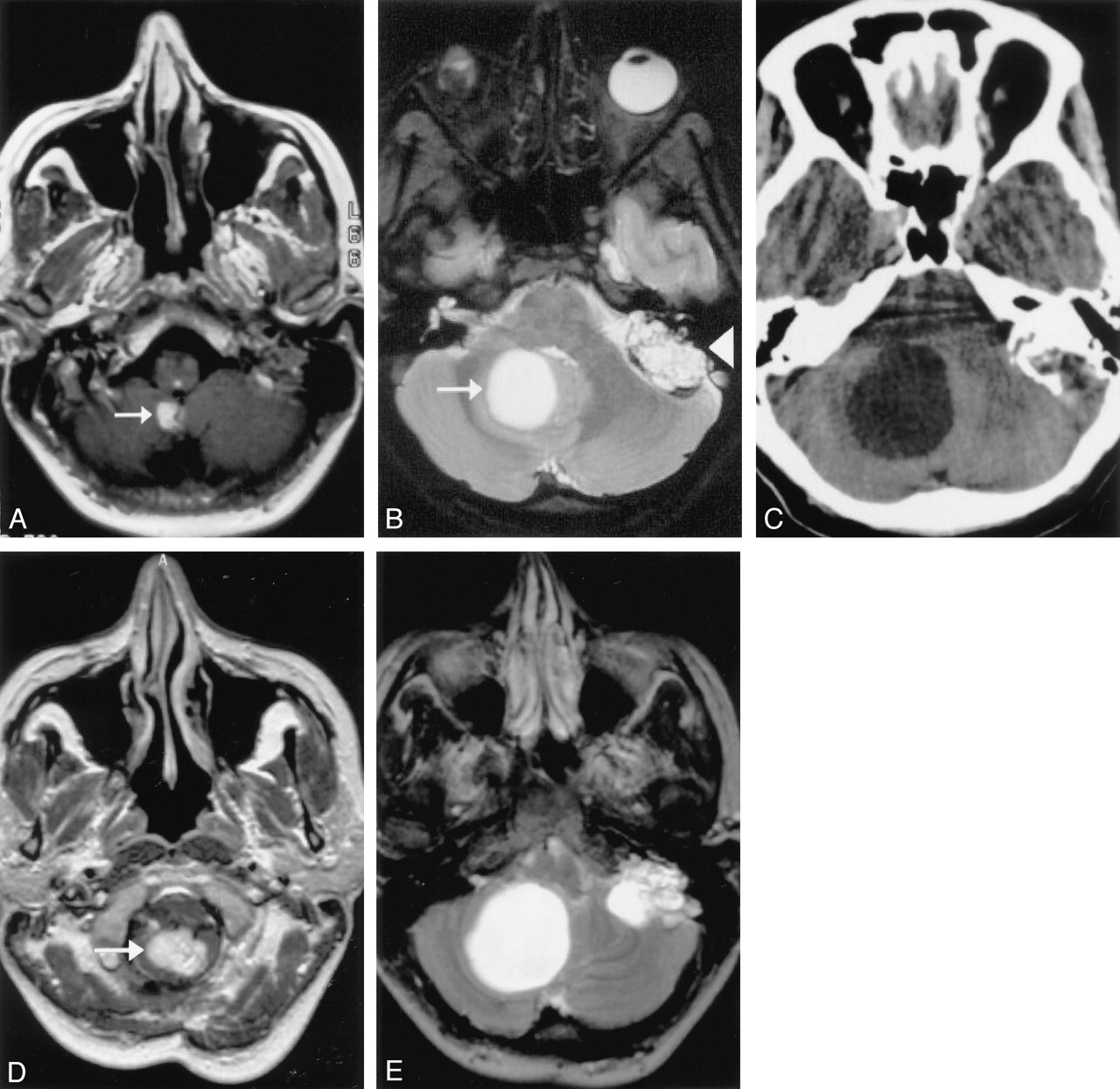
- Fig 1.
Images in a patient with a solid cerebellar tumor that progressed to enlarging cysts with the subsequent development of symptoms.
A, Contrast-enhanced T1-weighted MR image obtained at presentation demonstrates a 12-mm, enhancing, solid tumor in the right cerebellar hemisphere (arrow) (720/13 [TR/TE]).
B, Axial short-tau inversion recovery (STIR) image obtained 17 months later shows a large cyst associated with this solid tumor (arrow). An incidental left endolymphatic-sac tumor is also depicted on this image (arrowhead). (5660/29.7; TI, 150)
C, CT image shows further enlargement of the cyst 21 months after the initial study. The patient experienced posterior fossa symptoms at this time but declined surgical excision of this tumor, instead opting for craniotomy and cyst aspiration. (943/17)
D and E, MR images obtained 29 months after the initial examination. Contrast-enhanced axial T1-weighted image (D) (943/17) demonstrates enlargement of the solid part of the tumor (arrow). T2-weighted image (E) (4400/96) shows that the cyst has recurred, as had the patient’s symptoms. Surgical excision was performed shortly after this image was obtained.
- Fig 2.
Images in second patient with a solid cerebellar tumors that progressed to enlarging cysts with the subsequent development of symptoms.
A, Contrast-enhanced T1-weighted MR image shows an enhancing nodule in the right cerebellar hemisphere (arrow). (500/11 [TR/TE])
B, Image obtained 15 months later shows a large cyst associated with this nodule. Cerebellar symptoms necessitated surgical excision of this tumor. (460/13)
C, Follow-up contrast-enhanced MR image obtained 44 months after the initial study shows a new, small, enhancing nodule (arrow) and postsurgical change. (360/13)
D, Three years later, another contrast-enhanced MR image (D) shows that this nodule has increased in size. Also shown are two new, smaller nodules. (360/13)
E, Two years later, a STIR image reveals that a large cyst is now associated with this tumor. The patient experienced additional cerebellar symptoms, and further surgery was performed to remove this second tumor. (4400/96)
- Fig 3.
Images in a third patient with a solid cerebellar tumor that progressed to enlarging cyst with the subsequent development of symptoms.
A, Contrast-enhanced T1-weighted MR image obtained at presentation shows a tiny enhancing nodule in the left cerebellar hemisphere (arrow). (400/8 [TR/TE])
B, Image acquired 19 months later demonstrates a tiny cyst adjacent to this tumor. (300/9)
C, A T2-weighted image obtained 24 months after the initial study shows that the cyst has enlarged. It continued to increase in size, as demonstrated on a CT scan (not shown) obtained 3 months later. Shortly after this examination, surgical excision was performed. (3720/91.5)
- Fig 4.
Graph of cyst sizes versus time.


Tables
- TABLE 1:
Sizes of Nodules and Cysts at Presentation and at Final Follow-up Imaging Before Surgery
Size, mm Case A Case B, First Tumor Case B, Second Tumor Case C Case D Case E Case F, First Tumor Case F, Second Tumor Nodule at presentation 12 10 3 10 5 5 10 10 Cyst at presentation 0 0 0 0 0 15 40 0 Nodule at follow-up 22 12 12 16 8 5 10 15 Cyst at follow-up 40 45 35 34 22 20 50 5 Note.—The time intervals (months) to the last imaging study before surgery were as follows: case A, 39; case B (first tumor), 15; case B (second tumor), 58; case C, 26; case D, 12; case E, 30; and case F (first tumor), 13. The second tumor in case F was not excised.
Involvement Patient (No.) Renal cancer 3 Renal cysts 7 Pancreatic tumors 5 Pancreatic cysts 5 Pheochromocytoma 1


