Advertisement

Main menu

  • Home
  • Content
    • Current Issue
    • Accepted Manuscripts
    • Article Preview
    • Past Issue Archive
    • Video Articles
    • AJNR Case Collection
    • Case of the Week Archive
    • Case of the Month Archive
    • Classic Case Archive
  • Special Collections
    • AJNR Awards
    • Low-Field MRI
    • Alzheimer Disease
    • ASNR Foundation Special Collection
    • Photon-Counting CT
    • View All
  • Multimedia
    • AJNR Podcasts
    • AJNR SCANtastic
    • Trainee Corner
    • MRI Safety Corner
    • Imaging Protocols
  • For Authors
    • Submit a Manuscript
    • Submit a Video Article
    • Submit an eLetter to the Editor/Response
    • Manuscript Submission Guidelines
    • Statistical Tips
    • Fast Publishing of Accepted Manuscripts
    • Graphical Abstract Preparation
    • Imaging Protocol Submission
    • Author Policies
  • About Us
    • About AJNR
    • Editorial Board
    • Editorial Board Alumni
  • More
    • Become a Reviewer/Academy of Reviewers
    • Subscribers
    • Permissions
    • Alerts
    • Feedback
    • Advertisers
    • ASNR Home

User menu

  • Alerts
  • Log in

Search

  • Advanced search
American Journal of Neuroradiology

American Journal of Neuroradiology

ASHNR American Society of Functional Neuroradiology ASHNR American Society of Pediatric Neuroradiology ASSR
  • Alerts
  • Log in

Advanced Search

  • Home
  • Content
    • Current Issue
    • Accepted Manuscripts
    • Article Preview
    • Past Issue Archive
    • Video Articles
    • AJNR Case Collection
    • Case of the Week Archive
    • Case of the Month Archive
    • Classic Case Archive
  • Special Collections
    • AJNR Awards
    • Low-Field MRI
    • Alzheimer Disease
    • ASNR Foundation Special Collection
    • Photon-Counting CT
    • View All
  • Multimedia
    • AJNR Podcasts
    • AJNR SCANtastic
    • Trainee Corner
    • MRI Safety Corner
    • Imaging Protocols
  • For Authors
    • Submit a Manuscript
    • Submit a Video Article
    • Submit an eLetter to the Editor/Response
    • Manuscript Submission Guidelines
    • Statistical Tips
    • Fast Publishing of Accepted Manuscripts
    • Graphical Abstract Preparation
    • Imaging Protocol Submission
    • Author Policies
  • About Us
    • About AJNR
    • Editorial Board
    • Editorial Board Alumni
  • More
    • Become a Reviewer/Academy of Reviewers
    • Subscribers
    • Permissions
    • Alerts
    • Feedback
    • Advertisers
    • ASNR Home
  • Follow AJNR on Twitter
  • Visit AJNR on Facebook
  • Follow AJNR on Instagram
  • Join AJNR on LinkedIn
  • RSS Feeds

AJNR Awards, New Junior Editors, and more. Read the latest AJNR updates

Research ArticlePediatric Neuroimaging

MR Imaging and Proton MR Spectroscopic Studies in Sjögren-Larsson Syndrome: Characterization of the Leukoencephalopathy

Michèl A. A. P. Willemsen, Marinette van der Graaf, Marjo S. van der Knaap, Arend Heerschap, Peter H. M. F. van Domburg, Fons J. M. Gabreëls and Jan J. Rotteveel
American Journal of Neuroradiology April 2004, 25 (4) 649-657;
Michèl A. A. P. Willemsen
  • Find this author on Google Scholar
  • Find this author on PubMed
  • Search for this author on this site
Marinette van der Graaf
  • Find this author on Google Scholar
  • Find this author on PubMed
  • Search for this author on this site
Marjo S. van der Knaap
  • Find this author on Google Scholar
  • Find this author on PubMed
  • Search for this author on this site
Arend Heerschap
  • Find this author on Google Scholar
  • Find this author on PubMed
  • Search for this author on this site
Peter H. M. F. van Domburg
  • Find this author on Google Scholar
  • Find this author on PubMed
  • Search for this author on this site
Fons J. M. Gabreëls
  • Find this author on Google Scholar
  • Find this author on PubMed
  • Search for this author on this site
Jan J. Rotteveel
  • Find this author on Google Scholar
  • Find this author on PubMed
  • Search for this author on this site
  • Article
  • Figures & Data
  • Info & Metrics
  • Responses
  • References
  • PDF
Loading

Article Figures & Data

Figures

  • Tables
  • Fig 1.
    • Download figure
    • Open in new tab
    • Download powerpoint
    Fig 1.

    FALDH catalyzes the oxidation of long-chain fatty aldehydes (here, octadecanal) to the corresponding carboxylic acid.
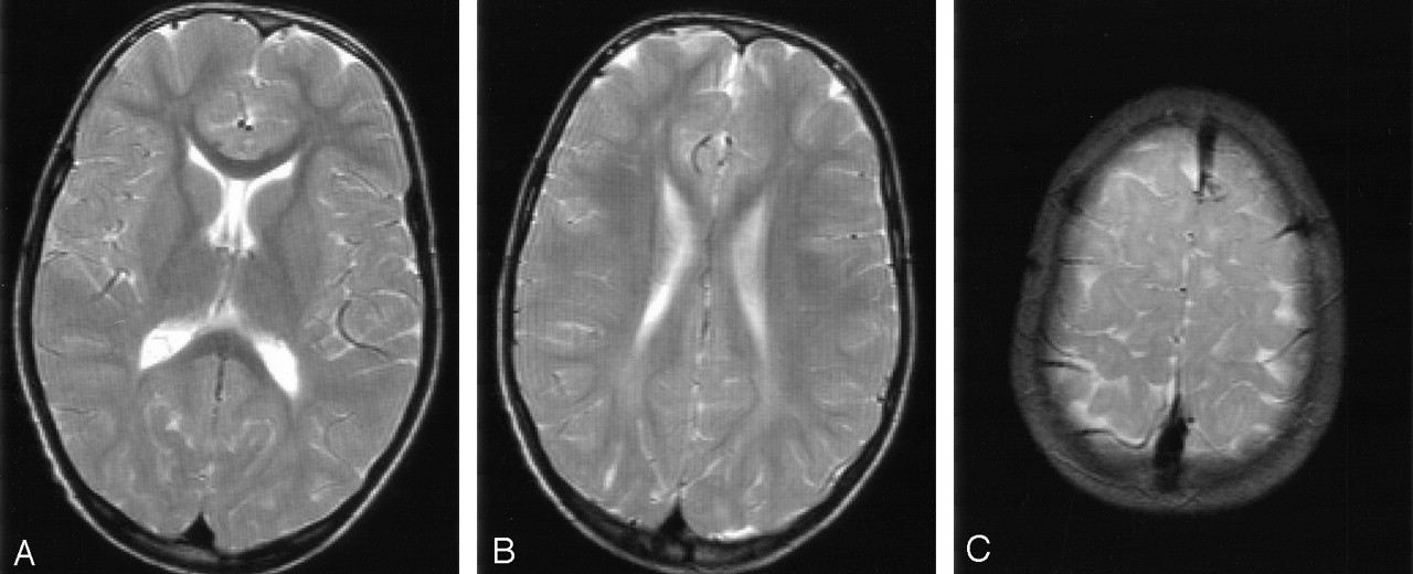
  • Fig 2.
    • Download figure
    • Open in new tab
    • Download powerpoint
    Fig 2.

    Patient 13 at 16 years of age. T2-weighted MR images (3100/98 [TR/TE]).

    A and B, Severe signal-intensity changes of the periventricular white matter with predominant involvement of the frontal trigones.

    C, Small areas of unmyelinated subcortical association fibers.

  • Fig 3.
    • Download figure
    • Open in new tab
    • Download powerpoint
    Fig 3.

    Patient 6 at 9 years of age. T2-weighted MR images (3100/98 [TR/TE]).

    A and B, Mild signal-intensity changes of the periventricular white matter with predominant involvement of the occipital trigones.

    C, Small areas of unmyelinated subcortical association fibers.

  • Fig 4.
    • Download figure
    • Open in new tab
    • Download powerpoint
    Fig 4.

    Patient 2. T2-weighted MR images (3100/98 [TR/TE]). There is a delay in the maturation of the white matter on all three images.

    A, At 5 months of age, the unmyelinated periventricular white matter shows no abnormal signal intensities.

    B and C, Images obtained at 16 (B) and 35 (C) months of age show nonprogressive, slight signal-intensity abnormalities in the periventricular white matter that mainly involve the occipital trigones.

  • Fig 5.
    • Download figure
    • Open in new tab
    • Download powerpoint
    Fig 5.

    Patient 3 at 5 years of age.

    A, Image shows voxel locations in the occipital trigone (box A) and in the central occipital gray matter (box B).

    B, Proton MR spectra (TE = 20 msec) obtained from cerebral white matter (spectrum A) and gray matter (spectrum B). Note the presence of the high, sharp lipid peak at 1.3 ppm and a small peak at 0.8–0.9 ppm in the spectrum obtained from the white matter.

  • Fig 6.
    • Download figure
    • Open in new tab
    • Download powerpoint
    Fig 6.

    Serial proton MR spectra (TE = 20 msec) from cerebral white matter of patients 1 and 2 demonstrate a gradual emergence of the lipid peak at 1.3 ppm during the first years of life.

  • Fig 7.
    • Download figure
    • Open in new tab
    • Download powerpoint
    Fig 7.

    Metabolite map derived from MRSI data of patient 13 shows the spatial distribution of the lipid peak over the cerebral white matter. The peak has its maximum height around the anterior and posterior trigones.

Tables

  • Figures
    • View popup
    TABLE 1:

    Results of cerebral MR imaging studies in 18 patients with SLS

    PatientSexNo. of StudiesAge Range, yearMyelin DeficitPeriventricular Zone of Abnormal Signal IntensityAtrophy
    DegreePredominance
    1F30.8–111PO*†0
    2M50.4–411PO0
    3M23–511PO0
    4M31–811PO0
    5M1811PO0
    6M25–911PO0
    7F26–911PO0
    8M28–912FP*0
    9F73–1011PO0
    10F11011FP*1
    11F11311PO†0
    12F311–1411PO0
    13M412–1612FP1
    14F312–1711PO1
    15F315–1712FP*†0
    16F316–2111PO1
    17M13801PO1
    18F241–4511PO1
    • Note.—The results reflect findings from each patient’s latest MR imaging study. The degree of myelin deficit, periventricular signal-intensity abnormalities, and atrophy were scored as follows: 0 = none, 1 = mild, 2 = severe. PO = parieto-occipital, FP = frontoparietal.

    • * Corpus callosum involved.

    • † Patchy white matter lesions.

    • View popup
    TABLE 2:

    Results of proton MR spectroscopy in parieto-occipital white matter and gray matter

    A: Patients’ last investigations
    PatientNo. of StudiesAge Range, yearWhite Matter Lipid Peak, auWhite Matter Metabolites, mmol/L
    t-NAACrChoInsGlx
    130.8–15.79.54.92.33.011.6
    250.4–43.09.54.71.54.69.8
    323–527.811.15.42.48.110.5
    41823.411.16.02.15.313.6
    51811.28.54.82.04.08.6
    625–915.510.16.11.85.113.4
    726–91.911.36.12.15.811.3
    81921.39.35.82.46.610.6
    947–1011.511.45.21.94.014.3
    10110Present—————
    1111311.29.44.71.85.98.7
    12311–1413.69.96.32.16.013.0
    13412–1619.811.96.02.06.310.2
    14217–1720.310.56.61.86.716.0
    15315–178.612.76.81.95.415.3
    16221–216.911.07.62.45.514.5
    181451.68.96.01.83.69.8
    B: Group means and standard deviations
    GroupLipid Peak, auMetabolites, mmol/L
    t-NAACrChoInsGlx
    White matter
     Patients (n = 16)12.7 (8.1)*10.4 (1.2)5.8 (0.8)†2.0 (0.3)‡5.4 (1.3)§12.0 (2.4)
     Control subjects (n = 9)0.6 (0.6)9.6 (0.9)5.1 (0.3)1.7 (0.3)3.5 (1.0)12.4 (0.9)
    Gray matter
     Patients (n = 15)0.9 (0.8)11.2 (1.6)7.1 (1.0)1.5 (0.3)3.9 (1.6)17.0 (2.7)
     Control subjects (n = 9)1.1 (0.8)11.1 (0.5)6.5 (0.5)1.4 (0.1)4.3 (0.8)15.6 (2.0)
    • Note.—au = arbitrary units, t-NAA = total NAA (N-acetylaspartate and N-acetylaspartylglutamate), Cr = creatine and creatine phosphate, Cho = free choline and the choline-containing compounds phosphocholine and glycerophosphorylcholine, Ins = myo-inositol, Glx = glutamine and glutamate, dashes = no data available. P values are given when P < .05 for SLS patients compared with healthy control subjects (unpaired two-tailed t test).

    • * P = .0002.

    • † P = .02.

    • ‡ P = .005.

    • § P = .001.

PreviousNext
Back to top

In this issue

American Journal of Neuroradiology: 25 (4)
American Journal of Neuroradiology
Vol. 25, Issue 4
1 Apr 2004
  • Table of Contents
  • Index by author
Advertisement
Print
Download PDF
Email Article

Thank you for your interest in spreading the word on American Journal of Neuroradiology.

NOTE: We only request your email address so that the person you are recommending the page to knows that you wanted them to see it, and that it is not junk mail. We do not capture any email address.

Enter multiple addresses on separate lines or separate them with commas.
MR Imaging and Proton MR Spectroscopic Studies in Sjögren-Larsson Syndrome: Characterization of the Leukoencephalopathy
(Your Name) has sent you a message from American Journal of Neuroradiology
(Your Name) thought you would like to see the American Journal of Neuroradiology web site.
CAPTCHA
This question is for testing whether or not you are a human visitor and to prevent automated spam submissions.
Cite this article
Michèl A. A. P. Willemsen, Marinette van der Graaf, Marjo S. van der Knaap, Arend Heerschap, Peter H. M. F. van Domburg, Fons J. M. Gabreëls, Jan J. Rotteveel
MR Imaging and Proton MR Spectroscopic Studies in Sjögren-Larsson Syndrome: Characterization of the Leukoencephalopathy
American Journal of Neuroradiology Apr 2004, 25 (4) 649-657;

Citation Manager Formats

  • BibTeX
  • Bookends
  • EasyBib
  • EndNote (tagged)
  • EndNote 8 (xml)
  • Medlars
  • Mendeley
  • Papers
  • RefWorks Tagged
  • Ref Manager
  • RIS
  • Zotero
0 Responses
Respond to this article
Share
Bookmark this article
MR Imaging and Proton MR Spectroscopic Studies in Sjögren-Larsson Syndrome: Characterization of the Leukoencephalopathy
Michèl A. A. P. Willemsen, Marinette van der Graaf, Marjo S. van der Knaap, Arend Heerschap, Peter H. M. F. van Domburg, Fons J. M. Gabreëls, Jan J. Rotteveel
American Journal of Neuroradiology Apr 2004, 25 (4) 649-657;
del.icio.us logo Twitter logo Facebook logo Mendeley logo
  • Tweet Widget
  • Facebook Like
  • Google Plus One

Jump to section

  • Article
    • Abstract
    • Methods
    • Results
    • Discussion
    • Conclusions
    • Acknowledgments
    • References
  • Figures & Data
  • Info & Metrics
  • Responses
  • References
  • PDF

Related Articles

  • No related articles found.
  • PubMed
  • Google Scholar

Cited By...

  • Child Neurology: Sjo&#x0308;gren-Larsson syndrome
  • Sjo&#x0308;gren-Larsson syndrome: a rare disease of the skin and central nervous system
  • Magnetic resonance spectroscopy of the brain
  • Lipid metabolism in myelinating glial cells: lessons from human inherited disorders and mouse models
  • MR Spectroscopy Detects Lipid Peaks in Cerebrotendinous Xanthomatosis
  • Crossref
  • Google Scholar

This article has not yet been cited by articles in journals that are participating in Crossref Cited-by Linking.

More in this TOC Section

  • FRACTURE MR in Congenital Vertebral Anomalies
  • Comparing MRI Perfusion in Pediatric Brain Tumors
  • Sodium MRI in Pediatric Brain Tumors
Show more Pediatric Neuroimaging

Similar Articles

Advertisement

Indexed Content

  • Current Issue
  • Accepted Manuscripts
  • Article Preview
  • Past Issues
  • Editorials
  • Editor's Choice
  • Fellows' Journal Club
  • Letters to the Editor
  • Video Articles

Cases

  • Case Collection
  • Archive - Case of the Week
  • Archive - Case of the Month
  • Archive - Classic Case

More from AJNR

  • Trainee Corner
  • Imaging Protocols
  • MRI Safety Corner
  • Book Reviews

Multimedia

  • AJNR Podcasts
  • AJNR Scantastics

Resources

  • Turnaround Time
  • Submit a Manuscript
  • Submit a Video Article
  • Submit an eLetter to the Editor/Response
  • Manuscript Submission Guidelines
  • Statistical Tips
  • Fast Publishing of Accepted Manuscripts
  • Graphical Abstract Preparation
  • Imaging Protocol Submission
  • Evidence-Based Medicine Level Guide
  • Publishing Checklists
  • Author Policies
  • Become a Reviewer/Academy of Reviewers
  • News and Updates

About Us

  • About AJNR
  • Editorial Board
  • Editorial Board Alumni
  • Alerts
  • Permissions
  • Not an AJNR Subscriber? Join Now
  • Advertise with Us
  • Librarian Resources
  • Feedback
  • Terms and Conditions
  • AJNR Editorial Board Alumni

American Society of Neuroradiology

  • Not an ASNR Member? Join Now

© 2025 by the American Society of Neuroradiology All rights, including for text and data mining, AI training, and similar technologies, are reserved.
Print ISSN: 0195-6108 Online ISSN: 1936-959X

Powered by HighWire