
Article Figures & Data
Figures
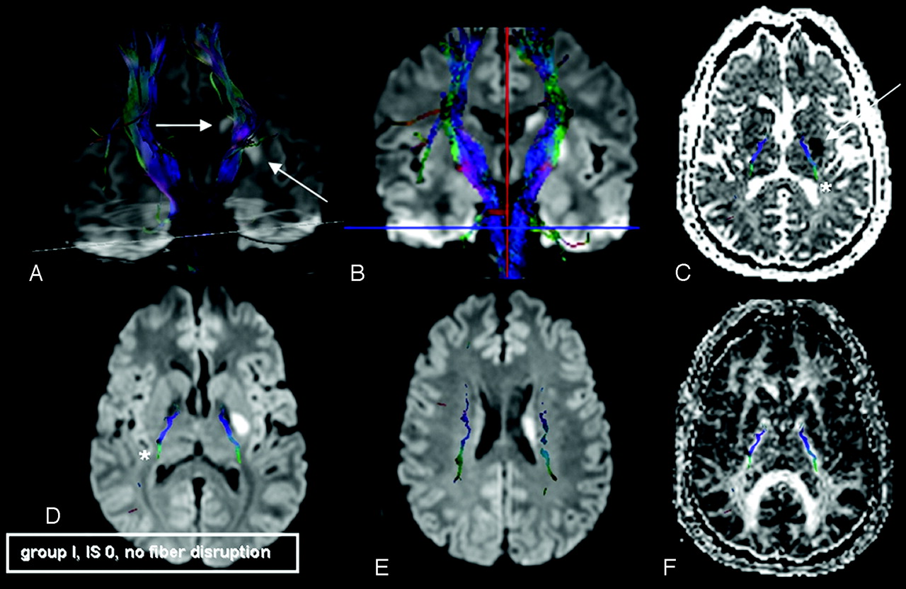
- Fig 1.
Sample patient, group I (favorable outcome), scanned on day 3 after symptom onset. IS indicates involvement scale. Stroke onset zones are marked with arrows. A, 3D volume rendering (50% opacity) of DWI and superimposed bilateral CSTs. B, FiberTracking coregistered with coronal 2D DWI sections. C, Axial ADC map; D and E, axial DWI; F, axial FA map coregistered with 2D overlay of CST fibers (*, C and D). In this case, the ischemic lesion is lateral of the CST to its major extent; there is no CST involvement (IS = 0) and no fiber disruption. This 46-year-old man suffered from dysarthria and decreased fine motor skills of his right hand at the time of admittance. He was hospitalized for 5 days and symptom free in re-evaluation after 3 months.
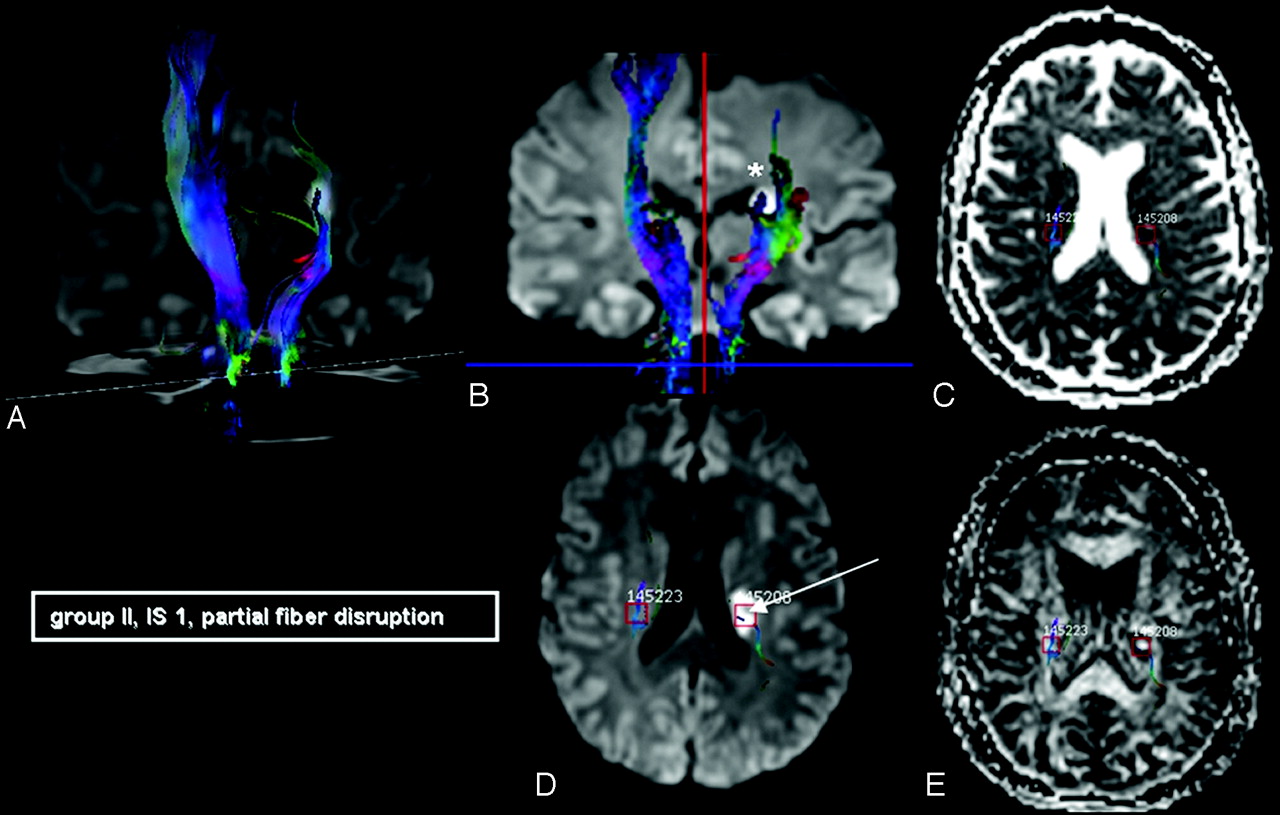
- Fig 2.
AchoA stroke with partial CST involvement (IS = 1, arrow) and partial fiber disruption (*). IS indicates involvement scale. A 56-year-old woman scanned on day 5 after symptom onset (initially with dysarthria and weakness of the cranial nerve VII [facial] buccal branch) who developed hemiparesis of the right arm and leg during her 10-day hospital course. After 3 months, she still had a lower extremity MS of 4 (“moderate” outcome, group II).
- Fig 3.
A 61-year-old man with left-sided hemiparesis (MS 4+) at hospital admittance. Paresis was progressive in the course of his 12-day stay (“unfavorable” outcome, group III). He was examined with DTI on the third day after symptom onset and had a complete CST disruption in DTT (open arrow).
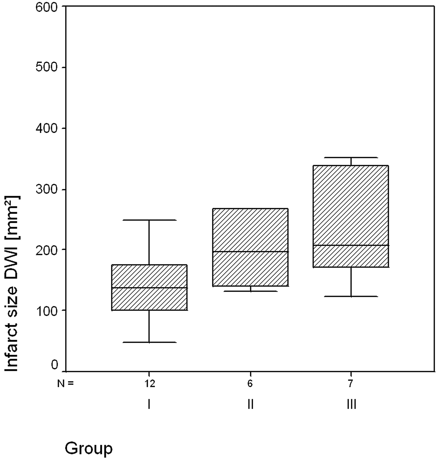
- Fig 4.
Median AchoA infarct sizes (millimeters squared) for patient subgroups, measured at the level of the largest stroke extents, resided closely together. There were no statistically significant differences between stroke dimensions of patients with more favorable versus patients with unfavorable functional outcome.
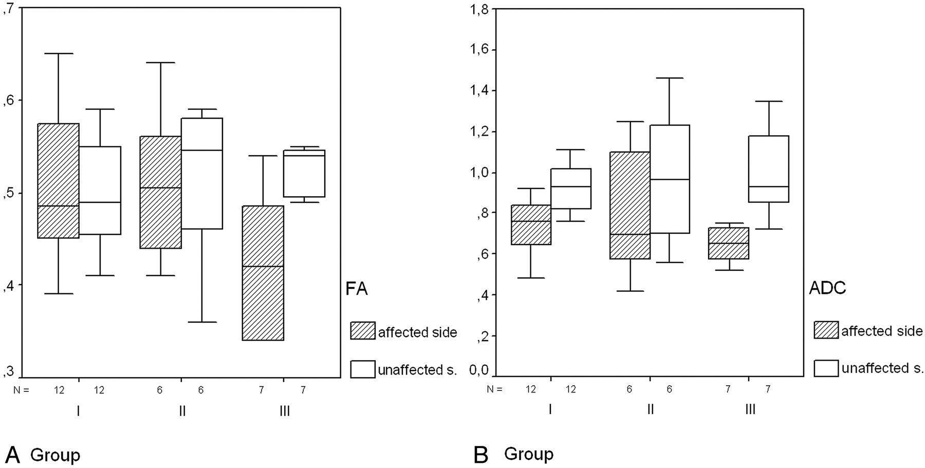
- Fig 5.
Whisker plots of FA (A) and ADCs (B) illustrate that there is higher FA asymmetry (P = .03) between the affected and unaffected sides in AchoA stroke in patients with unfavorable outcome (III). B, ADC is reduced in all of the cases with no significant difference between single subgroups (see text).
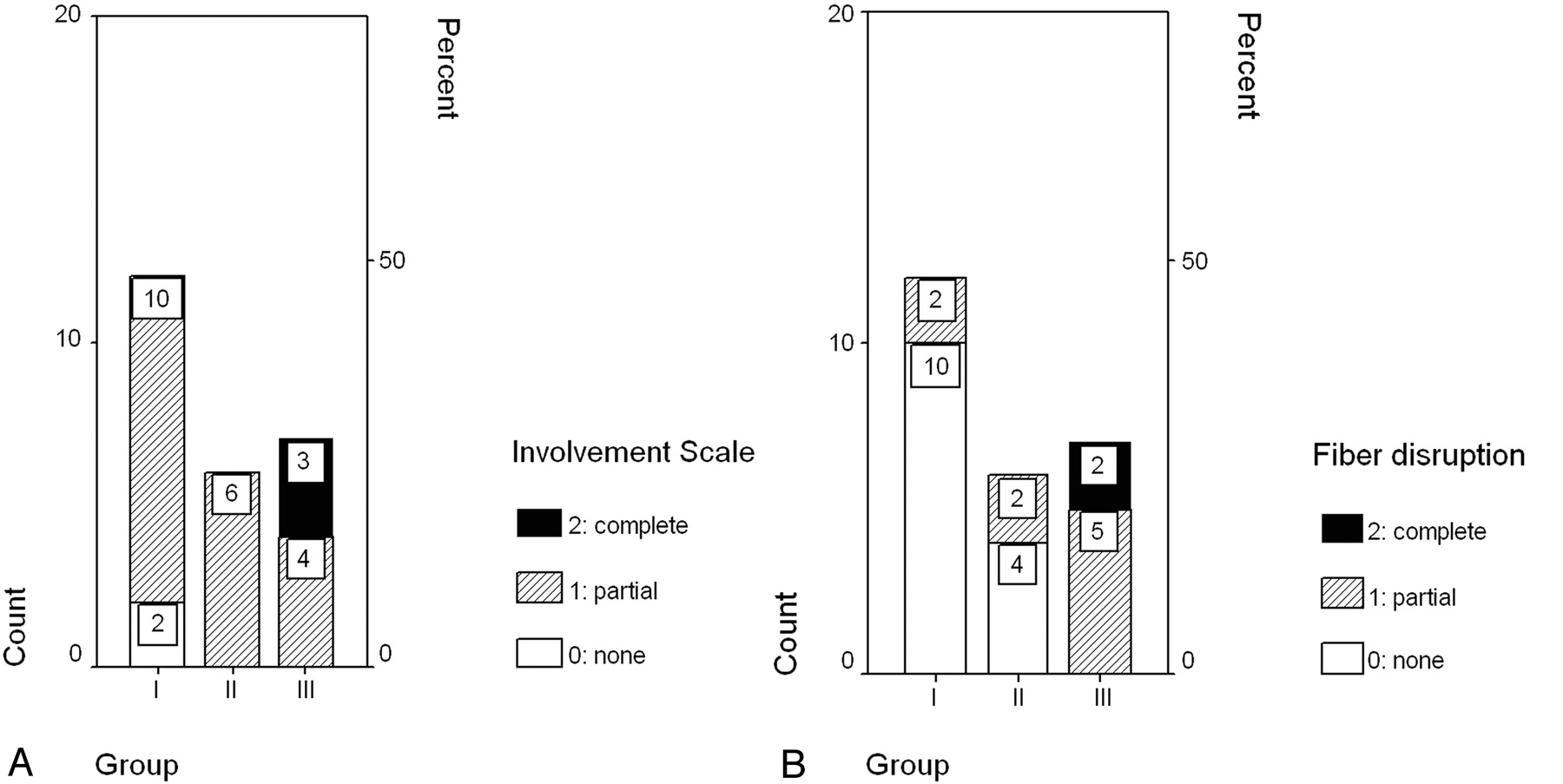
- Fig 6.
Case numbers of different subgroups regarding complete/partial/no CST involvement (A) or CST fiber disruption (B). There is complete CST involvement and complete fiber disruption in group III only.


Tables
Group Outcome n Mean Infarct Size, mm2 Fiber Ratio I, “favorable”: MS=5 (maximum of 1 deficit) 12 139.42 ± 57.77 1.14 II, “moderate”: MS ≥ 4− (and maximum of 2 deficits) 6 248.00 ± 161.04 0.55 III, “unfavorable”: MS ≤ 3 (and further deficits) 7 243.43 ± 98.02 1.05 Note:—Inclusion criteria for different subgroups (long-term outcome) are detailed in the first column. Group I had a motor score (MS) of 5 and a maximum of 1 neurologic deficit. Group II had at most 2 deficits and a minimum score of 4−. Group III had an MS ≤ 3 and further deficits. Fiber ratios were smaller for patients with lower MS, though confounders with higher amounts of ipsilateral voxels, partly due to high fractional anisotropy in acute stroke phases, were present in 3 of 7 cases in group III.
FA/ADC Groups I and II FA Group III FA Groups I and II ADC Group III ADC n 18 7 18 7 Mean 0.51 0.42 0.75 0.63 SD 0.08 0.08 0.20 0.24 P(ttest) .03 >.2 Note:—FA indicates fractional anisotropy; ADC, apparent diffusion coefficient. FA of patients with unfavorable outcome was significantly lower, whereas all of the subjects exhibited reduced ADC without significant differences between groups.
In this issue


Jump to section
Related Articles
Cited By...
- Geniculocalcarine Tract Disintegration after Ischemic Stroke: A Diffusion Tensor Imaging Study
- Anterior choroidal artery ischaemic patterns predict outcome of carotid occlusion
- Acute Damage to the Posterior Limb of the Internal Capsule on Diffusion Tensor Tractography as an Early Imaging Predictor of Motor Outcome after Stroke
- Anatomy of Stroke Injury Predicts Gains From Therapy
- Tract-Specific and Region of Interest Analysis of Corticospinal Tract Integrity in Subcortical Ischemic Stroke: Reliability and Correlation with Motor Function of Affected Lower Extremity
- Structural integrity of corticospinal motor fibers predicts motor impairment in chronic stroke
- Diffusion tensor imaging may help the determination of time at onset in cerebral ischaemia