Advertisement

Main menu

  • Home
  • Content
    • Current Issue
    • Accepted Manuscripts
    • Article Preview
    • Past Issue Archive
    • AJNR Case Collection
    • Case of the Week Archive
    • Classic Case Archive
    • Case of the Month Archive
  • Special Collections
    • Spinal CSF Leak Articles (Jan 2020-June 2024)
    • 2024 AJNR Journal Awards
    • Most Impactful AJNR Articles
  • Multimedia
    • AJNR Podcast
    • AJNR Scantastics
    • Video Articles
  • For Authors
    • Submit a Manuscript
    • Author Policies
    • Fast publishing of Accepted Manuscripts
    • Graphical Abstract Preparation
    • Manuscript Submission Guidelines
    • Imaging Protocol Submission
    • Submit a Case for the Case Collection
  • About Us
    • About AJNR
    • Editorial Board
  • More
    • Become a Reviewer/Academy of Reviewers
    • Subscribers
    • Permissions
    • Alerts
    • Feedback
    • Advertisers
    • ASNR Home
  • Other Publications
    • ajnr

User menu

  • Alerts
  • Log in

Search

  • Advanced search
American Journal of Neuroradiology

American Journal of Neuroradiology

ASHNR American Society of Functional Neuroradiology ASHNR American Society of Pediatric Neuroradiology ASSR
  • Alerts
  • Log in

Advanced Search

  • Home
  • Content
    • Current Issue
    • Accepted Manuscripts
    • Article Preview
    • Past Issue Archive
    • AJNR Case Collection
    • Case of the Week Archive
    • Classic Case Archive
    • Case of the Month Archive
  • Special Collections
    • Spinal CSF Leak Articles (Jan 2020-June 2024)
    • 2024 AJNR Journal Awards
    • Most Impactful AJNR Articles
  • Multimedia
    • AJNR Podcast
    • AJNR Scantastics
    • Video Articles
  • For Authors
    • Submit a Manuscript
    • Author Policies
    • Fast publishing of Accepted Manuscripts
    • Graphical Abstract Preparation
    • Manuscript Submission Guidelines
    • Imaging Protocol Submission
    • Submit a Case for the Case Collection
  • About Us
    • About AJNR
    • Editorial Board
  • More
    • Become a Reviewer/Academy of Reviewers
    • Subscribers
    • Permissions
    • Alerts
    • Feedback
    • Advertisers
    • ASNR Home
  • Follow AJNR on Twitter
  • Visit AJNR on Facebook
  • Follow AJNR on Instagram
  • Join AJNR on LinkedIn
  • RSS Feeds

Welcome to the new AJNR, Updated Hall of Fame, and more. Read the full announcements.


AJNR is seeking candidates for the position of Associate Section Editor, AJNR Case Collection. Read the full announcement.


Getting new auth cookie, if you see this message a lot, tell someone!
Research ArticleBRAIN

Noncontrast CT in Deep Cerebral Venous Thrombosis and Sinus Thrombosis: Comparison of its Diagnostic Value for Both Entities

J. Linn, T. Pfefferkorn, K. Ivanicova, S. Müller-Schunk, S. Hartz, M. Wiesmann, M. Dichgans and H. Brückmann
American Journal of Neuroradiology April 2009, 30 (4) 728-735; DOI: https://doi.org/10.3174/ajnr.A1451
J. Linn
  • Find this author on Google Scholar
  • Find this author on PubMed
  • Search for this author on this site
T. Pfefferkorn
  • Find this author on Google Scholar
  • Find this author on PubMed
  • Search for this author on this site
K. Ivanicova
  • Find this author on Google Scholar
  • Find this author on PubMed
  • Search for this author on this site
S. Müller-Schunk
  • Find this author on Google Scholar
  • Find this author on PubMed
  • Search for this author on this site
S. Hartz
  • Find this author on Google Scholar
  • Find this author on PubMed
  • Search for this author on this site
M. Wiesmann
  • Find this author on Google Scholar
  • Find this author on PubMed
  • Search for this author on this site
M. Dichgans
  • Find this author on Google Scholar
  • Find this author on PubMed
  • Search for this author on this site
H. Brückmann
  • Find this author on Google Scholar
  • Find this author on PubMed
  • Search for this author on this site
  • Article
  • Figures & Data
  • Info & Metrics
  • Responses
  • References
  • PDF
Loading

Article Figures & Data

Figures

  • Tables
  • Fig 1.
    • Download figure
    • Open in new tab
    • Download powerpoint
    Fig 1.

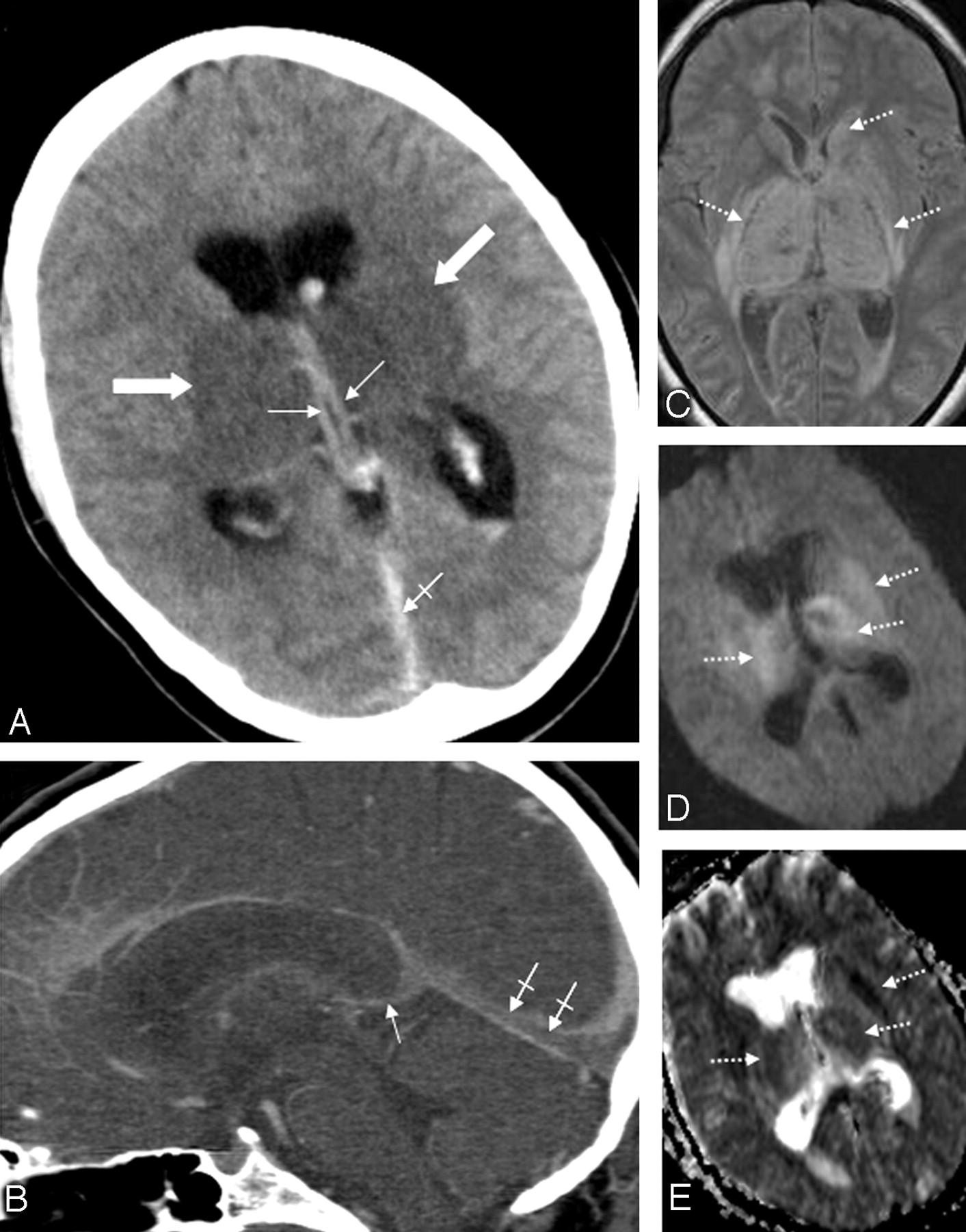
    Patient 5. A 31-year-old woman with combined deep cerebral venous thrombosis of the TSV, the ICVs, the BVR, the VG, the SS, and the LTS. A, The NCCT scan demonstrates an attenuated vein sign in both ICVs (thin arrows), in the SS (crossed arrow) as well as bilateral edema in the thalami and in the putamen (thick arrows). B, Multiplanar sagittal reconstruction of an MDCTA. The thrombosis is visualized indirectly by demonstration of contrast-filling defects in the ICVs (thin arrow) and the SS (crossed arrows). C, An axial proton-attenuation-weighted MR image (acquired as a dual-echo acquisition) also depicts the bilateral edema in the thalamus and the putamen as well as edema in the left caudate nucleus as hyperintense areas (dotted arrows). D, Diffusion-weighted image (b-value = 1000) and the apparent diffusion coefficient map (E) show a restriction of diffusion in the respective areas (dotted arrows).

  • Fig 2.
    • Download figure
    • Open in new tab
    • Download powerpoint
    Fig 2.

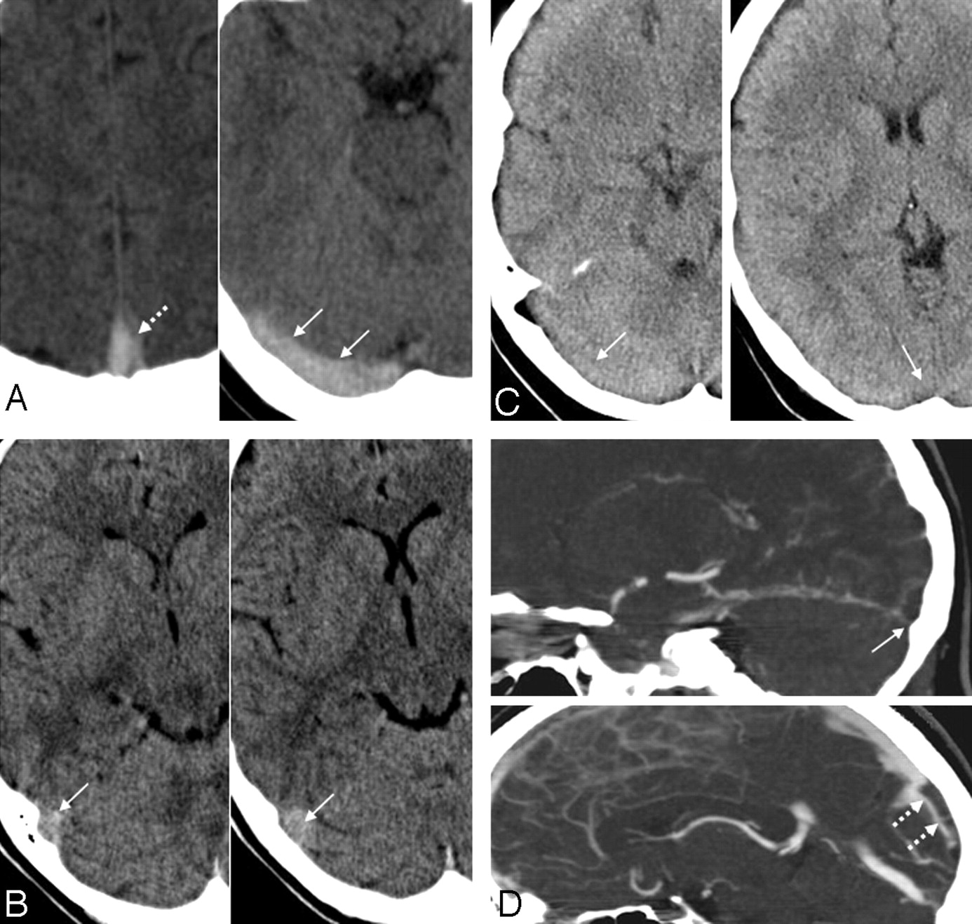
    Examples for true-positive, false-positive, and false-negative cord signs in the evaluation of sinus thrombosis. A, A 35-year-old woman with an SVT of the SSS and the RTS, who presented with headache and nausea. The NCCT scan shows a true-positive cord sign in the SSS (dotted arrow) and the RTS (arrows). The case of this patient was judged as positive with regard to the presence of a cord sign by all 3 readers. B, A 27-year-old woman in the control group, who initially presented with headache, but all other available imaging modalities showed no pathologic findings and clinical follow-up was uneventful. The RTS appeared hyperattenuated on NCCT (arrows). This finding was interpreted as a cord sign by all readers, resulting in the false-positive diagnosis of a SVT in this patient. C and D, A 45-year old woman with headache and nausea and a SVT involving the RTS and the SSS. The NCCT scan (C) shows no hyperattenuated signal intensity (ie, no cord sign) in the RTS or the SSS: The case of this patient was judged as false-negative regarding the presence of a cord sign by all readers. D, Multiplanar sagittal reconstruction of a MDCTA demonstrates contrast-filling defects in the RTS (arrow) and in the SSS (dotted arrow), indicating an SVT.

Tables

  • Figures
    • View popup
    Table 1:

    Characteristics of patients with DVT

    Patient No.Age/SexDiagnosisAvailable ImagingAffected Venous StructuresParenchymal ChangesDiagnostic Delay (d)#
    139/fIDVTNCCT, MRI, MRARICV, SSEdema right thalamus, caudate nucleus, putamen11
    239/fIDVTNCCT, CTA, MRI, MRALTSV, both ICV, both BVR, VG, SSEdema thalami, caudate nuclei, putamen,  bilaterally14
    347/fIDVTNCCT, CTA, MRI, MRA,  DSAboth ICV, VG, SSHemorrhage left thalamus, edema both thalami21
    449/mIDVTNCCT, MRI, MRA, DSALTSV, both ICV, both BVR, VG, SSEdema both thalami1
    531/fCDVTNCCT, CTA, MRI, MRA,  DSABoth TSV, both ICV, both BVR, VG,  SS, LTSEdema both thalami, left caudate nucleus,  putamen, bilaterally1
    627/mIDVTNCCT, CTA, MRI, MRALTSV, both ICV, LBVR, VG, SSEdema both thalami, posterior part of left  hippocampus28
    741/fCDVTNCCT, CTA, MRI, MRABoth ICV, RBVR, VG, SS, RTSNone5
    838/fCDVTNCCT, CTA, MRI, MRABoth ICV, RBVR, VG, SS, LTS, LSS  SSSEdema both thalami, left parietal lobe, left  occipital lobe, hemorrhage both thalami6
    • Note:—DVT indicates deep cerebral venous thrombosis; f, female; m, male; IDVT, isolated deep venous thrombosis; CDVT, combined deep venous thrombosis; NCCT, noncontrast cranial CT; CTA, CT angiography; MRI, MR imaging; MRA, MR angiography; DSA, digital subtraction angiography; ICV, internal cerebral veins; RICV, right internal cerebral vein; SS, straight sinus; TSV, thalamostriate vein; LTSV, left thalamostriate vein; BVR, basal veins of Rosenthal; LBVR, left basal vein of Rosenthal; RBVR, right basal vein of Rosenthal; VG, great vein of Galen; diagnostic delay #, time between onset of initial symptoms and definite diagnosis of DVT; LTS, left transverse sinus; RTS, right transverse sinus; LSS, left sigmoid sinus; SSS, superor sagittal sinus.

    • View popup
    Table 2:

    Characteristics of patients with SVT

    Patient No.Age/SexAvailable ImagingAffected SinusesParenchymal ChangesDiagnostic Delay (d)#
    137/fNCCT, CTA, MRI, MRASSS, LTS, LSSNone7
    245/fNCCT, CTASSS, RTSNone5
    327/fNCCT, CTA, MRI, MRASSSNone3
    444/fNCCT, MRI, MRA, DSASSS, LTS, LSSNone1
    535/fNCCT, CTA, MRI, MRASSS, RTSNone4
    663/mNCCT, CTA, MRI, MRASSS, RTSEdema and hemorrhage, right occipital lobe2
    723/fNCCT, CTA, MRI, MRASSS, RSS, CVEdema and hemorrhage, right frontal lobe1
    878/fNCCT, CTA, MRI, MRALTSEdema, left frontal lobe1
    943/fNCCT, MRI, MRA, DSASSS, LTS, LSS, CVEdema and hemorrhage, right parietal lobe3
    1052/mNCCT, CTA, MRI, MRA, DSASSS, RTSEdema and hemorrhage, right parietal lobe1
    1123/fNCCT, CTA, MRI, MRALTS, LSS, RSSEdema, left parietal and temporal lobes2
    1230/fNCCT, CTA, MRI, MRA, DSASSS, LTS, LSSEdema and hemorrhage, left occipital lobe2
    1320/fNCCT, MRI, MRA, DSASSS, RTS, RSSNone6
    1421/mNCCT, CTA, MRI, MRALTS, RTSNone2
    1582/fNCCT, CTA, MRI, MRASSS, RTS, CVEdema and hemorrhage, left frontal and occipital lobes0
    1664/fNCCT, CTASSS, LTS, LSSEdema, left parietal lobe2
    1722/fNCCT, CTA, MRI, MRALTS, LSSNone8
    1852/mNCCT, MRI, MRA, DSASSS, LTS, LSSEdema and hemorrhage, right parietal lobe1
    1966/mNCCT, MRI, MRASSS, LTS, LSSEdema, left frontal lobe0
    2053/fNCCT, CTA, MRI, MRALSSNone5
    2160/fNCCT, CTA, MRI, MRASSS, LTS, CVNone3
    2233/mNCCT, CTA, MRI, MRALTS, LSSNone3
    2365/fNCCT, CTA, MRI, MRALTS, LSSEdema and hemorrhage, left occipital lobe1
    2432/fNCCT, CTA, MRI, MRASSSEdema and hemorrhage, right frontal lobe2
    2518/mNCCT, MRI, MRA, DSASSS, LTS, LSSEdema and hemorrhage, right frontal lobe0
    • Note:—SVT indicates sinus thrombosis; RSS, right sigmoid sinus; CV, cortical veins.

    • View popup
    Table 3:

    Available imaging modalities*

    Patient GroupTotalMRI, DSA, MDCTAMRI, DSAMRI, MDCTAMRI aloneMDCTA alone
    DVT8 (100)2 (25)1 (12.5)4 (50)1 (12.5)0 (0)
    SVT25 (100)2 (8)5 (20)15 (60)1 (4)2 (8)
    Control36 (100)0 (0)5 (13.9)9 (25)5 (13.9)17 (47.2)
    Total69 (100)4 (5.8)11 (15.9)28 (40.6)7 (10.1)19 (27.5)
    • Note:—MRI indicates magnetic resonance imaging including venous MR angiography and T2*-weighted images; DSA, digital subtraction angiography; MDCTA, multi-detector row CT angiography; DVT, deep venous thrombosis; SVT, sinus thrombosis.

    • * Data are given as numbers (percentages).

    • View popup
    Table 4:

    Results of the readings–veins

    Venous StructureBlinded ReadingsConsensus ReadingIntraobserver Agreement (κ values)Sensitivity (95% CI)*Specificity (95% CI)*
    Dense Vein Signtpfptnfn
    SS2423118510.9695.8 (85.6–98.9)99.5 (98.1–99.9)
    LICV1918118230.9285.7 (72.7–89.6)99.5 (98–99.9)
    RICV2423118210.9695.8 (85.6–98.9)99.5 (98.1–99.8)
    VG2423118210.9695.8 (85.6–98.9)99.5 (98.1–99.8)
    LBVR1111019510.9891.7 (74.9–91.7)100 (99–100)
    RBVR1414019210.9893.3 (79.6–93.3)100 (98.9–100)
    LTSV1312119400.98100 (83–100)99.5 (98.4–99.5)
    RTSV95419710.9083.3 (47.6–96.9)98 (96.9–98.4)
    Total1381299150990.95893.5 (89.9–95.9)99.4 (99.1–99.6)
    • Note:—tp indicates true-positive; fp, false-positive; tn, true-negative; fn, false-negative; 95% CI, 95% confidence interval; LICV, left internal cerebral vein; RTSV, right thalamostriate vein.

    • * Data are given in percentages.

    • View popup
    Table 5:

    Results of the readings–sinus

    SinusBlinded ReadingConsensus ReadingInterobserver Agreement (κ values)Sensitivity (95% CI)*Specificity (95% CI)*
    Cord Signtpfptnfn
    SSS45369138240.7060 (51.4–66.3)93.9 (90.4–96.4)
    ISS80819630.840 (0–53.3)96.1 (96.1–96.9)
    LTS18117146430.7620.4 (13.3–26.4)95.4 (92.9–97.5)
    RTS20812171160.833.3 (19.3–48.4)93.4 (91.6–95.4)
    LSS1477164290.7819.4 (10.8–28.1)95.9 (94.1–97.7)
    RSS1111018880.8411.1 (2–39.1)94.9 (94.5–96.2)
    Total116635310031230.8033.9 (28.8–38.8)95 (94.1–95.8)
    • Note:—ISS indicates inferior sagittal sinus.

    • * Data are given in percentages.

PreviousNext
Back to top

In this issue

American Journal of Neuroradiology: 30 (4)
American Journal of Neuroradiology
Vol. 30, Issue 4
April 2009
  • Table of Contents
  • Index by author
Advertisement
Print
Download PDF
Email Article

Thank you for your interest in spreading the word on American Journal of Neuroradiology.

NOTE: We only request your email address so that the person you are recommending the page to knows that you wanted them to see it, and that it is not junk mail. We do not capture any email address.

Enter multiple addresses on separate lines or separate them with commas.
Noncontrast CT in Deep Cerebral Venous Thrombosis and Sinus Thrombosis: Comparison of its Diagnostic Value for Both Entities
(Your Name) has sent you a message from American Journal of Neuroradiology
(Your Name) thought you would like to see the American Journal of Neuroradiology web site.
CAPTCHA
This question is for testing whether or not you are a human visitor and to prevent automated spam submissions.
Cite this article
J. Linn, T. Pfefferkorn, K. Ivanicova, S. Müller-Schunk, S. Hartz, M. Wiesmann, M. Dichgans, H. Brückmann
Noncontrast CT in Deep Cerebral Venous Thrombosis and Sinus Thrombosis: Comparison of its Diagnostic Value for Both Entities
American Journal of Neuroradiology Apr 2009, 30 (4) 728-735; DOI: 10.3174/ajnr.A1451

Citation Manager Formats

  • BibTeX
  • Bookends
  • EasyBib
  • EndNote (tagged)
  • EndNote 8 (xml)
  • Medlars
  • Mendeley
  • Papers
  • RefWorks Tagged
  • Ref Manager
  • RIS
  • Zotero
0 Responses
Respond to this article
Share
Bookmark this article
Noncontrast CT in Deep Cerebral Venous Thrombosis and Sinus Thrombosis: Comparison of its Diagnostic Value for Both Entities
J. Linn, T. Pfefferkorn, K. Ivanicova, S. Müller-Schunk, S. Hartz, M. Wiesmann, M. Dichgans, H. Brückmann
American Journal of Neuroradiology Apr 2009, 30 (4) 728-735; DOI: 10.3174/ajnr.A1451
del.icio.us logo Twitter logo Facebook logo Mendeley logo
  • Tweet Widget
  • Facebook Like
  • Google Plus One
Purchase

Jump to section

  • Article
    • Abstract
    • Materials and Methods
    • Results
    • Discussion
    • Conclusions
    • Acknowledgments
    • Footnotes
    • References
  • Figures & Data
  • Info & Metrics
  • Responses
  • References
  • PDF

Related Articles

  • No related articles found.
  • PubMed
  • Google Scholar

Cited By...

  • Isolated unilateral thalamic venous infarct in a toddler
  • Diagnostic accuracy of noncontrast CT imaging markers in cerebral venous thrombosis
  • CT Density Measurement and H:H Ratio Are Useful in Diagnosing Acute Cerebral Venous Sinus Thrombosis
  • Frequency of Adequate Contrast Opacification of the Major Intracranial Venous Structures with CT Angiography in the Setting of Intracerebral Hemorrhage: Comparison of 16- and 64-Section CT Angiography Techniques
  • Crossref (102)
  • Google Scholar

This article has been cited by the following articles in journals that are participating in Crossref Cited-by Linking.

  • Thrombin-activatable fluorescent peptide incorporated gold nanoparticles for dual optical/computed tomography thrombus imaging
    Sung-Pil Kwon, Sangmin Jeon, Sung-Hoon Lee, Hong Yeol Yoon, Ju Hee Ryu, Dayil Choi, Jeong-Yeon Kim, Jiwon Kim, Jae Hyung Park, Dong-Eog Kim, Ick Chan Kwon, Kwangmeyung Kim, Cheol-Hee Ahn
    Biomaterials 2018 150
  • CT Density Measurement and H:H Ratio Are Useful in Diagnosing Acute Cerebral Venous Sinus Thrombosis
    P.-J. Buyck, F. De Keyzer, D. Vanneste, G. Wilms, V. Thijs, P. Demaerel
    American Journal of Neuroradiology 2013 34 8
  • Dural Sinus Thrombosis: Sources of Error in Image Interpretation
    James M. Provenzale, Peter G. Kranz
    American Journal of Roentgenology 2011 196 1
  • Direct thrombolysis for cerebral venous sinus thrombosis
    Maryam Rahman, Gregory J. Velat, Brian L. Hoh, J Mocco
    Neurosurgical Focus 2009 27 5
  • Cortical vein thrombosis: the diagnostic value of different imaging modalities
    Jennifer Linn, Stefan Michl, Bochmann Katja, Thomas Pfefferkorn, Martin Wiesmann, Sabine Hartz, Martin Dichgans, Hartmut Brückmann
    Neuroradiology 2010 52 10
  • Bilateral lesions of the basal ganglia and thalami (central grey matter)—pictorial review
    Sofie Van Cauter, Mariasavina Severino, Rosamaria Ammendola, Brecht Van Berkel, Hrvoje Vavro, Luc van den Hauwe, Zoran Rumboldt
    Neuroradiology 2020 62 12
  • Current imaging modalities for diagnosing cerebral vein thrombosis – A critical review
    Lisette F. van Dam, Marianne A.A. van Walderveen, Lucia J.M. Kroft, Nyika D. Kruyt, Marieke J.H. Wermer, Matthias J.P. van Osch, Menno V. Huisman, Frederikus A. Klok
    Thrombosis Research 2020 189
  • Hemorrhagic Stroke
    Scott D. Smith, Clifford J. Eskey
    Radiologic Clinics of North America 2011 49 1
  • Nontraumatic Intracranial Hemorrhage
    Nancy J. Fischbein, Christine A.C. Wijman
    Neuroimaging Clinics of North America 2010 20 4
  • Cerebral sinovenous thrombosis in pediatric practice
    Gary L. Hedlund
    Pediatric Radiology 2013 43 2

More in this TOC Section

  • Fast Contrast-Enhanced 4D MRA and 4D Flow MRI Using Constrained Reconstruction (HYPRFlow): Potential Applications for Brain Arteriovenous Malformations
  • Quiet PROPELLER MRI Techniques Match the Quality of Conventional PROPELLER Brain Imaging Techniques
  • Predictors of Reperfusion in Patients with Acute Ischemic Stroke
Show more Brain

Similar Articles

Advertisement

Indexed Content

  • Current Issue
  • Accepted Manuscripts
  • Article Preview
  • Past Issues
  • Editorials
  • Editors Choice
  • Fellow Journal Club
  • Letters to the Editor

Cases

  • Case Collection
  • Archive - Case of the Week
  • Archive - Case of the Month
  • Archive - Classic Case

Special Collections

  • Special Collections

Resources

  • News and Updates
  • Turn around Times
  • Submit a Manuscript
  • Author Policies
  • Manuscript Submission Guidelines
  • Evidence-Based Medicine Level Guide
  • Publishing Checklists
  • Graphical Abstract Preparation
  • Imaging Protocol Submission
  • Submit a Case
  • Become a Reviewer/Academy of Reviewers
  • Get Peer Review Credit from Publons

Multimedia

  • AJNR Podcast
  • AJNR SCANtastic
  • Video Articles

About Us

  • About AJNR
  • Editorial Board
  • Not an AJNR Subscriber? Join Now
  • Alerts
  • Feedback
  • Advertise with us
  • Librarian Resources
  • Permissions
  • Terms and Conditions

American Society of Neuroradiology

  • Not an ASNR Member? Join Now

© 2025 by the American Society of Neuroradiology All rights, including for text and data mining, AI training, and similar technologies, are reserved.
Print ISSN: 0195-6108 Online ISSN: 1936-959X

Powered by HighWire