Advertisement

Main menu

  • Home
  • Content
    • Current Issue
    • Accepted Manuscripts
    • Article Preview
    • Past Issue Archive
    • AJNR Case Collection
    • Case of the Week Archive
    • Classic Case Archive
    • Case of the Month Archive
  • Special Collections
    • Spinal CSF Leak Articles (Jan 2020-June 2024)
    • 2024 AJNR Journal Awards
    • Most Impactful AJNR Articles
  • Multimedia
    • AJNR Podcast
    • AJNR Scantastics
    • Video Articles
  • For Authors
    • Submit a Manuscript
    • Author Policies
    • Fast publishing of Accepted Manuscripts
    • Graphical Abstract Preparation
    • Manuscript Submission Guidelines
    • Imaging Protocol Submission
    • Submit a Case for the Case Collection
  • About Us
    • About AJNR
    • Editorial Board
  • More
    • Become a Reviewer/Academy of Reviewers
    • Subscribers
    • Permissions
    • Alerts
    • Feedback
    • Advertisers
    • ASNR Home
  • Other Publications
    • ajnr

User menu

  • Alerts
  • Log in

Search

  • Advanced search
American Journal of Neuroradiology

American Journal of Neuroradiology

ASHNR American Society of Functional Neuroradiology ASHNR American Society of Pediatric Neuroradiology ASSR
  • Alerts
  • Log in

Advanced Search

  • Home
  • Content
    • Current Issue
    • Accepted Manuscripts
    • Article Preview
    • Past Issue Archive
    • AJNR Case Collection
    • Case of the Week Archive
    • Classic Case Archive
    • Case of the Month Archive
  • Special Collections
    • Spinal CSF Leak Articles (Jan 2020-June 2024)
    • 2024 AJNR Journal Awards
    • Most Impactful AJNR Articles
  • Multimedia
    • AJNR Podcast
    • AJNR Scantastics
    • Video Articles
  • For Authors
    • Submit a Manuscript
    • Author Policies
    • Fast publishing of Accepted Manuscripts
    • Graphical Abstract Preparation
    • Manuscript Submission Guidelines
    • Imaging Protocol Submission
    • Submit a Case for the Case Collection
  • About Us
    • About AJNR
    • Editorial Board
  • More
    • Become a Reviewer/Academy of Reviewers
    • Subscribers
    • Permissions
    • Alerts
    • Feedback
    • Advertisers
    • ASNR Home
  • Follow AJNR on Twitter
  • Visit AJNR on Facebook
  • Follow AJNR on Instagram
  • Join AJNR on LinkedIn
  • RSS Feeds

Welcome to the new AJNR, Updated Hall of Fame, and more. Read the full announcements.


AJNR is seeking candidates for the position of Associate Section Editor, AJNR Case Collection. Read the full announcement.


Case ReportSpine

A Case of Spinal Cord Infarction Following Lumbar Transforaminal Epidural Steroid Injection: MR Imaging and Angiographic Findings

E.M. Lyders and P.P. Morris
American Journal of Neuroradiology October 2009, 30 (9) 1691-1693; DOI: https://doi.org/10.3174/ajnr.A1567
E.M. Lyders
  • Find this author on Google Scholar
  • Find this author on PubMed
  • Search for this author on this site
P.P. Morris
  • Find this author on Google Scholar
  • Find this author on PubMed
  • Search for this author on this site
  • Article
  • Figures & Data
  • Info & Metrics
  • Responses
  • References
  • PDF
Loading

Abstract

SUMMARY: Spinal cord infarction following lumbar transforaminal epidural steroid injection is a rare and devastating complication. We describe the case of a 55-year-old woman who developed spinal cord infarction following right L2-3 transforaminal epidural injection, diagnosed on the basis of clinical and MR imaging findings. Spinal angiography demonstrated occlusion of the right L2 segmental artery with reconstitution of the radicular branch from collaterals. The artery of Adamkeiwicz could not be demonstrated and was presumably occluded by the steroid injection.

Transforaminal epidural steroid injection (TF ESI) is a common procedure in the nonsurgical management of lumbosacral radiculopathy. The incidence of significant complications is low. Spinal cord infarction (SCI) following lumbar TF ESI is one of the rarest yet most devastating complications, with 5 cases reported in the literature.1–3 Inadvertent vascular penetration of a radiculomedullary artery by the procedure needle has been implicated as the likely etiology. Cord ischemia could result from vascular injury with thrombosis or embolism involving the artery of Adamkiewicz or from occlusion secondary to intra-arterial injection of steroid suspension. In this case, we describe the MR imaging and angiographic findings in a case of SCI following lumbar TF ESI. To our knowledge, the angiographic findings have never been reported.

Case Report

A 55-year-old woman with an acute-on-chronic disk herniation at L1–2 presented for TF ESI at L2–3 on the right. Initial placement of a 22-gauge spinal needle reportedly showed backbleeding on removal of the stylet, which apparently resolved once the needle was repositioned. Epidural placement of the needle tip was reportedly confirmed following injection of 2 mL of myelographic contrast (Fig 1), followed by therapeutic injection of an additional 1 mL of triamcinolone acetonide (Kenalog) and 0.25% bupivacaine. Within minutes of the injection, the patient developed bilateral lower extremity weakness, which progressed to flaccid paralysis.

Fig 1.
  • Download figure
  • Open in new tab
  • Download powerpoint
Fig 1.

Prone fluoroscopic image during right L2–3 TF ESI demonstrates epidural opacification before therapeutic injection.

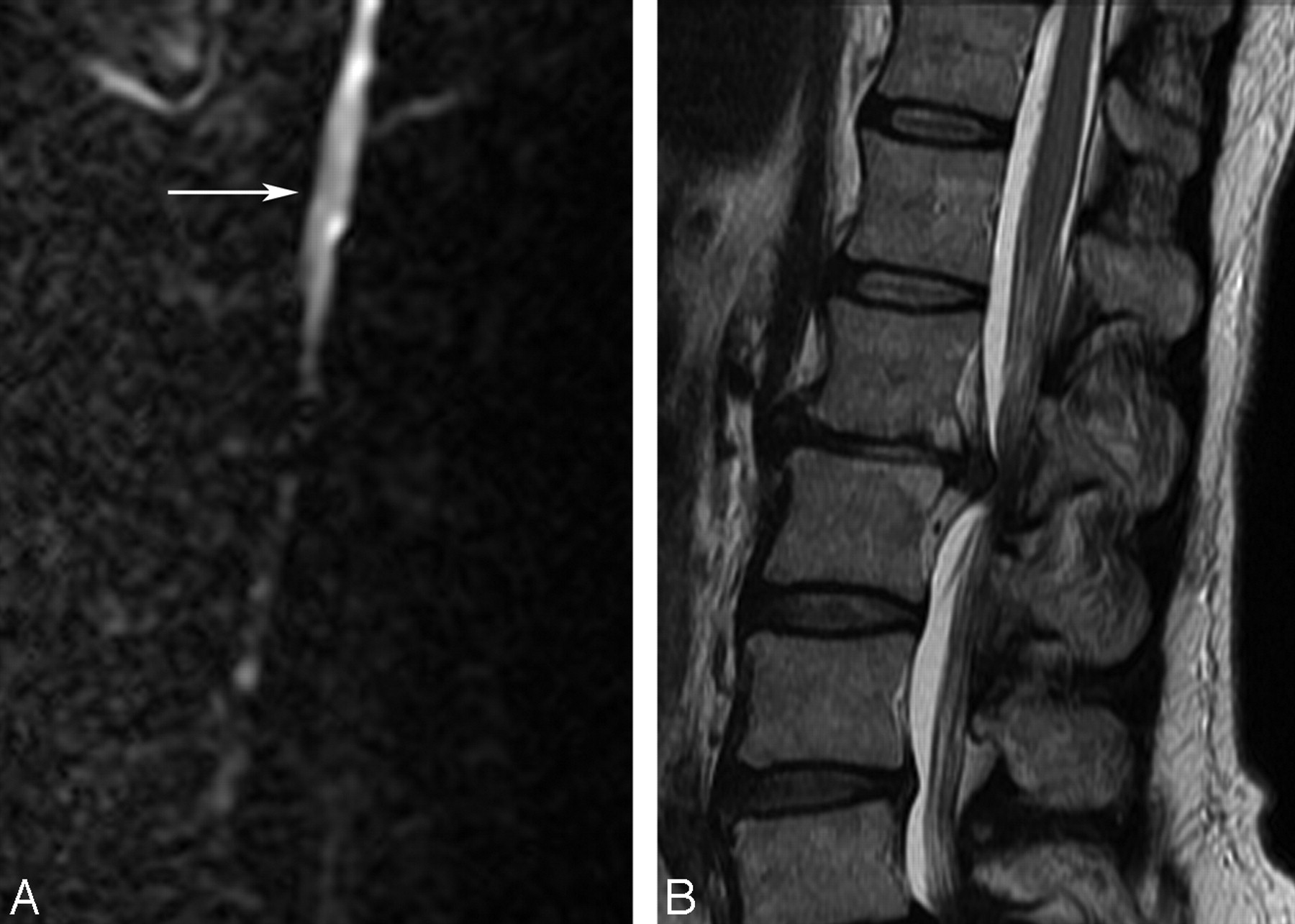
The patient underwent CT of the thoracolumbar spine within 1.5-hours of the injection, which demonstrated no evidence of epidural hematoma. MR imaging of the spine was performed approximately 4 hours after the injection, including sagittal and axial T1 (TR, 350; TE, 15.7), T2 (TR, 3950; TE, 105), and echo-planar diffusion-weighted imaging (b = 1000). T2-weighted images demonstrated subtly increased signal intensity within the central gray matter of the distal thoracic cord with restricted water diffusion in the same area (Fig 2).

Fig 2.
  • Download figure
  • Open in new tab
  • Download powerpoint
Fig 2.

Sagittal DWI (left) and T2-weighted images of the thoracolumbar spine demonstrate restricted water diffusion in the distal cord and conus (white arrow). Note a large disk herniation at L1–2, for which the TF ESI was being performed.

Catheter-directed spinal angiography was performed to evaluate potentially treatable causes of spinal cord ischemia, including spasm or dissection. The ostium of the right L2 segmental artery could not be selected and was presumed to be occluded. Injection of the right L3 segmental artery demonstrated collateral vessels coursing toward the right L2–3 foramen, with irregular attenuated reconstitution of the distal L2 segmental branch and radicular artery (Fig 3). Additionally, a thoracolumbar spinal angiogram failed to demonstrate the origin of the artery of Adamkiewicz.

Fig 3.
  • Download figure
  • Open in new tab
  • Download powerpoint
Fig 3.

Angiogram of the right L3 segmental artery (L3) demonstrates prominent collateral vessels (short black arrows) in the region of the right L2 pedicle. The distal right L2 segmental (L2) and radicular (long black arrow) arteries are irregular and attenuated. The ostium of the right L2 segmental lumbar artery could not be found and was presumably occluded.

Acute spinal cord ischemia was diagnosed on the basis of the clinical and radiographic findings. The patient's symptoms have not improved, and she remains paraplegic.

Discussion

TF ESI is a common procedure used in the nonsurgical management of lumbosacral radiculopathy.4,5 The transforaminal approach allows concentrated delivery of medication to the ventral neural foramen and anterior epidural space, the areas most likely affected by disk pathology. However, needle placement via a transforaminal approach makes inadvertent vascular penetration of the radicular artery possible.

Minor complications include headache, back pain, worsening leg pain, facial flushing, and vasovagal reaction and have a reported incidence of 9.6%.6 Serious complications (eg, epidural hematoma, abscess, arachnoiditis) are uncommon, with an incidence of 0.9%.7 One of the rarest, yet most devastating, complications is spinal cord ischemia, which has been reported 5 times in the literature following lumbosacral TF ESI.1–3

The blood supply to the thoracic spinal cord is predominantly from the anterior spinal artery, and a poor collateral network with the smaller posterior spinal arteries places the cord at high risk of ischemia if the dominant blood supply is compromised. At every level in the thoracolumbar spine, radicular branches off the segmental intercostal or lumbar arteries supply the exiting nerve root and foraminal structures. At as many as 5–6 levels, the radicular arteries also contribute to the anterior spinal artery, termed “radiculomedullary arteries.” The dominant radiculomedullary artery is called the “artery of Adamkiewicz” and is the primary source of blood for the thoracic cord. The artery of Adamkiewicz originates between T9 and L2 on the left in 85% of people, though aberrant origins from every lumbar level and S1 have been reported.

Theories regarding the cause of the SCI after TF ESI implicate inadvertent penetration of the radicular artery within the neural foramen in combination with a low origin of the artery of Adamkiewicz. Penetration of the radicular artery could result in focal dissection and distal emboli. If the injured radicular artery was, in fact, the artery of Adamkiewicz, this could result in spinal cord ischemia. Another proposed mechanism of injury suggests that intra-arterial injection of steroid suspension results in a shower of thrombogenic particulates to the distal cord with resultant ischemia.8

The incidence of vascular penetration with transforaminal needle placement is reportedly 8.1% at lumbar levels.9 Additionally, the use of a “flash” or backbleeding is not a reliable predictor of inadvertent vascular penetration.9 In this case, a small amount of backbleeding was reported on initial needle placement, which apparently resolved with repositioning. Contrast injection before therapeutic injection reportedly demonstrated epidural opacification.

CT and MR imaging are primarily used to exclude surgically correctable pathology that may mimic cord ischemia clinically. In this case, CT demonstrated no evidence of epidural collection or explanation for the flaccid paralysis. Findings on T2-weighted MR images are often normal but may demonstrate cord expansion and hyperintense signal intensity in the central cord and/or gray matter.10 Diffusion-weighted imaging (DWI) can also be useful in the setting of acute cord ischemia, demonstrating restricted diffusion.10,11 In this case, MR imaging of the spine performed 4 hours after injection demonstrated increased T2 signal intensity in the gray matter and restricted diffusion in the lower thoracic cord. These findings are clinically useful because transient paralysis following subarachnoid injection of anesthetic has been reported.12

Catheter-directed spinal angiography in the setting of acute SCI following TF ESI has not been previously reported and was performed to evaluate potentially treatable lesions. In this case, the steroid injection presumably penetrated the right L2 radicular artery. The right L2 segmental artery origin could not be selected; however, if it was thrombosed back close to its origin, its presence would not have been detectable due to the inability to opacify the lumen. Irregular attenuated opacification of the distal right L2 segmental and radicular arteries via collaterals suggests that there was also spasm and/or thrombosis involving these vessels. The artery of Adamkiewicz was not demonstrated and putatively arose from the right L2 and was thrombosed. With such widespread vascular irregularity, we believe the SCI was likely secondary to inadvertent intra-arterial injection of thrombogenic particulate steroid. Although a focal dissection could theoretically result in distal emboli to the cord, it is unlikely to cause such widespread alteration in the appearance of the branches of the L2 segmental artery.

Conclusions

TF ESI is a common procedure in the nonsurgical management of chronic radicular pain. Although the incidence of serious complications is exceeding small, practitioners who perform this procedure should be aware of the potentially devastating complication of spinal cord ischemia. On the basis of the angiographic findings in our patient, the underlying etiology is likely related to inadvertent vascular penetration with intra-arterial injection of particulate steroid into the artery of Adamkiewicz, rather than a focal vascular injury.

References

  1. 1.↵
    1. Houten J,
    2. Errico T
    . Paraplegia after lumbosacral nerve root block: report of three cases. Spine J 2002;2:70–75
    PubMed
  2. 2.↵
    1. Huntoon M,
    2. Martin D
    . Paralysis after transforaminal epidural injection and previous spinal surgery. Reg Anesth Pain Med 2004;29:494–95
    PubMed
  3. 3.↵
    1. Somayaji H,
    2. Saifuddin A,
    3. Cassey A,
    4. et al
    . Spinal cord infarction following therapeutic computed tomography guided left L2 nerve root injection. Spine 2005;30:106–08
    CrossRef
  4. 4.↵
    1. Lutz G,
    2. Vad V,
    3. Wisneski R
    . Fluoroscopic transforaminal lumbar epidural steroids: an outcome study. Arch Phys Med Rehabil 1998;79:1362–66
    CrossRefPubMed
  5. 5.↵
    1. Vad V,
    2. Bhat A,
    3. Lutz G,
    4. et al
    . Transforaminal epidural steroid injections in lumbosacral radiculopathy. Spine 2002;27:11–16
    CrossRefPubMed
  6. 6.↵
    1. Botwin K,
    2. Gruber R,
    3. Bouchlas C
    . Complications of fluoroscopically guided transforaminal lumbar epidural injections. Arch Phys Med Rehabil 2000;81:1045–50
    CrossRefPubMed
  7. 7.↵
    1. Slipman C,
    2. Meyers J,
    3. Chou L,
    4. et al
    . Complications of fluoroscopically guided spinal injections. Arch Phys Med Rehabil 1995;76:1032
  8. 8.↵
    1. Tiso R,
    2. Cutler T,
    3. Catania J,
    4. et al
    . Adverse central nervous system sequelae after selective transforaminal block: the role of corticosteroids. Spine J 2004;4:468–74
    CrossRefPubMed
  9. 9.↵
    1. Furman M,
    2. O'Brien E,
    3. Zgleszewski T
    . Incidence of intravascular penetration in transforaminal lumbosacral epidural steroid injections. Spine 2000;25:2628–32
    CrossRefPubMed
  10. 10.↵
    1. Kuker W,
    2. Weller M,
    3. Klose U,
    4. et al
    . Diffusion-weighted MRI of spinal cord infarction: high-resolution imaging and time course of diffusion abnormality. J Neurol 2004;251:818–24
    PubMed
  11. 11.↵
    1. Sibon I,
    2. Menegon P,
    3. Moonen C,
    4. et al
    . Early diagnosis of spinal cord infarct using magnetic resonance diffusion imaging. Neurology 2003;61:1622
    FREE Full Text
  12. 12.↵
    1. McLain R,
    2. Fry M,
    3. Hecht S
    . Transient paralysis associated with epidural steroid injection. J Spinal Disord 1997;10:441–44
    PubMed
  • Received January 20, 2009.
  • Accepted after revision January 28, 2009.
  • Copyright © American Society of Neuroradiology
View Abstract
PreviousNext
Back to top

In this issue

American Journal of Neuroradiology: 30 (9)
American Journal of Neuroradiology
Vol. 30, Issue 9
1 Oct 2009
  • Table of Contents
  • Index by author
Advertisement
Print
Download PDF
Email Article

Thank you for your interest in spreading the word on American Journal of Neuroradiology.

NOTE: We only request your email address so that the person you are recommending the page to knows that you wanted them to see it, and that it is not junk mail. We do not capture any email address.

Enter multiple addresses on separate lines or separate them with commas.
A Case of Spinal Cord Infarction Following Lumbar Transforaminal Epidural Steroid Injection: MR Imaging and Angiographic Findings
(Your Name) has sent you a message from American Journal of Neuroradiology
(Your Name) thought you would like to see the American Journal of Neuroradiology web site.
CAPTCHA
This question is for testing whether or not you are a human visitor and to prevent automated spam submissions.
Cite this article
E.M. Lyders, P.P. Morris
A Case of Spinal Cord Infarction Following Lumbar Transforaminal Epidural Steroid Injection: MR Imaging and Angiographic Findings
American Journal of Neuroradiology Oct 2009, 30 (9) 1691-1693; DOI: 10.3174/ajnr.A1567

Citation Manager Formats

  • BibTeX
  • Bookends
  • EasyBib
  • EndNote (tagged)
  • EndNote 8 (xml)
  • Medlars
  • Mendeley
  • Papers
  • RefWorks Tagged
  • Ref Manager
  • RIS
  • Zotero
0 Responses
Respond to this article
Share
Bookmark this article
A Case of Spinal Cord Infarction Following Lumbar Transforaminal Epidural Steroid Injection: MR Imaging and Angiographic Findings
E.M. Lyders, P.P. Morris
American Journal of Neuroradiology Oct 2009, 30 (9) 1691-1693; DOI: 10.3174/ajnr.A1567
del.icio.us logo Twitter logo Facebook logo Mendeley logo
  • Tweet Widget
  • Facebook Like
  • Google Plus One
Purchase

Jump to section

  • Article
    • Abstract
    • Case Report
    • Discussion
    • Conclusions
    • References
  • Figures & Data
  • Info & Metrics
  • Responses
  • References
  • PDF

Related Articles

  • No related articles found.
  • PubMed
  • Google Scholar

Cited By...

  • Intraforaminal Location of Thoracolumbar Radicular Arteries Providing an Anterior Radiculomedullary Artery Using Flat Panel Catheter Angiotomography
  • Sildenafil-Induced Cervical Spinal Cord Infarction
  • Reducing the Risk of Spinal Cord Infarction during Transforaminal Steroid Injections
  • Adjacent Double-Nerve Root Contributions in Unilateral Lumbar Radiculopathy
  • Crossref (95)
  • Google Scholar

This article has been cited by the following articles in journals that are participating in Crossref Cited-by Linking.

  • Safeguards to Prevent Neurologic Complications after Epidural Steroid Injections
    James P. Rathmell, Honorio T. Benzon, Paul Dreyfuss, Marc Huntoon, Mark Wallace, Ray Baker, K. Daniel Riew, Richard W. Rosenquist, Charles Aprill, Natalia S. Rost, Asokumar Buvanendran, D. Scott Kreiner, Nikolai Bogduk, Daryl R. Fourney, Eduardo Fraifeld, Scott Horn, Jeffrey Stone, Kevin Vorenkamp, Gregory Lawler, Jeffrey Summers, David Kloth, David O’Brien, Sean Tutton
    Anesthesiology 2015 122 5
  • Comparative Effectiveness of Lumbar Transforaminal Epidural Steroid Injections with Particulate Versus Nonparticulate Corticosteroids for Lumbar Radicular Pain due to Intervertebral Disc Herniation: A Prospective, Randomized, Double-Blind Trial
    David J. Kennedy, Christopher Plastaras, Ellen Casey, Christopher J. Visco, Joshua D. Rittenberg, Bryan Conrad, James Sigler, Paul Dreyfuss
    Pain Medicine 2014 15 4
  • 11. Lumbosacral Radicular Pain
    Koen Van Boxem, Jianguo Cheng, Jacob Patijn, Maarten Van Kleef, Arno Lataster, Nagy Mekhail, Jan Van Zundert
    Pain Practice 2010 10 4
  • Spinal cord ischemia: aetiology, clinical syndromes and imaging features
    Stefan Weidauer, Michael Nichtweiß, Elke Hattingen, Joachim Berkefeld
    Neuroradiology 2015 57 3
  • The Noninferiority of the Nonparticulate Steroid Dexamethasone vs the Particulate Steroids Betamethasone and Triamcinolone in Lumbar Transforaminal Epidural Steroid Injections
    Christine El-Yahchouchi, Jennifer R. Geske, Rickey E. Carter, Felix E. Diehn, John T. Wald, Naveen S. Murthy, Timothy J. Kaufmann, Kent R. Thielen, Jonathan M. Morris, Kimberly K. Amrami, Timothy P. Maus
    Pain Medicine 2013 14 11
  • Intraforaminal Location of the Great Anterior Radiculomedullary Artery (Artery of Adamkiewicz): A Retrospective Review
    Naveen S. Murthy, Timothy P. Maus, Curt L. Behrns
    Pain Medicine 2010 11 12
  • Safe Use of Epidural Corticosteroid Injections: Recommendations of the WIP Benelux Work Group
    Koen Van Boxem, Mienke Rijsdijk, Guy Hans, Jasper de Jong, Jan Willem Kallewaard, Kris Vissers, Maarten van Kleef, James P. Rathmell, Jan Van Zundert
    Pain Practice 2019 19 1
  • Lumbar Transforaminal Epidural Dexamethasone
    Farshad M. Ahadian, Kai McGreevy, Gerhard Schulteis
    Regional Anesthesia and Pain Medicine 2011 36 6
  • Particulate versus non-particulate steroids for lumbar transforaminal or interlaminar epidural steroid injections: an update
    Tobias J. Dietrich, Reto Sutter, Johannes M. Froehlich, Christian W. A. Pfirrmann
    Skeletal Radiology 2015 44 2
  • Systematic Review of the Efficacy of Particulate Versus Nonparticulate Corticosteroids in Epidural Injections
    Priyesh Mehta, Isaac Syrop, Jaspal Ricky Singh, Jonathan Kirschner
    PM&R 2017 9 5

More in this TOC Section

  • Bern Score Validity for SIH
  • MP2RAGE 7T in MS Lesions of the Cervical Spine
  • Deep Learning for STIR Spine MRI Quality
Show more Spine

Similar Articles

Advertisement

Indexed Content

  • Current Issue
  • Accepted Manuscripts
  • Article Preview
  • Past Issues
  • Editorials
  • Editors Choice
  • Fellow Journal Club
  • Letters to the Editor

Cases

  • Case Collection
  • Archive - Case of the Week
  • Archive - Case of the Month
  • Archive - Classic Case

Special Collections

  • Special Collections

Resources

  • News and Updates
  • Turn around Times
  • Submit a Manuscript
  • Author Policies
  • Manuscript Submission Guidelines
  • Evidence-Based Medicine Level Guide
  • Publishing Checklists
  • Graphical Abstract Preparation
  • Imaging Protocol Submission
  • Submit a Case
  • Become a Reviewer/Academy of Reviewers
  • Get Peer Review Credit from Publons

Multimedia

  • AJNR Podcast
  • AJNR SCANtastic
  • Video Articles

About Us

  • About AJNR
  • Editorial Board
  • Not an AJNR Subscriber? Join Now
  • Alerts
  • Feedback
  • Advertise with us
  • Librarian Resources
  • Permissions
  • Terms and Conditions

American Society of Neuroradiology

  • Not an ASNR Member? Join Now

© 2025 by the American Society of Neuroradiology All rights, including for text and data mining, AI training, and similar technologies, are reserved.
Print ISSN: 0195-6108 Online ISSN: 1936-959X

Powered by HighWire