
Article Figures & Data
Figures
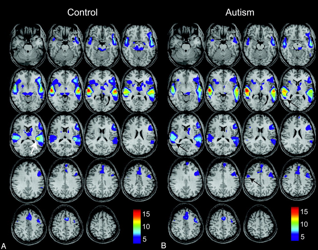
- Fig 1.
Group-level activation maps for an auditory phrase-recognition task for 15 control subjects (A) and for 26 high-functioning autistic subjects (B). Results for each group represent P < .05, false discovery rate (FDR), and color bars represent values for t-scores. Arrows show left posterior insular, right lateral premotor, and right Wernicke homolog areas where differences in activity are observed.
- Fig 2.
Group-level activation maps for 15 control subjects for auditory > visual tasks (blue) and auditory tasks (red) after masking the auditory > visual tasks. Both results show P < .05, FDR.
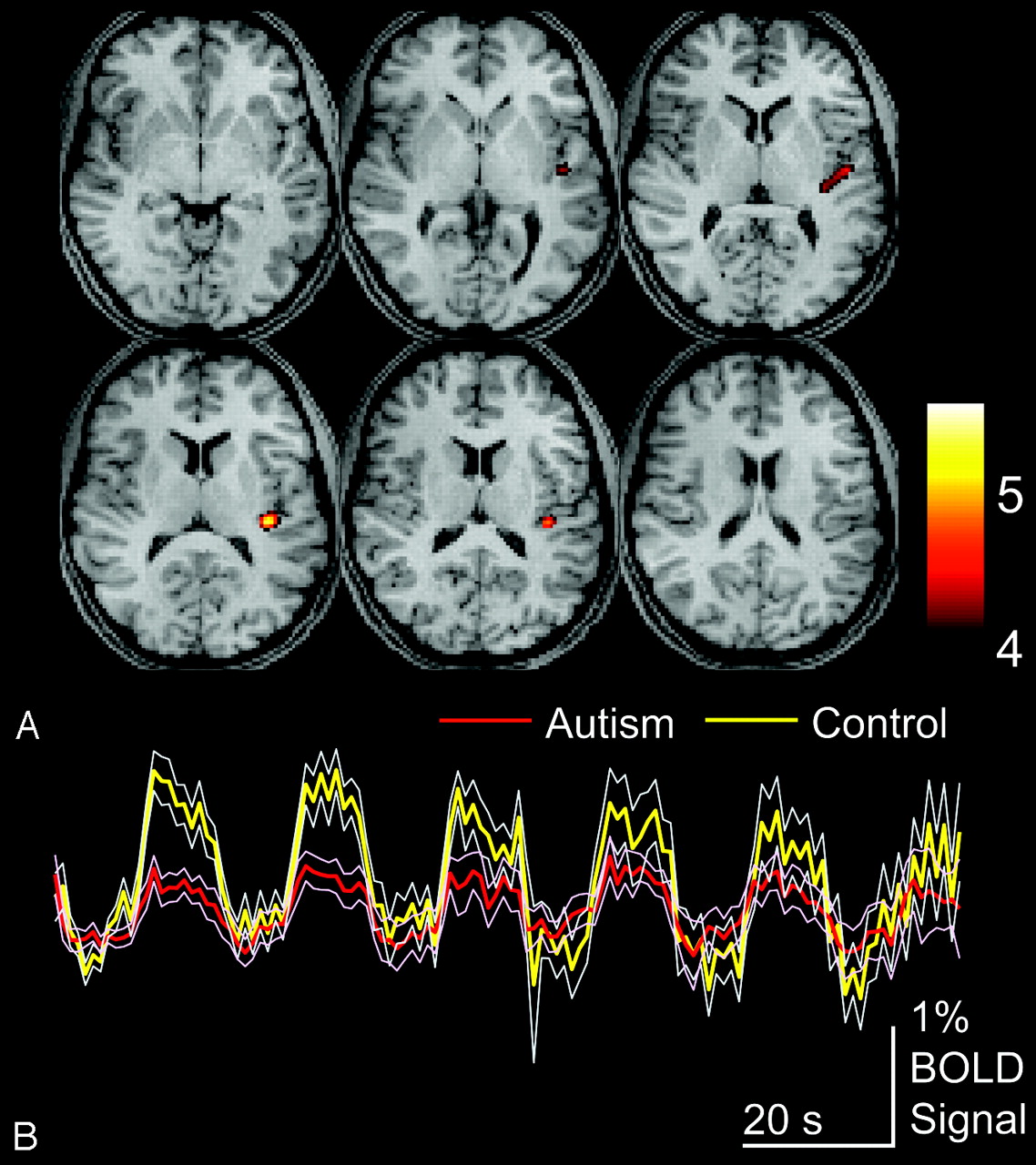
- Fig 3.
Areas of greater activation for control than autism subjects for auditory language tasks. A, Control > autism activation for P < .05, FDR. Color bar shows t-scores. B, Blood oxygen level–dependent time series data for clusters are shown above for the entire auditory language task in autism and control populations, averaged across subjects for each group. Thin traces show standard error of the mean across subjects for each group.
- Fig 4.
Hand preference and language laterality for autism and control subjects. Histograms show the number of subjects exhibiting scores between −100 and 100 (Edinburgh Handedness Inventory) or between −1 and 1 (functional MR imaging [fMRI] laterality index), in which 100 represents strong right-handedness and 1 represents strong left-hemispheric language dominance.
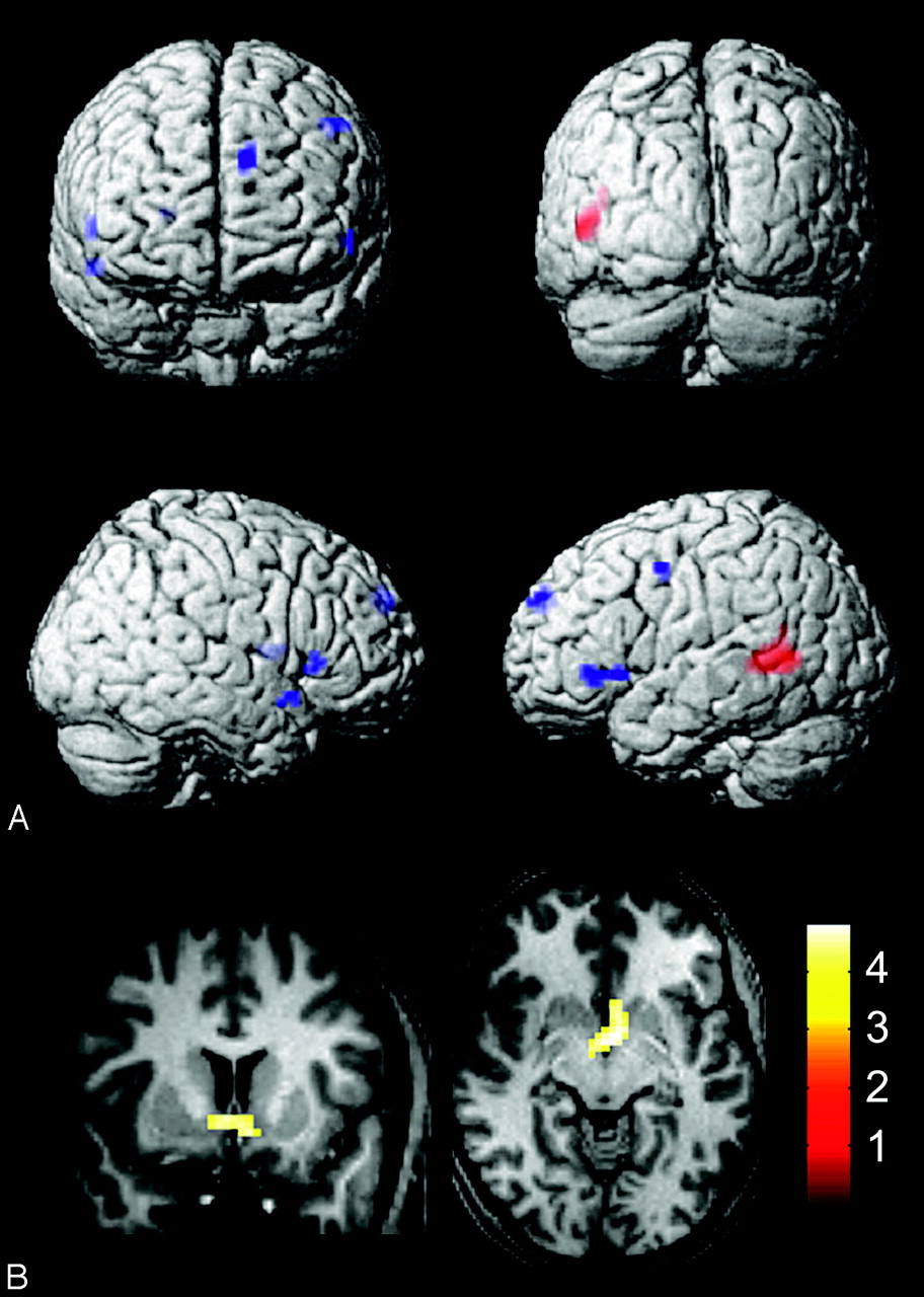
- Fig 5.
Associations between auditory language activation and covariables for all subjects. A, Activity associated with higher receptive-language (Clinical Evaluation of Language Fundamentals, 3rd ed) scores (red, P < .05, FDR) and higher verbal intelligence quotients (blue, P < .001, uncorrected). B, Activity associated with younger age (P < .05, FDR).


Tables
Age (yr) Edinburgh Handedness Inventory Verbal IQ (WAIS III or WASI) Performance IQ (WAIS III or WASI) CELF-3 fMRI Laterality Index (L − R)/(L + R) Autism mean (n = 26) 21.7 67.7 106.0 102.8 83.6 0.23 Autism SD 6.4 38.5 22.2 16.7 27.1 0.56 Autism range 12–35 −73–100 69–139 67–135 50–125 −1.0–1.0 Control mean 22.5 78.3 121.8 116.3 114.2 0.59 Control SD 6.3 28.7 12.7 16.4 9.4 0.33 Control range 9–32 7–100 97–140 90–155 92–122 −0.11–0.95 P value (2-tailed t-test) 0.70 0.38 0.017 0.027 0.0012 0.030 -
Note:—IQ indicates intelligence quotient; CELF-3, Clinical Evaluation of Language Fundamentals, 3rd ed; fMRI, functional MR imaging; R, right; L, left; WAIS, Wechsler Adult Intelligence Scale; WASI, Wechsler Abbreviated Scale of Intelligence.
-
Region MNI Coordinates, Statistical t-Score Control Group Autism Group X Y Z t X Y Z t Left primary auditory cortex −57 −16 1 17.3 −60 −19 1 13.5 Left posterior insula −39 −31 16 12.5 −39 −31 16 6.9 Left anterior temporal pole −51 11 −11 10.9 −51 14 −11 6.3 Left posterior superior temporal gyrus (Wernicke) −63 −40 13 9.4 −63 −40 13 10.2 Left dorsolateral prefrontal cortex −54 23 22 8.7 −54 20 22 7.5 Left posterior inferior frontal gyrus (Broca) −45 23 −2 8.5 −45 23 −2 6.0 Left lateral premotor cortex −45 −1 55 6.5 −45 −1 55 8.0 Left thalamus −6 −7 10 6.5 −6 −7 10 4.2 Left inferior colliculus −12 −31 −5 6.4 −9 −34 −5 5.2 Left caudate head −18 5 16 5.5 −12 −4 16 4.7 Bilateral supplementary motor area −3 11 61 8.2 −3 8 61 7.0 Right primary auditory cortex 57 −16 1 17.3 57 −19 1 16.2 Right anterior temporal pole 54 11 −11 8.3 54 11 −11 4.7 Right frontoinsular cortex 36 20 10 6.0 33 23 1 3.9 Right inferior colliculus 12 −31 −8 6.0 6 −37 −8 3.6 Right posterior superior temporal gyrus (Wernicke) 51 −40 13 5.7 51 −40 13 8.9 Right posterior inferior frontal gyrus (Broca) 48 20 4 5.0 45 25 0 3.0 Right lateral premotor cortex 48 −4 55 3.5 54 −4 49 5.8 -
Note:—MNI indicates Montreal Neurological Institute.
-
Test Region X Y Z t-Score P (uncorrected) Voxels CELF-3 Left posterior middle temporal gyrus −45 −43 4 4.67 1.09E-05 53 VIQ Left dorsomedial prefrontal cortex −12 56 34 4.18 5.60E-05 14 VIQ Left posterior inferior frontal gyrus (Broca) −54 26 −2 3.64 3.14E-04 13 VIQ Left lateral premotor cortex −48 −1 46 3.58 3.70E-04 12 VIQ Right putamen 24 2 10 3.72 2.50E-04 7 VIQ Right posterior inferior frontal gyrus (Broca) 54 23 4 3.55 4.20E-04 5 VIQ Right anterior temporal pole 54 11 −11 3.49 4.90E-04 5 -
Note:—VIQ indicates verbal IQ.
-
In this issue


Jump to section
Related Articles
Cited By...
- Parsing Brain Network Specialization: A Replication and Expansion of Wang et al. (2014)
- Reduced Lateralization of Multiple Functional Brain Networks in Autistic Males
- Maladaptive Laterality in Cortical Networks Related to Social Communication in Autism Spectrum Disorder
- Maladaptive laterality in cortical networks related to social communication in autism spectrum disorder
- Reduced language lateralization in autism and the broader autism phenotype as assessed with robust individual-subjects analyses
- The small and efficient language network of polyglots and hyper-polyglots