
Article Figures & Data
Figures
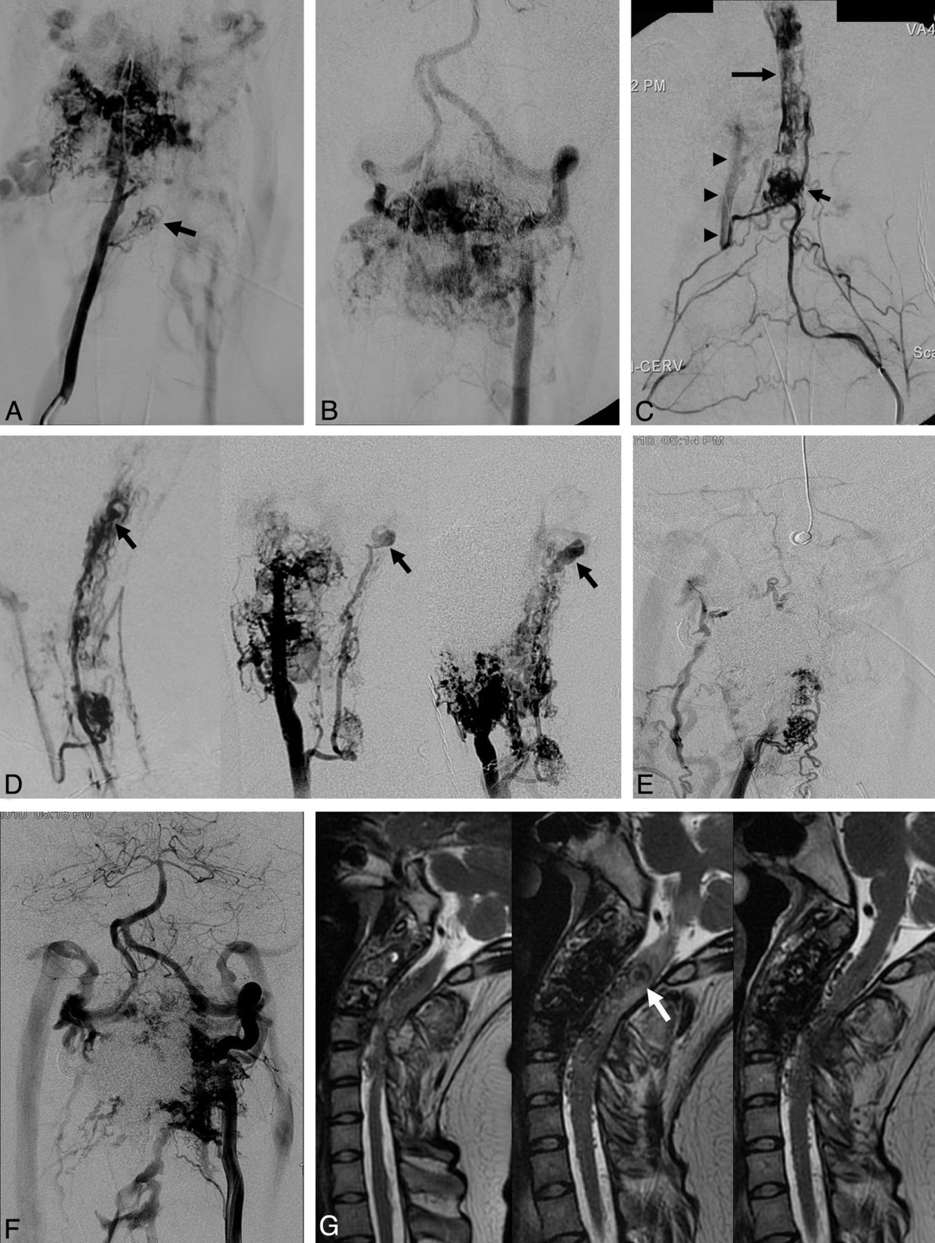
- Fig 1.
Case 1. A, Right vertebral artery angiogram before the first treatment demonstrates an intramedullary nidus at the C5 level (arrow) and paravertebral/vertebral fistulas at the C2 and C3 levels. B, Left vertebral artery angiogram demonstrates paravertebral/vertebral fistulas at the C2 and C3 levels. The distal right vertebral artery is opacified in a retrograde fashion due to steal phenomenon. C, Left dorsocervical artery angiogram at the time of the second treatment demonstrates a radiculomedullary artery opacifying the C5 level nidus (short arrow) as well as a second discrete nidus at the C2 and C3 levels (long arrow). There is anastomotic visualization of the right vertebral artery (arrowheads) (compare with A). D, Series of angiographic appearances of the spinal cord AVMs in the lateral view. Right: At the time of the second treatment (lateral view of C) showing 2 discrete nidi. There is a small ASA aneurysm (arrow). Middle: Right vertebral artery injection 21 months later showing enlargement of the ASA aneurysm (arrow). Left: Right vertebral artery angiogram 18 months later from the middle when the patient developed acute left-sided weakness showing further enlargement of the ASA aneurysm (arrow). The distal right vertebral artery is occluded by previous Onyx embolization. E, Right vertebral artery angiogram after the last (eighth) embolization shows minimal residual paravertebral/vertebral fistulas. Spinal cord AVMs are persistently opacified. F, Left vertebral artery angiogram after the last (eighth) embolization shows residual paravertebral/vertebral fistulas. G, Serial T2-weighted sagittal MR images of the cervical spine. Left: After the fourth n-BCA embolization showing resolution of T2 signal abnormality seen before the treatment (not shown). Middle: Before the seventh embolization after development of acute left-sided weakness. There is increased T2 high-signal abnormality involving the upper cervical cord and medulla surrounding the aneurysm (arrow). Signal voids obscuring the C2 and C3 vertebral bodies are due to Onyx. Left: Three months after n-BCA embolization of the anterior spinal artery aneurysm, showing disappearance of the aneurysm and resolution of the T2 high-signal abnormality. ASA indicates anterior spinal artery.
- Fig 2.
Case 2. A, Right internal iliac artery angiogram in 1995 at the time of presentation demonstrates S2 radicular arteriovenous shunt draining to the perimedullary veins. B, Left T10 intercostal artery angiogram in 1995 demonstrates 2 nidi of spinal cord AVMs (arrows) at the lower spinal cord and conus supplied by the ASA and the left PSA. There is a small ASA aneurysm (arrowhead). C, Left T10 intercostal artery angiogram in 2004 after the patient developed recurrent SAH demonstrates enlargement of the ASA aneurysm with a pseudoaneurysm at the tip (arrow). The AVM at the conus is fistulous and is supplied by the ASA. The left PSA feeder is smaller than that in 1995. D, Left T10 intercostal artery angiogram after coil embolization of the aneurysm demonstrates occlusion of most of the aneurysm, particularly the pseudoaneurysm. E, Left T10 intercostal artery angiogram in 2007 shows remodeling and progressive occlusion of the ASA aneurysm. There is increased caliber irregularity of the radiculomedullary artery and the ASA (arrowheads). F, Left T10 intercostal artery angiogram in 2011 shows spontaneous occlusion of the ASA. The size of the left PSA has increased. The ASA is partially reconstituted by the right PSA (not shown). ASA indicates anterior spinal artery; PSA, posterior spinal artery.


In this issue


Jump to section
Related Articles
Cited By...
- Arteriovenous shunts of the cervical spine: patient demographics, presentation, patterns of high-risk venous drainage, and updated classification
- A proposed grading system for spinal cord arteriovenous shunts
- Clinical features and outcomes of perimedullary arteriovenous fistulas: comparison between micro- and macro-type lesions
- Combined endovascular and surgical management of a case of Cobb syndrome