
Article Figures & Data
Figures
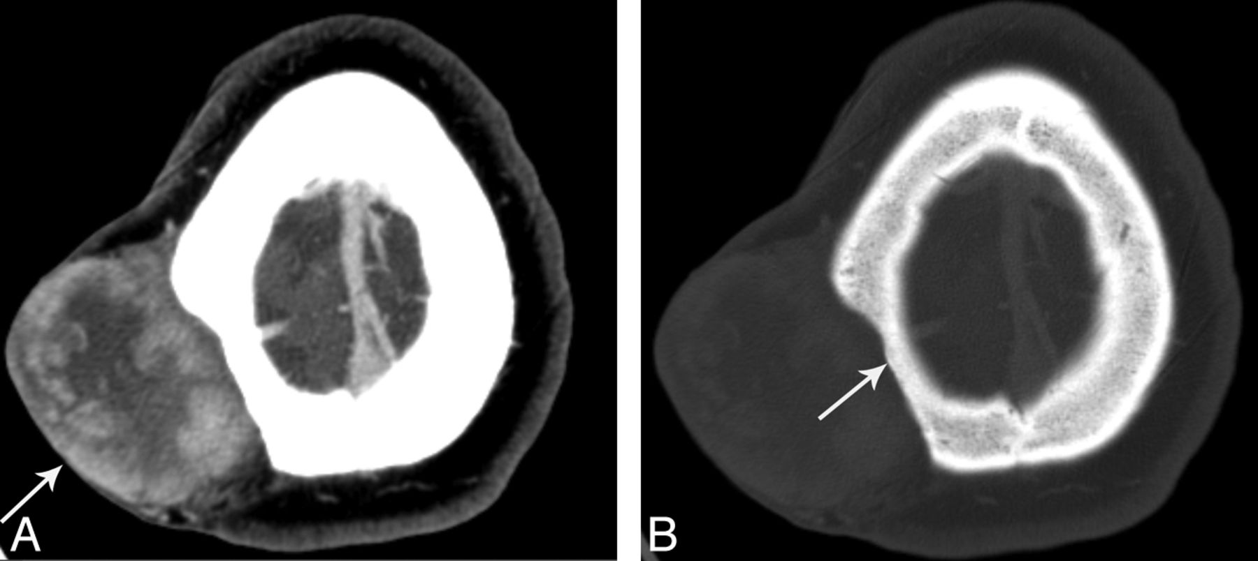
- Fig 1.
A 31-year-old woman (patient 10) with DFSP of the right parietal scalp. Contrast-enhanced axial CT in soft tissue (A) and bone window (B) shows a 6.1 × 4.8-cm heterogeneously enhancing mass (A, arrow) that bulges the skin surface outward and thins underlying calvaria (B, arrow).
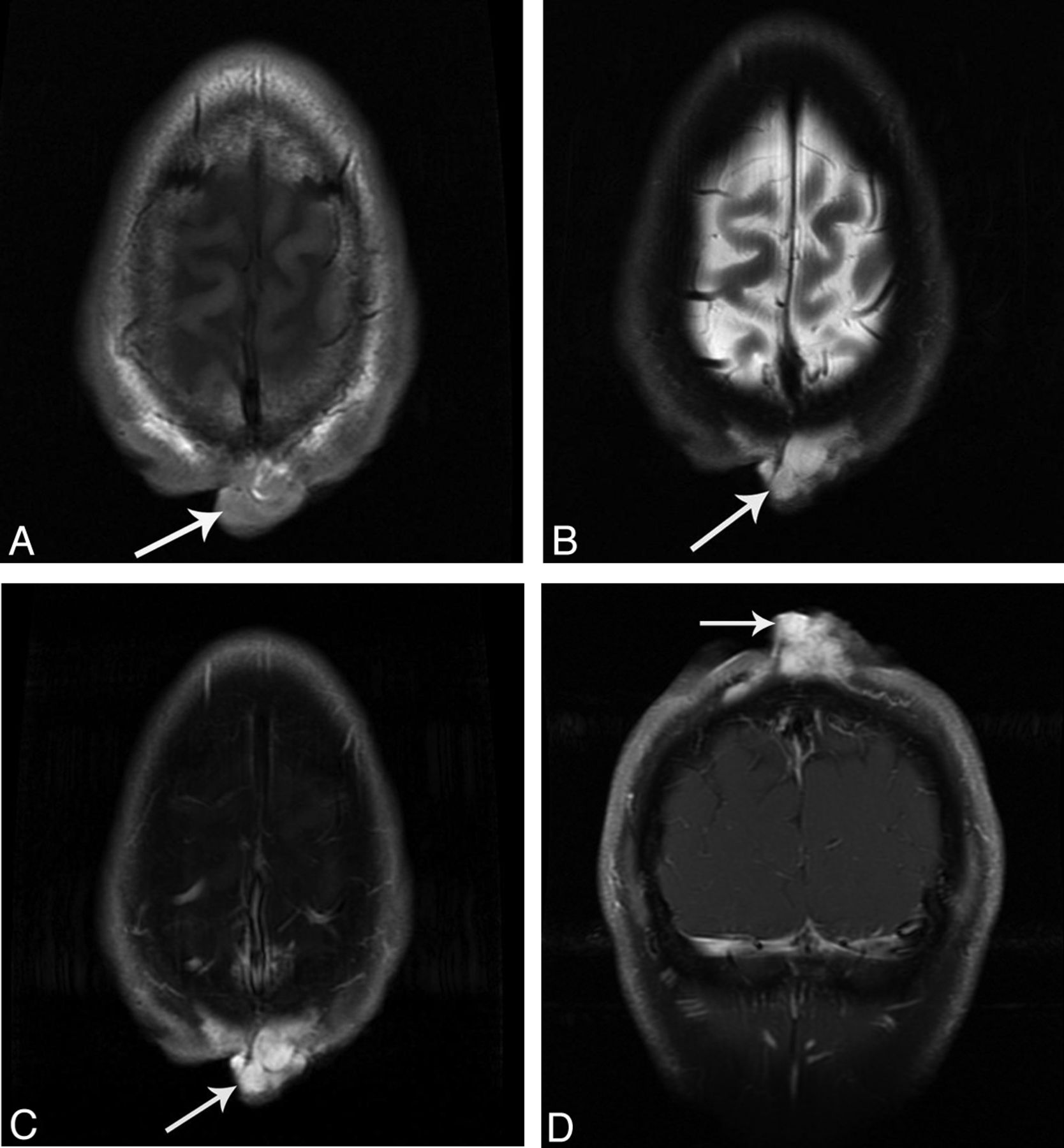
- Fig 2.
A 31-year-old man (patient 1) with DFSP of the parietal midline scalp. MR imaging shows a 3.5 × 1.8-cm T1-isointense, T2-hyperintense, coalescing nodular mass with marked homogeneous enhancement in the parietal midline scalp (arrows). A, Axial noncontrast T1. B, Fast spin-echo T2 with fat saturation. C, T1 postcontrast axial with fat saturation. D, T1 postcontrast coronal with fat saturation.
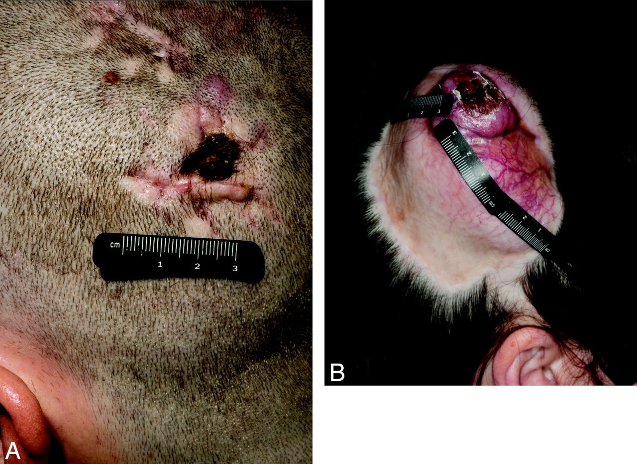
- Fig 3.
Photographs of 2 patients with DFSP. A, Patient 16, a 25-year-old man with a large DFSP of the scalp extending from the midline near the vertex down to the left lower parietal region with subcutaneous infiltration. B, Patient 10, a 31-year-old woman with a large DFSP of the right paramedian posterior parietal scalp.
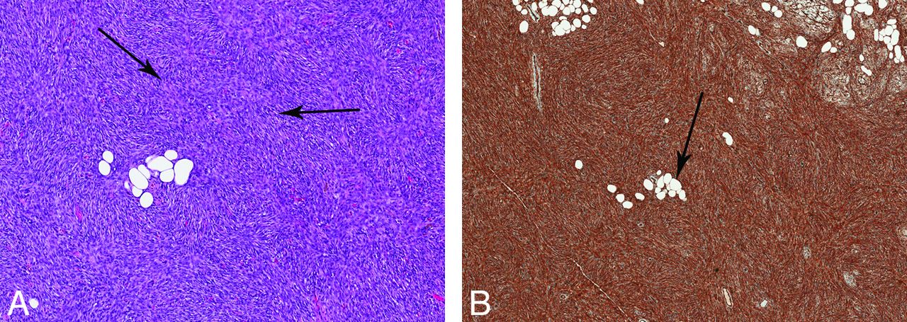
- Fig 4.
Pathologic specimen of DFSP. A, Hematoxylin and eosin stain. Elongated spindle cells with a storiform (whorled) pattern arranged around foci of collagen or vascular spaces, radiating outward (arrows). B, CD34 stain. The specimen is positive for CD34 staining. Fat entrapment is noted with a honeycomb pattern (arrow).


Tables
Patient demographics and image appearance
Patient Age, Years/Sex Location CT Enhancement Pattern MR Appearance (T2) MR T1 Enhancement 1 31/M Parietal midline scalp NA Hyperintense Marked homogeneous 2 68/M Right cheek Homogeneous NA NA 3 37/M Frontal paramedian scalp Homogeneous NA NA 4 66/M Frontal midline scalp Heterogeneous Hyperintense Marked homogeneous 5 39/F Frontal midline scalp NA Hyperintense Mild 6 46/F Frontal midline scalp NA Hyperintense Marked homogeneous 7 25/F Right parietal paramedian scalp Minimal NA NA 8 29/F Right pre-auricular region Homogeneous NA NA 9 58/F Frontal midline scalp NA Hyperintense Marked homogeneous 10 31/F Right parietal scalp near midline Heterogeneous Hyperintense Marked heterogeneous 11 45/M Left parietal paramedian scalp Minimal NA NA 12 42/M Parietal scalp near midline NA Hyperintense Marked heterogeneous 13 48/F Parietal midline scalp Homogeneous NA NA 14 15/F Frontal paramedian scalp NA Iso-/hypointense Marked homogeneous 15 40/M Frontal paramedian scalp Homogeneous NA NA 16 25/M Parietal scalp to midline Heterogeneous NA NA 17 64/F Midline chin Minimal NA NA 18 36/M Midline occipital scalp NA Hyperintense Marked heterogeneous 19 34/M Right frontal scalp NA Hypointense Marked homogeneous 20 72/F Right posterior neck Homogeneous Hyperintense Marked heterogeneous 21 55/M Left face to midline NA Iso-/hypointense Marked homogeneous 22 42/M Parieto-occipital scalp Homogeneous Hypointense Marked homogeneous 23 31/F Parietal scalp to midline Homogeneous NA NA 24 40/F Parietal scalp NA Hyperintense Marked homogeneous Note:—NA indicates not applicable.


Jump to section
Related Articles
Cited By...
- No citing articles found.