Advertisement

Main menu

  • Home
  • Content
    • Current Issue
    • Accepted Manuscripts
    • Article Preview
    • Past Issue Archive
    • Video Articles
    • AJNR Case Collection
    • Case of the Week Archive
    • Case of the Month Archive
    • Classic Case Archive
  • Special Collections
    • AJNR Awards
    • Low-Field MRI
    • Alzheimer Disease
    • ASNR Foundation Special Collection
    • Photon-Counting CT
    • View All
  • Multimedia
    • AJNR Podcasts
    • AJNR SCANtastic
    • Trainee Corner
    • MRI Safety Corner
    • Imaging Protocols
  • For Authors
    • Submit a Manuscript
    • Submit a Video Article
    • Submit an eLetter to the Editor/Response
    • Manuscript Submission Guidelines
    • Statistical Tips
    • Fast Publishing of Accepted Manuscripts
    • Graphical Abstract Preparation
    • Imaging Protocol Submission
    • Author Policies
  • About Us
    • About AJNR
    • Editorial Board
    • Editorial Board Alumni
  • More
    • Become a Reviewer/Academy of Reviewers
    • Subscribers
    • Permissions
    • Alerts
    • Feedback
    • Advertisers
    • ASNR Home

User menu

  • Alerts
  • Log in

Search

  • Advanced search
American Journal of Neuroradiology

American Journal of Neuroradiology

ASHNR American Society of Functional Neuroradiology ASHNR American Society of Pediatric Neuroradiology ASSR
  • Alerts
  • Log in

Advanced Search

  • Home
  • Content
    • Current Issue
    • Accepted Manuscripts
    • Article Preview
    • Past Issue Archive
    • Video Articles
    • AJNR Case Collection
    • Case of the Week Archive
    • Case of the Month Archive
    • Classic Case Archive
  • Special Collections
    • AJNR Awards
    • Low-Field MRI
    • Alzheimer Disease
    • ASNR Foundation Special Collection
    • Photon-Counting CT
    • View All
  • Multimedia
    • AJNR Podcasts
    • AJNR SCANtastic
    • Trainee Corner
    • MRI Safety Corner
    • Imaging Protocols
  • For Authors
    • Submit a Manuscript
    • Submit a Video Article
    • Submit an eLetter to the Editor/Response
    • Manuscript Submission Guidelines
    • Statistical Tips
    • Fast Publishing of Accepted Manuscripts
    • Graphical Abstract Preparation
    • Imaging Protocol Submission
    • Author Policies
  • About Us
    • About AJNR
    • Editorial Board
    • Editorial Board Alumni
  • More
    • Become a Reviewer/Academy of Reviewers
    • Subscribers
    • Permissions
    • Alerts
    • Feedback
    • Advertisers
    • ASNR Home
  • Follow AJNR on Twitter
  • Visit AJNR on Facebook
  • Follow AJNR on Instagram
  • Join AJNR on LinkedIn
  • RSS Feeds

AJNR Awards, New Junior Editors, and more. Read the latest AJNR updates

Research ArticleHead and Neck Imaging
Open Access

MRI Texture Analysis Predicts p53 Status in Head and Neck Squamous Cell Carcinoma

M. Dang, J.T. Lysack, T. Wu, T.W. Matthews, S.P. Chandarana, N.T. Brockton, P. Bose, G. Bansal, H. Cheng, J.R. Mitchell and J.C. Dort
American Journal of Neuroradiology January 2015, 36 (1) 166-170; DOI: https://doi.org/10.3174/ajnr.A4110
M. Dang
bDepartment of Radiology (M.D., J.T.L.), University of Calgary, Calgary, Alberta, Canada
  • Find this author on Google Scholar
  • Find this author on PubMed
  • Search for this author on this site
J.T. Lysack
bDepartment of Radiology (M.D., J.T.L.), University of Calgary, Calgary, Alberta, Canada
  • Find this author on Google Scholar
  • Find this author on PubMed
  • Search for this author on this site
T. Wu
eSchool of Computing, Informatics, Decision Systems Engineering (G.B., T.W.), Arizona State University, Tempe, Arizona.
  • Find this author on Google Scholar
  • Find this author on PubMed
  • Search for this author on this site
T.W. Matthews
aFrom the Section of Otolaryngology–Head and Neck Surgery (T.W.M., S.P.C., P.B., J.C.D.)
  • Find this author on Google Scholar
  • Find this author on PubMed
  • Search for this author on this site
S.P. Chandarana
aFrom the Section of Otolaryngology–Head and Neck Surgery (T.W.M., S.P.C., P.B., J.C.D.)
  • Find this author on Google Scholar
  • Find this author on PubMed
  • Search for this author on this site
N.T. Brockton
dDepartment of Population Health Research (N.T.B.), Alberta Health Services, Calgary, Alberta, Canada
  • Find this author on Google Scholar
  • Find this author on PubMed
  • Search for this author on this site
P. Bose
aFrom the Section of Otolaryngology–Head and Neck Surgery (T.W.M., S.P.C., P.B., J.C.D.)
  • Find this author on Google Scholar
  • Find this author on PubMed
  • Search for this author on this site
G. Bansal
eSchool of Computing, Informatics, Decision Systems Engineering (G.B., T.W.), Arizona State University, Tempe, Arizona.
  • Find this author on Google Scholar
  • Find this author on PubMed
  • Search for this author on this site
H. Cheng
cDepartment of Radiology (H.C., J.R.M.), Mayo Clinic College of Medicine, Scottsdale, Arizona
  • Find this author on Google Scholar
  • Find this author on PubMed
  • Search for this author on this site
J.R. Mitchell
cDepartment of Radiology (H.C., J.R.M.), Mayo Clinic College of Medicine, Scottsdale, Arizona
  • Find this author on Google Scholar
  • Find this author on PubMed
  • Search for this author on this site
J.C. Dort
aFrom the Section of Otolaryngology–Head and Neck Surgery (T.W.M., S.P.C., P.B., J.C.D.)
  • Find this author on Google Scholar
  • Find this author on PubMed
  • Search for this author on this site
  • Article
  • Figures & Data
  • Info & Metrics
  • Responses
  • References
  • PDF
Loading

Article Figures & Data

Figures

  • Tables
  • Fig 1.
    • Download figure
    • Open in new tab
    • Download powerpoint
    Fig 1.

    Sample of the FTFT-2D input data for texture analysis. A, Original DICOM MR image (T2WI). B, Region-of-interest mask of the tumor.
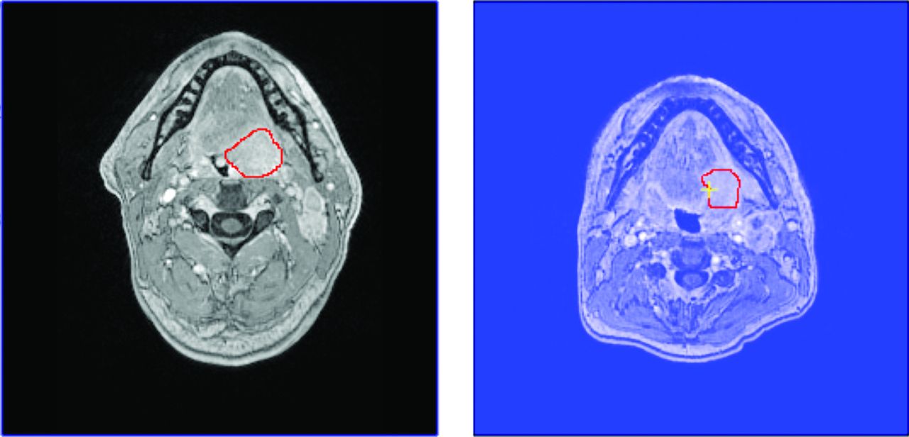
  • Fig 2.
    • Download figure
    • Open in new tab
    • Download powerpoint
    Fig 2.

    Tumors outlined on source MR images (Post-Gad T1WI) of 256 × 256 image size. The tumor in the left image is p53-negative. The tumor in the right image is p53-positive.

  • Fig 3.
    • Download figure
    • Open in new tab
    • Download powerpoint
    Fig 3.

    Graphic display of the average local spectrum for a p53-negative tumor (left image, black shades) and for a p53-positive tumor (right image, blue shades). The average amplitudes of ST at individual pixels were obtained by using FTFT-2D by using the same intensity range and scale for the 2 plots. Darker shades indicate higher ST amplitudes.

  • Fig 4.
    • Download figure
    • Open in new tab
    • Download powerpoint
    Fig 4.

    Radial ST amplitude plots for a p53-negative (black line) tumor and a p53-positive (blue line) tumor (the same tumor as in Fig 3).

Tables

  • Figures
    • View popup
    Table 1:

    Clinical characteristicsa

    Characteristicp53-Positivep53-Negative
    Female11
    Male77
    Age (yr) (mean)56 ± 1356 ± 11
    T213
    T321
    T454
    N111
    N277
    • ↵a All P values are not significant.

    • View popup
    Table 2:

    Discriminatory texture features

    Texture AttributeMRI SequenceRadial Frequency Band (cycles/mm)
    Average value of the local spectrumADC map0.189–0.231
    0.269–0.307
    0.307–0.344
    0.344–0.382
    Post-Gad T1WI0.382–0.420
    SD of the local spectrumADC map0.307–0.344
    0.496–0.533
    Post-Gad T1WI0.420–0.458
    0.458–0.496
    0.496–0.533
    Maximum value of the local spectrumADC map0.231–0.269
    0.269–0.307
    Post-Gad T1WI0.382–0.420
    0.496–0.533
    T2WI0.496–0.533
    • Note:—SD indicates standard deviation.

    • View popup
    Table 3:

    BN classification of p53 status

    TP RateFP RatePrecisionRecallF-MeasureROC Area
    p53-Positive0.6250.3750.6250.6250.6250.680
    p53-Negative0.6250.3750.6250.6250.6250.680
    Weighted average0.6250.3750.6250.6250.6250.680
    • Note:—BN indicates Bayesian network; FP, false-positive; TP, true-positive; ROC, receiver operating characteristic.

    • View popup
    Table 4:

    Discriminatory texture features

    Texture AttributeMRI SequenceRadial Frequency Band (cycles/mm)
    Average value of the local spectrumADC map0.269–0.307
    SD of the local spectrumPost-Gad T1WI0.458–0.496
    0.496–0.533
    Maximum value of the local spectrumADC map0.269–0.307
    Post-Gad T1WI0.382–0.420
    0.496–0.533
    T2WI0.496–0.533
    • View popup
    Table 5:

    BN classification results

    TP RateFP RatePrecisionRecallF-MeasureROC Areap53 Status
    p53-Positive0.8750.2500.7780.8750.8240.742Positive
    p53-Negative0.7500.1250.8570.7500.8000.742Negative
    Weighted average0.8130.1880.8170.8130.8120.742
    • Note:—BN indicates Bayesian network; FP, false-positive; ROC, receiver operating characteristic; TP, true-positive.

PreviousNext
Back to top

In this issue

American Journal of Neuroradiology: 36 (1)
American Journal of Neuroradiology
Vol. 36, Issue 1
1 Jan 2015
  • Table of Contents
  • Index by author
  • Complete Issue (PDF)
Advertisement
Print
Download PDF
Email Article

Thank you for your interest in spreading the word on American Journal of Neuroradiology.

NOTE: We only request your email address so that the person you are recommending the page to knows that you wanted them to see it, and that it is not junk mail. We do not capture any email address.

Enter multiple addresses on separate lines or separate them with commas.
MRI Texture Analysis Predicts p53 Status in Head and Neck Squamous Cell Carcinoma
(Your Name) has sent you a message from American Journal of Neuroradiology
(Your Name) thought you would like to see the American Journal of Neuroradiology web site.
CAPTCHA
This question is for testing whether or not you are a human visitor and to prevent automated spam submissions.
Cite this article
M. Dang, J.T. Lysack, T. Wu, T.W. Matthews, S.P. Chandarana, N.T. Brockton, P. Bose, G. Bansal, H. Cheng, J.R. Mitchell, J.C. Dort
MRI Texture Analysis Predicts p53 Status in Head and Neck Squamous Cell Carcinoma
American Journal of Neuroradiology Jan 2015, 36 (1) 166-170; DOI: 10.3174/ajnr.A4110

Citation Manager Formats

  • BibTeX
  • Bookends
  • EasyBib
  • EndNote (tagged)
  • EndNote 8 (xml)
  • Medlars
  • Mendeley
  • Papers
  • RefWorks Tagged
  • Ref Manager
  • RIS
  • Zotero
0 Responses
Respond to this article
Share
Bookmark this article
MRI Texture Analysis Predicts p53 Status in Head and Neck Squamous Cell Carcinoma
M. Dang, J.T. Lysack, T. Wu, T.W. Matthews, S.P. Chandarana, N.T. Brockton, P. Bose, G. Bansal, H. Cheng, J.R. Mitchell, J.C. Dort
American Journal of Neuroradiology Jan 2015, 36 (1) 166-170; DOI: 10.3174/ajnr.A4110
del.icio.us logo Twitter logo Facebook logo Mendeley logo
  • Tweet Widget
  • Facebook Like
  • Google Plus One
Purchase

Jump to section

  • Article
    • Abstract
    • ABBREVIATIONS:
    • Materials and Methods
    • Results
    • Discussion
    • Conclusions
    • Footnotes
    • References
  • Figures & Data
  • Info & Metrics
  • Responses
  • References
  • PDF

Related Articles

  • No related articles found.
  • PubMed
  • Google Scholar

Cited By...

  • Prediction of Tumor Grade and Nodal Status in Oropharyngeal and Oral Cavity Squamous-cell Carcinoma Using a Radiomic Approach
  • MRI texture analysis as a predictor of tumor recurrence or progression in patients with clinically non-functioning pituitary adenomas
  • Additional Clinical Value for PET/MRI in Oncology: Moving Beyond Simple Diagnosis
  • MRI-Based Texture Analysis to Differentiate Sinonasal Squamous Cell Carcinoma from Inverted Papilloma
  • Crossref
  • Google Scholar

This article has not yet been cited by articles in journals that are participating in Crossref Cited-by Linking.

More in this TOC Section

  • Hydrops Herniation into the Semicircular Canals
  • Post SRS Peritumoral Hyperintense Signal of VSs
  • DIVr of NPC Predicted Outcome
Show more HEAD AND NECK IMAGING

Similar Articles

Advertisement

Indexed Content

  • Current Issue
  • Accepted Manuscripts
  • Article Preview
  • Past Issues
  • Editorials
  • Editor's Choice
  • Fellows' Journal Club
  • Letters to the Editor
  • Video Articles

Cases

  • Case Collection
  • Archive - Case of the Week
  • Archive - Case of the Month
  • Archive - Classic Case

More from AJNR

  • Trainee Corner
  • Imaging Protocols
  • MRI Safety Corner
  • Book Reviews

Multimedia

  • AJNR Podcasts
  • AJNR Scantastics

Resources

  • Turnaround Time
  • Submit a Manuscript
  • Submit a Video Article
  • Submit an eLetter to the Editor/Response
  • Manuscript Submission Guidelines
  • Statistical Tips
  • Fast Publishing of Accepted Manuscripts
  • Graphical Abstract Preparation
  • Imaging Protocol Submission
  • Evidence-Based Medicine Level Guide
  • Publishing Checklists
  • Author Policies
  • Become a Reviewer/Academy of Reviewers
  • News and Updates

About Us

  • About AJNR
  • Editorial Board
  • Editorial Board Alumni
  • Alerts
  • Permissions
  • Not an AJNR Subscriber? Join Now
  • Advertise with Us
  • Librarian Resources
  • Feedback
  • Terms and Conditions
  • AJNR Editorial Board Alumni

American Society of Neuroradiology

  • Not an ASNR Member? Join Now

© 2025 by the American Society of Neuroradiology All rights, including for text and data mining, AI training, and similar technologies, are reserved.
Print ISSN: 0195-6108 Online ISSN: 1936-959X

Powered by HighWire