Advertisement

Main menu

  • Home
  • Content
    • Current Issue
    • Accepted Manuscripts
    • Article Preview
    • Past Issue Archive
    • AJNR Case Collection
    • Case of the Week Archive
    • Classic Case Archive
    • Case of the Month Archive
  • Special Collections
    • Spinal CSF Leak Articles (Jan 2020-June 2024)
    • 2024 AJNR Journal Awards
    • Most Impactful AJNR Articles
  • Multimedia
    • AJNR Podcast
    • AJNR Scantastics
    • Video Articles
  • For Authors
    • Submit a Manuscript
    • Author Policies
    • Fast publishing of Accepted Manuscripts
    • Graphical Abstract Preparation
    • Manuscript Submission Guidelines
    • Imaging Protocol Submission
    • Submit a Case for the Case Collection
  • About Us
    • About AJNR
    • Editorial Board
  • More
    • Become a Reviewer/Academy of Reviewers
    • Subscribers
    • Permissions
    • Alerts
    • Feedback
    • Advertisers
    • ASNR Home
  • Other Publications
    • ajnr

User menu

  • Alerts
  • Log in

Search

  • Advanced search
American Journal of Neuroradiology

American Journal of Neuroradiology

ASHNR American Society of Functional Neuroradiology ASHNR American Society of Pediatric Neuroradiology ASSR
  • Alerts
  • Log in

Advanced Search

  • Home
  • Content
    • Current Issue
    • Accepted Manuscripts
    • Article Preview
    • Past Issue Archive
    • AJNR Case Collection
    • Case of the Week Archive
    • Classic Case Archive
    • Case of the Month Archive
  • Special Collections
    • Spinal CSF Leak Articles (Jan 2020-June 2024)
    • 2024 AJNR Journal Awards
    • Most Impactful AJNR Articles
  • Multimedia
    • AJNR Podcast
    • AJNR Scantastics
    • Video Articles
  • For Authors
    • Submit a Manuscript
    • Author Policies
    • Fast publishing of Accepted Manuscripts
    • Graphical Abstract Preparation
    • Manuscript Submission Guidelines
    • Imaging Protocol Submission
    • Submit a Case for the Case Collection
  • About Us
    • About AJNR
    • Editorial Board
  • More
    • Become a Reviewer/Academy of Reviewers
    • Subscribers
    • Permissions
    • Alerts
    • Feedback
    • Advertisers
    • ASNR Home
  • Follow AJNR on Twitter
  • Visit AJNR on Facebook
  • Follow AJNR on Instagram
  • Join AJNR on LinkedIn
  • RSS Feeds

Welcome to the new AJNR, Updated Hall of Fame, and more. Read the full announcements.


AJNR is seeking candidates for the position of Associate Section Editor, AJNR Case Collection. Read the full announcement.


Getting new auth cookie, if you see this message a lot, tell someone!
Research ArticleAdult Brain

Impact of Skull Defects on the Role of CTA for Brain Death Confirmation

D.M. Nunes, A.C.M. Maia, R.C. Boni and A.J. da Rocha
American Journal of Neuroradiology July 2019, 40 (7) 1177-1183; DOI: https://doi.org/10.3174/ajnr.A6100
D.M. Nunes
aFrom the Section of Neuroradiology (D.M.N., A.C.M.M.Jr., A.J.d.R.)
  • Find this author on Google Scholar
  • Find this author on PubMed
  • Search for this author on this site
  • ORCID record for D.M. Nunes
A.C.M. Maia Jr
aFrom the Section of Neuroradiology (D.M.N., A.C.M.M.Jr., A.J.d.R.)
  • Find this author on Google Scholar
  • Find this author on PubMed
  • Search for this author on this site
  • ORCID record for A.C.M. Maia Jr
R.C. Boni
bSection of Organ Procurement Organization (R.C.B.), Santa Casa de Misericórdia de São Paulo, São Paulo, SP, Brazil.
  • Find this author on Google Scholar
  • Find this author on PubMed
  • Search for this author on this site
  • ORCID record for R.C. Boni
A.J. da Rocha
aFrom the Section of Neuroradiology (D.M.N., A.C.M.M.Jr., A.J.d.R.)
  • Find this author on Google Scholar
  • Find this author on PubMed
  • Search for this author on this site
  • ORCID record for A.J. da Rocha
  • Article
  • Figures & Data
  • Info & Metrics
  • Responses
  • References
  • PDF
Loading

Article Figures & Data

Figures

  • Tables
  • Fig 1.
    • Download figure
    • Open in new tab
    • Download powerpoint
    Fig 1.

    Flow chart of patients with a clinical diagnosis of brain death who underwent a CTA protocol proposed by Frampas et al,12 according to the presence and type of skull defect. Application of the interpretation criteria of the images proposed by Frampas et al.
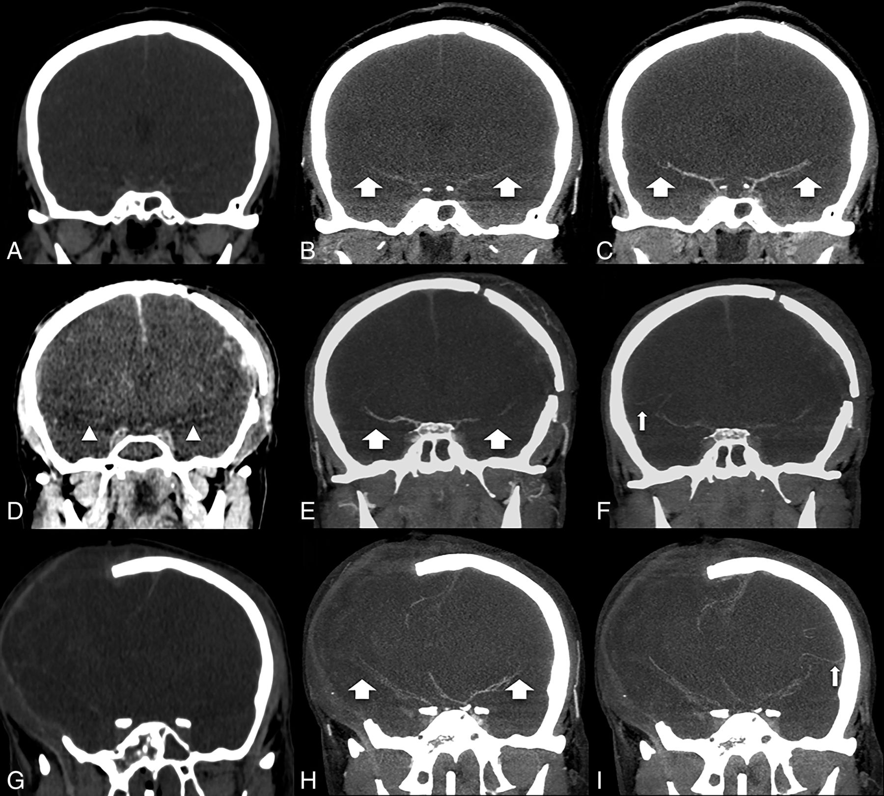
  • Fig 2.
    • Download figure
    • Open in new tab
    • Download powerpoint
    Fig 2.

    Comparison of CT images without contrast medium (A, D, and G) in the arterial (B, E, and H) and venous (C, F, and I) phases of CTA (MIP reformations in coronal plane) from 3 distinct patients with a brain death clinical diagnosis. In a patient with an intact skull (upper row, A–C), opacification was observed in the M1–M2 branches (broad arrows) in the arterial (B) and venous (C) phases and was more intense in the late phase. In a patient with craniotomy (middle row, D–F), one can appreciate the relative hyperattenuation of both M1 branches mimicking vascular opacification in noncontrast CT (arrowheads in D). Opacification was observed in both M2 branches (broad arrows in the arterial phase, E) and the right M3 branch in the venous phase (thin arrow in F), a phenomenon known as stasis filling. In a patient presenting with craniectomy (lower row, G–I), opacification was observed in both M2 branches (broad arrows in the arterial phase, H), the right M3 branch, and the left M4 branch in the venous phase (thin arrow in I) due stasis filling, a false-negative case of BD.
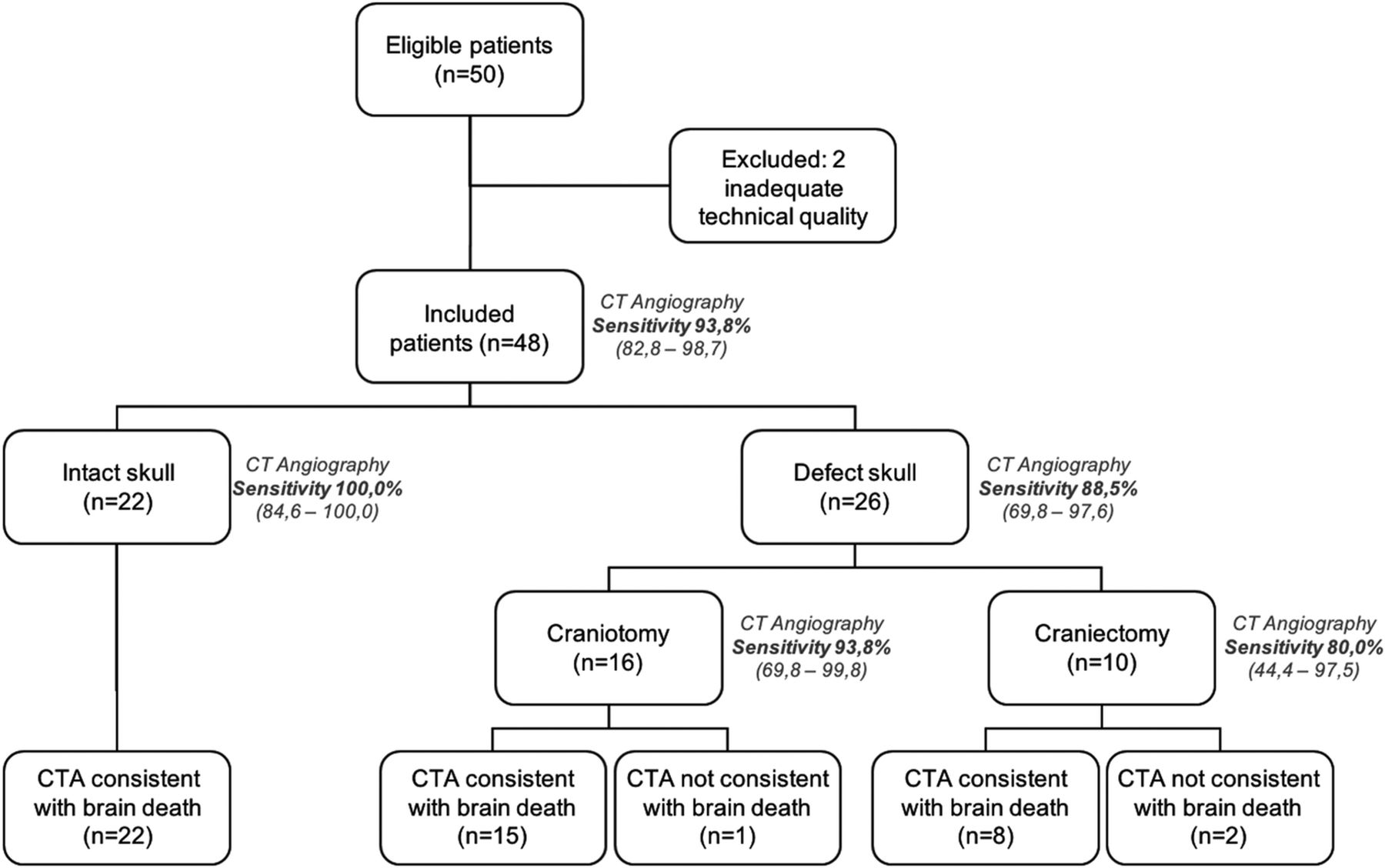
  • Fig 3.
    • Download figure
    • Open in new tab
    • Download powerpoint
    Fig 3.

    Flow chart of patients with a clinical diagnosis of brain death who underwent the CT angiography protocol proposed by Frampas et al, according to the presence and type of skull defect. Application of the interpretation criteria of the images of the modified Frampas criteria.

Tables

  • Figures
  • Characterization of the groups according to the presence and type of skull defecta

    VariableControls, ISk (n = 22)Patients with BDP Value
    ISk (n = 22)Craniotomy (n = 16)Craniectomy (n = 10)
    Age (yr)57.6 ± 16.442.8 ± 21.943.7 ± 18.847.4 ± 9.2.811
    Male sex12 (54.5)13 (59.1)8 (50.0)7 (70.0).600
    Etiology.287
        SAH0 (0.0)6 (27.3)8 (50.0)3 (30.0)
        TBI0 (0.0)4 (18.2)4 (25.0)5 (50.0)
        HS0 (0.0)4 (18.2)1 (6.3)0 (0.0)
        IS22 (100)4 (18.2)0 (0.0)0 (0.0)
        Tumor0 (0.0)1 (4.5)2 (12.5)1 (10.0)
        GW0 (0.0)1 (4.5)1 (6.3)0 (0.0)
        Other0 (0.0)2 (9.1)0 (0.0)1 (10.0)
    Time interval from BD to CTA (min)–659670483.618
    (274–964)(269–1013)(236–713)
    • Note:—SAH indicates subarachnoid hemorrhage; TBI, traumatic brain injury; HS, hemorrhagic stroke; IS, ischemic stroke; GW, gunshot wound; ISk, intact skull; BD, brain death; -, no data.

    • ↵a Data are presented as No. (%), except for age (mean ± SD) and time (median and quartile). P values are for comparison of the subgroups (ISk, craniotomy, and craniectomy).

PreviousNext
Back to top

In this issue

American Journal of Neuroradiology: 40 (7)
American Journal of Neuroradiology
Vol. 40, Issue 7
1 Jul 2019
  • Table of Contents
  • Index by author
  • Complete Issue (PDF)
Advertisement
Print
Download PDF
Email Article

Thank you for your interest in spreading the word on American Journal of Neuroradiology.

NOTE: We only request your email address so that the person you are recommending the page to knows that you wanted them to see it, and that it is not junk mail. We do not capture any email address.

Enter multiple addresses on separate lines or separate them with commas.
Impact of Skull Defects on the Role of CTA for Brain Death Confirmation
(Your Name) has sent you a message from American Journal of Neuroradiology
(Your Name) thought you would like to see the American Journal of Neuroradiology web site.
CAPTCHA
This question is for testing whether or not you are a human visitor and to prevent automated spam submissions.
Cite this article
D.M. Nunes, A.C.M. Maia, R.C. Boni, A.J. da Rocha
Impact of Skull Defects on the Role of CTA for Brain Death Confirmation
American Journal of Neuroradiology Jul 2019, 40 (7) 1177-1183; DOI: 10.3174/ajnr.A6100

Citation Manager Formats

  • BibTeX
  • Bookends
  • EasyBib
  • EndNote (tagged)
  • EndNote 8 (xml)
  • Medlars
  • Mendeley
  • Papers
  • RefWorks Tagged
  • Ref Manager
  • RIS
  • Zotero
0 Responses
Respond to this article
Share
Bookmark this article
Impact of Skull Defects on the Role of CTA for Brain Death Confirmation
D.M. Nunes, A.C.M. Maia, R.C. Boni, A.J. da Rocha
American Journal of Neuroradiology Jul 2019, 40 (7) 1177-1183; DOI: 10.3174/ajnr.A6100
del.icio.us logo Twitter logo Facebook logo Mendeley logo
  • Tweet Widget
  • Facebook Like
  • Google Plus One
Purchase

Jump to section

  • Article
    • Abstract
    • ABBREVIATIONS:
    • Materials and Methods
    • Results
    • Discussion
    • Conclusions
    • Acknowledgments
    • REFERENCES
  • Figures & Data
  • Info & Metrics
  • Responses
  • References
  • PDF

Related Articles

  • No related articles found.
  • PubMed
  • Google Scholar

Cited By...

  • No citing articles found.
  • Crossref (16)
  • Google Scholar

This article has been cited by the following articles in journals that are participating in Crossref Cited-by Linking.

  • Assessment of Cerebral Circulatory Arrest via CT Angiography and CT Perfusion in Brain Death Confirmation
    Asli Irmak Akdogan, Yeliz Pekcevik, Hilal Sahin, Ridvan Pekcevik
    Korean Journal of Radiology 2021 22 3
  • Computed tomography angiography scoring systems and the role of skull defects in the confirmation of brain death
    Petros Zampakis, Vasilios Panagiotopoulos, Christina Kalogeropoulou, Maria Karachaliou, Diamanto Aretha, Nektarios Sioulas, Sofia Dimoulia, Dimitrios Karnabatidis, Fotini Fligou
    Scientific Reports 2021 11 1
  • Computed tomography angiography in the diagnosis of brain death: Implementation and results in Germany
    Olaf Hoffmann, Farid Salih, Florian Masuhr
    European Journal of Neurology 2024 31 4
  • Brain death in children: is computed tomography angiography reliable as an ancillary test?
    Eda Almus, Erhan Bıyıklı, Özge Yapıcı, Ferdağ Almus, Feyza İnceköy Girgin, Nilüfer Öztürk
    Pediatric Radiology 2023 53 1
  • Comparison of early- and late-phase CT angiography findings in brain death
    Ethem Murat Arsava, Cansu Ayvacioglu Cagan, Ekim Gumeler, Safak Parlak, Kader Karli Oguz, Mehmet Akif Topcuoglu
    Journal of Neurology 2022 269 11
  • Cerebral blood vessels and perfusion in the pediatric brain death: five cases studied by neuroimaging
    Quan Wang, Tong Yu, Jie Wu, Jun Liu, Hengmiao Gao, Xiaojuan Tao, Huanyu Luo, Suyun Qian, Yun Peng
    Neuroradiology 2022 64 8
  • Le scanner de mort encéphalique
    F. Clarençon, V. Degos, D. Leclercq, E. Shotar, S. Lenck, K. Premat, D. Galanaud, N. Sourour, D. Dormont
    Journal d'imagerie diagnostique et interventionnelle 2020 3 3
  • Reliability of CT angiography scoring systems used for brain death and the effect of cranial interventions on the results
    Rifat Ozpar, Mehmet Tonkaz, Nermin Kelebek Girgin, Muhittin Bodur, Yasemin Dinc, Hasan Kocaeli, Bahattin Hakyemez
    Clinical Imaging 2021 79
  • Self-harm, suicide and brain death: the role of the radiologist
    G. Lambe, P. Dempsey, M. Bolger, F. Bolster
    Clinical Radiology 2024 79 4
  • CT-Angiographie zum Nachweis des zerebralen Zirkulationsstillstandes: Aktualisierung der Kriterien
    Heinrich Lanfermann, Stephan A. Brandt
    Clinical Neuroradiology 2022 32 4

More in this TOC Section

  • Diagnostic Neuroradiology of Monoclonal Antibodies
  • Segmentation of Brain Metastases with BLAST
  • NCCT vs. MRI for Brain Atrophy in Acute Stroke
Show more Adult Brain

Similar Articles

Advertisement

Indexed Content

  • Current Issue
  • Accepted Manuscripts
  • Article Preview
  • Past Issues
  • Editorials
  • Editors Choice
  • Fellow Journal Club
  • Letters to the Editor

Cases

  • Case Collection
  • Archive - Case of the Week
  • Archive - Case of the Month
  • Archive - Classic Case

Special Collections

  • Special Collections

Resources

  • News and Updates
  • Turn around Times
  • Submit a Manuscript
  • Author Policies
  • Manuscript Submission Guidelines
  • Evidence-Based Medicine Level Guide
  • Publishing Checklists
  • Graphical Abstract Preparation
  • Imaging Protocol Submission
  • Submit a Case
  • Become a Reviewer/Academy of Reviewers
  • Get Peer Review Credit from Publons

Multimedia

  • AJNR Podcast
  • AJNR SCANtastic
  • Video Articles

About Us

  • About AJNR
  • Editorial Board
  • Not an AJNR Subscriber? Join Now
  • Alerts
  • Feedback
  • Advertise with us
  • Librarian Resources
  • Permissions
  • Terms and Conditions

American Society of Neuroradiology

  • Not an ASNR Member? Join Now

© 2025 by the American Society of Neuroradiology All rights, including for text and data mining, AI training, and similar technologies, are reserved.
Print ISSN: 0195-6108 Online ISSN: 1936-959X

Powered by HighWire