
Article Figures & Data
Figures
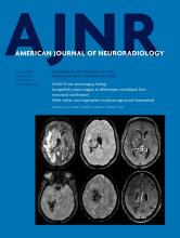
- Fig 1.
Tractography findings in selected patients with lissencephaly and polymicrogyria. T1WI, direction-encoded color maps, and tractography reconstructions in 2 patients with LIS (A and B) and 2 with PMG (C and D) are shown. For each patient, a comparable tractography reconstruction from the age-matched healthy control template is shown in the last column for comparison. A, An irregular (shorter and smaller) bilateral cingulum (arrows on DEC map) in a patient with pachygyria and thick subcortical band heterotopia. B, A bilateral, irregular (smaller and distorted) superior longitudinal fasciculus (arrows on DEC map) in a patient with posterior-quadrant pachygyria and a thick SBH. C, Bilateral irregular (smaller and thinner) CG (arrows on DEC map) in a patient with generalized PMG. D, An irregular (shorter and distorted) right inferior fronto-occipital fasciculus (IFOF; arrows) in a patient with unilateral right peri-Sylvian PMG. The left IFOF (arrowheads) has a normal appearance. r. indicates right.
- Fig 2.
The superior longitudinal fasciculus involvement in peri-Sylvian polymicrogyria. T1WI, direction-encoded color maps, and tractography reconstructions of the SLF in 3 patients with exemplary peri-Sylvian PMG (A–C). Sagittal T1-weighted images and tractography refer to the right hemisphere. A comparable SLF tractography reconstruction from the age-matched healthy control template is shown in the last row for comparison (D). Patient A has bilateral focal peri-Sylvian PMG in the opercular regions (arrows on T1WI). Both SLFs look normal on both the DEC map (arrows on DEC map) and tractography reconstruction. Patient B has bilateral diffuse peri-Sylvian PMG (arrows on T1WI). Both SLFs (arrows on DEC maps) are irregular in appearance. The right SLF tractography is notably shorter and smaller compared with the right SLF from a control. Patient C has bilateral diffuse peri-Sylvian PMG extending to adjacent cortical regions. The right SLF is absent and could not be reconstructed (symbolized by the X), and the left SLF is irregular in appearance (smaller compared with the left SLF from a control).
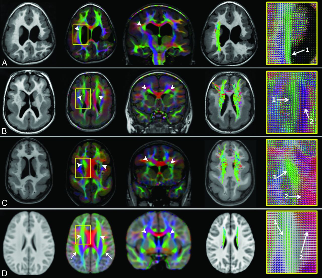
- Fig 3.
Thickened superior fronto-occipital fasciculus. T1WI, direction-encoded color maps with associated fiber orientation distribution glyph profiles, and tractography reconstructions in 3 patients with thickened SFOFs (A–C) and in the age-matched healthy control template for comparison (D). Patient A has bilateral peri-Sylvian PMG plus a left schizencephaly and a thickened right SFOF (arrowheads). The left SFOF cannot be recognized. Patients B and C demonstrate pachygyria and subcortical band heterotopia and bilateral thickened SFOF (arrowheads). On FOD glyphs maps, 1 indicates the SFOF (green, predominately anterior-posterior oriented fibers), and 2, the corpus callosum (red, predominately left-right oriented fibers).


Tables
- Table 1:
Study patients by the MCD subgroups and their conventional structural MR imaging findings
MCD Subgroups/Variants No. of Cases Unilateral MCD Prenatal CMV Infection Associated Brain Anomalies Ventricles BGT Hippocampus Brain Stem Cerebellum CC PMG Peri-Sylvian 16 4 1 9 3 5 2 4 5 Frontoparietal 7 2 4 2 0 0 2 2 2 Generalized 4 0 0 4 1 4 0 2 3 Focala 7 2 0 4 2 3 2 3 5 Parieto-occipital 1 1 0 1 0 0 0 0 1 Multifocal 6 2 0 3 0 3 0 1 5 With schizencephaly 1 0 0 1 1 1 1 1 1 Total 42 11 5 24 7 16 7 13 22 LISb Pachygyria with SBH 5 0 0 5 2 3 2 2 4 Pachygyria without SBH 1 0 0 0 1 2 1 2 2 SBH without pachygyria 2 0 0 1 0 0 0 0 2 Total 8 0 0 6 3 5 3 4 8 - Table 2:
Summary of the white matter tract appearance in all study patients presented by each MCD subgroupa
MCD Subgroups No. of Patients SLF CG SFOF IFOF OR-PCR ILF Peri-Sylvian PMG 16 8/16/0/8 27/5/0/0 31/1/0/0 27/5/0/0 20/12/0/0 21/11/0/0 Frontoparietal PMG 7 4/6/0/4 13/1/0/0 12/2/0/0 12/2/0/0 6/8/0/0 7/7/0/0 Frontal PMG 7 3/10/0/1 5/9/0/0 12/1/0/1 12/2/0/0 10/4/0/0 11/3/0/0 Generalized PMG 4 0/2/0/6 1/5/0/2 2/2/0/4 2/6/0/0 0/8/0/0 0/8/0/0 Multifocal PMG 6 9/3/0/0 7/5/0/0 10/0/0/2 12/0/0/0 9/3/0/0 11/1/0/0 Parieto-occipital PMG 1 2/0/0/0 2/0/0/0 2/0/0/0 2/0/0/0 1/1/0/0 2/0/0/0 PMG and schizenchephaly 1 0/0/0/2 0/2/0/0 0/0/1/1 0/2/0/0 0/2/0/0 0/2/0/0 Pachygyria with SBH 5 0/6/0/4 2/8/0/0 2/4/4/0 2/8/0/0 0/10/0/0 0/10/0/0 SBH (no pachygyria) 2 0/4/0/0 2/2/0/0 4/0/0/0 2/2/0/0 0/4/0/0 0/4/0/0 Pachygyria (no SBH) 1 0/2/0/0 0/2/0/0 2/0/0/0 0/2/0/0 2/0/0/0 2/0/0/0 All PMG 42 27/36/0/21 55/27/0/2 69/6/1/8 67/17/0/0 46/38/0/0 52/32/0/0 All LIS 8 0/12/0/4 4/12/0/0 8/4/4/0 4/12/0/0 2/14/0/0 2/14/0/0 ↵a For each white matter tract, the number of tract appearances classified as grades I/IIA/IIB/III is reported.


