Abstract
Summary: We describe the adult radiographic shuntogram, a simple method to evaluate the function and patency of a ventriculoperitoneal or ventriculoatrial shunt. The procedure involves placing contrast material into the valve of a shunt system and following the flow for appropriate clearing of contrast agent from the shunt tubing. Twenty-three studies were obtained in 15 patients in whom shunt malfunction was suspected. The method can be used to establish valve malfunction, ventricular or distal catheter obstruction, and peritoneal encystment.
Ventriculoperitoneal or ventriculoatrial shunt malfunction is a common problem that is difficult to diagnose and manage. Clinical presentation in patients with shunt malfunction is typically nonspecific, and brain imaging often fails to show a change in ventricular size. The radiographic shuntogram is a simple procedure previously described in children that allows objective demonstration of shunt function and patency (1, 2). To our knowledge this is the first report describing use of the shuntogram technique in adults. The procedure is only minimally invasive and is rapid as well as easy to perform.
Technique and Patients
The radiographic shuntogram involves injection of a small quantity of nonionic contrast material into the valve of a ventricular shunt system. Serial filming is performed over a 15-minute period to document forward flow of contrast material and CSF. Pumping the shunt prior to obtaining the final radiographs ensures complete system function. A shuntogram is performed only after systemic infection is excluded as the cause of the patient's neurologic changes.
With the patient supine, the head is turned for optimal display of the shunt system and scout radiographs of the cranial, chest, and abdominal components of the shunt are obtained. The shunt valve is located and scalp hair is generously removed from the valve region. The skin overlying the valve is thoroughly cleansed with Betadine and a sterile drape is placed over the field. By palpation and with fluoroscopic guidance, the valve is entered by using a 25-gauge butterfly needle (Fig 1A), which is connected to adequate extension tubing. Free backflow of CSF confirms proper placement within the valve. A 5-mL syringe is connected to the butterfly system and a minimal amount (1–2 mL) of CSF is gently withdrawn. This provides fluid for analysis and confirms free flow in the ventricular component of the system. Resistance to CSF withdrawal suggests ventricular component obstruction or valve malfunction, and continued withdrawal should not be forced. Only a small amount of CSF should be removed, since excessive withdrawal of CSF would diminish the pressure gradient and can reduce CSF flow through the shunt system.

Fig 1. A and B, Patient 2.
A, A 25-gauge needle is placed in the valve component of a Cordis Unishunt ventriculoperitoneal shunt (closed arrow). A shunt reservoir is seen just proximal to the valve (open arrow).
B, Contrast material is injected into the valve (closed arrow). A shunt reservoir is again seen proximal to the valve (open arrow).
C–G, Patient 6.
C and D, 3-minute films show contrast material in the Cordis Unishunt system (arrow).
E and F, 12-minute films show forward motion of the contrast material in the tube (arrow) with slight early peritoneal spillage (arrowhead). Redundant coiling of the peritoneal component is present with occasional catheter overlap noted.
G, 15-minute film of the abdomen after pumping the valve shows clearing of contrast material from the shunt tube and free spillage into the peritoneal cavity (arrow).
Two to three milliliters of Iohexol 240 mg I/mL (Nycomed, Princeton, NJ) is then injected into the shunt valve (Fig 1B) and serial filming of the cranial, chest, and abdominal components of the shunt system are obtained at 3, 6, 9, and 12 minutes (Fig 1C–F). At 15 minutes, the shunt valve is pumped 30 times to effect complete clearing of the shunt system and final films of all regions are obtained (Fig 1G).
In children, a normal shunt system can be seen to clear contrast material partially or completely from the shunt tube on a 3-minute film (1, 2). Previous investigators used 9 to 10 minutes as the upper limits of normal for identifying spontaneous flow and complete clearing in an unobstructed shunt system (1, 2).
In adults, normal CSF production is 0.3 to 0.4 mL/min (approximately 18–24 mL/h, or 432–576 mL/day) (3). In vivo studies of adult indwelling shunts have exhibited a wide range of measured CSF flow rates in asymptomatic patients and in normally functioning systems (4–8). Slow flow rates of 2 to 5 mL/h (0.03–0.08 mL/min) have commonly been observed, and flow is not constant during the course of a day (5).
The diameter of the peritoneal component of the shunt system is 1.2 mm with a cross-sectional area of 1.13 mm2. At 20 mL/h, contrast material would travel 88 cm in 3 minutes, consistent with rapid shunt clearing observed in healthy children. At 2 mL/h, contrast would travel 8.7 cm in 3 minutes, or 34 cm in 12 minutes.
Guided by these observations in adults, along with the published shuntogram experience in children, we chose 12 minutes as the upper limit of normal for demonstrating spontaneous flow and a functioning shunt in our adult patients. Spontaneous shunt flow was observed when contrast material was seen flowing down the proximal end of the shunt tube, usually coupled with the observation of progressive dilution of contrast within the valve. The shuntogram was labeled functioning and normal if CSF could be withdrawn from the valve and if evidence of spontaneous forward flow of contrast was demonstrated within the first 12 minutes. Free peritoneal spillage should be identified in patients with functioning ventriculoperitoneal shunts.
The abnormal shuntogram can reveal several different shunt problems: 1) ventricular catheter obstruction or valve malfunction is suspected if CSF cannot be freely withdrawn from the valve; 2) valve malfunction is present when contrast material freely refluxes from the valve reservoir into the ventricular system; 3) valve malfunction with inappropriate valve resistance or incomplete obstruction of the shunt system is suggested when contrast material does not empty from the shunt spontaneously but the system can be cleared after pumping the valve; 4) peritoneal obstruction of the distal catheter is suspected if free spillage of contrast into the peritoneal cavity is not present or if inappropriate peritoneal or visceral accumulation of contrast material is identified; and 5) complete distal obstruction is present if contrast material cannot be cleared from the tube after pumping the valve.
The radiographic shuntogram was performed on 23 occasions in 15 adult patients to evaluate ventricular shunt flow and patency. Patients with ventriculoperitoneal shunts had either a Cordis Unishunt (Cordis, Miami Lakes, FL) or Holter valve (Codman, Johnson & Johnson, New Brunswick, NJ) in place for CSF diversion. In a single individual, a Delta valve (Medtronics-PS Medical, Goleta, CA) ventriculoatrial shunt was present. Eight of the patients were women and seven were men. The age range was 37 to 77 years (average age, 57 years). All patients presented with clinical problems compatible with shunt malfunction. Systemic infection was excluded in all patients before the shuntogram procedure was performed. Patient 2 had two shunts in place and both were studied.
Results
The results are summarized in the Table. In four instances, spontaneous shunt flow was documented and these shunts were not surgically revised. In all four patients, progressive dilution of contrast material was noted in the valve. Contrast agent was observed moving down the proximal shunt tube during the course of filming, with tapering and dilution of the contrast column secondary to mixing of inflowing CSF and residual contrast material in the valve. The tapered dilution effect typically extended from the valve overlying the skull to the neck. This tapered dilution allowed documentation of forward flow of CSF but did not allow an accurate quantification of flow rate. In two of the four patients with functioning shunts considered normal, the shunts have remained in place and have not required revision 2 years (patient 6) and 5 years (patient 8) after the shuntogram procedure. In one of the patients in whom the shuntogram was considered normal, shunt revision was subsequently required 3½ years after the procedure. In the other patient with a normal shuntogram, shunt revision was ultimately performed 2 years after the procedure. No complications were encountered from the shuntogram procedure.
Shunt type, imaging results, and surgical findings in patients undergoing a shuntogram
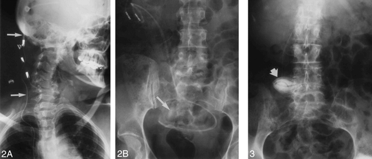
The shuntogram was abnormal with slow or absent flow into the peritoneal cavity in 19 studies. In all 19 instances, the shunt system was revised with excellent results. The most common shunt problem encountered was valve malfunction (11 studies) with the shuntogram demonstrating absence of spontaneous flow but free CSF withdrawal and shunt clearing after pumping at 15 minutes (Fig 2). Distal shunt occlusion with absent post pump clearing occurred in three instances and distal obstruction caused by peritoneal adhesions with an abnormal distal CSF collection was observed in two studies (Fig 3). In the ventriculoatrial shunt system, spontaneous anterograde flow did not occur on the shuntogram and elevated right atrial pressures were found at the time of shunt revision.

Fig 2. Patient 2 (with a Cordis Unishunt system).
A, 12-minute film at the level of the valve shows contrast material in one of two ventriculoperitoneal shunts but no forward motion of contrast agent (arrows). Shunt flow at this point is abnormal.
B, After pumping, the contrast material has cleared from the tube and is seen freely spilling into the peritoneal cavity (arrow). The shunt valve was functioning improperly in this patient and required replacement.
Patient 1 (with a Cordis Unishunt system). 15-minute post pump abdominal film shows encystment of contrast material at the peritoneal end of the drainage catheter (arrow). Abdominal adhesions were lysed and the peritoneal catheter repositioned
Discussion
A ventriculoperitoneal or ventriculoatrial shunt is placed for diversion of CSF in the presence of an obstructed ventricular system or normal pressure hydrocephalus. The shunt consists of several components: ventricular catheter, shunt valve, and distal peritoneal or atrial catheter. A ventricular reservoir is occasionally added for CSF access, and angled or straight connection hardware is used to connect the components. The shunt valves are unidirectional check valves, available in several degrees of resistance: low pressure (2–5 cm H2O), medium pressure (5–9 cm H2O), and high pressure (10–15 cm H2O). Medium-pressure systems are the most typical.
Ventriculoperitoneal shunt malfunction is a common complication of shunt placement (9). In one comprehensive actuarial study, the rate of shunt failure after 1 year was 30%, and 50% of shunts required some form of revision within 6 years of placement (10). This may depend on the type of shunt used as well as the location of the ventricular catheter (11). The confident diagnosis of ventriculoperitoneal shunt malfunction is a difficult and important problem.
Shunt malfunction is caused by ventricular catheter obstruction, valve malfunction, distal catheter obstruction, pressure mismatch, or component disconnection. Shunt infection also causes obstruction, most likely as a result of the accumulation of debris within the shunt system. Patients with shunt malfunction present with nonspecific neurologic changes, such as fever, headache, nausea, and vomiting. These symptoms clearly overlap with typical viral syndromes.
Ventricular catheter obstruction accounts for approximately 40% to 56% of shunt failures, with peritoneal obstruction accounting for approximately 14% to 33% (9). Multiple site obstruction also occurs. Valve obstruction is considered common, but statistics are lacking. Shunt infection is reported to range from 3% to 29%, with an average of 10% to 15%. The frequency of shunt infection appears to be lower in more recent studies, possibly owing to the use of perioperative antibiotics (9).
The radiographic shuntogram is a simple nonemergent examination that can easily be used to evaluate the flow characteristics of a ventriculoperitoneal or ventriculoatrial shunt. Problems such as ventricular catheter obstruction, valve pressure choice, and distal catheter obstruction can often be separated, aiding the neurosurgeon in targeting the part of the shunt requiring revision. Demonstration of normal shunt function directs the neurosurgeon to seek other causes for the patient's clinical changes.
The valvogram was originally introduced by Amador et al (12) in 1969 to assess shunt patency in children. With this technique, the ventricular end of the Holter valve system was assessed by gentle CSF aspiration, and shunt obstruction and distal catheter position were evaluated with contrast injection. In early descriptions of the valvogram or shuntogram technique, larger needles (22–23 gauge) were used to enter the valve, and a variable, often large (2–8 mL), volume of ionic contrast material was injected into the shunt system (12, 13). Nonionic contrast material was introduced and recommended with the availability of Dimer-X, since ventricular reflux was occasionally encountered with nonfunctioning incompetent valves (13, 14). Metrizamide has also been used (15).
Dewey et al (1) described a more standard approach to the shuntogram technique in children, using 25-gauge needles to avoid valve damage, a 2-mL contrast injection, and specific filming intervals to follow contrast progression in the shunt system. Aspiration of CSF after entering the valve was again used to assess flow from the ventricular component of the shunt. Filming was performed during contrast injection and at 3-minute intervals for up to 9 minutes to follow contrast progression in the distal tubing. In their series, normal shunts were easily aspirated and emptied in 3 minutes. Shunt systems were considered abnormal if aspiration was difficult, if contrast failed to clear within 9 minutes, if contrast failed to clear after pumping the valve, or if an obvious disconnection was present. Shunts were considered questionable if clearing required 6 minutes or longer or if clearing occurred in 3 minutes but other problems, such as kinking, mesothelial sleeve formation, or difficulty with aspiration or injection, were encountered.
Savoiardo et al (2) expanded this technique to include children with Pudenz valves as well as shunts with double-dome reservoirs. They advocated placing only 1.5 to 3 mL of contrast material into the shunt system and emphasized that excessive injection could overcome distal obstruction. Contrast progression was again observed with initial filming, followed by films at 3, 6, and 10 minutes, and by post pump films, if required. If clearing of the peritoneal catheter occurred within 10 minutes, the system was considered normal. If clearing was incomplete at 10 minutes but achieved after valve pumping, flow was defined as slow.
The shuntogram technique has been used in children, but its application in the adult has not been previously described. While the principles of injecting contrast material into the valve and serial filming are similar, we believe interpretation of the shuntogram in adults is different from that in children. Children usually require shunt diversion for obstructive hydrocephalus. In adults, shunts are more commonly placed for communicating hydrocephalus or normal pressure hydrocephalus. Normal CSF production is approximately 0.3 to 0.4 mL/min (18–24 mL/h, or 500 mL/day) (3). While this represents normal CSF production, typical flow rates in adult indwelling shunt systems in asymptomatic patients appear to be more variable (4–7). Harbert et al (4), using a nuclear medicine technique, recorded shunt flow rates of 0.04, 0.05, and 0.11 mL/min (2.4, 3.0, and 6.6 mL/h) in three adults with unobstructed shunts. Hara et al (5), using a unique electrolysis technique, encountered variation in shunt flow rates during the course of the day with low flow rates in three functioning shunts of 0.05, 0.10, and 0.12 mL/min (3.0, 6.0, and 7.2 mL/h). Martin et al (7), using MR imaging, showed slow flow rates in four of seven asymptomatic adult patients (4 mL/h, 5 mL/h), including two patients in whom flow rates were below the measurement threshold of 2 mL/h. These reports suggest that shunt flow rates in asymptomatic adults may be slow.
It would be ideal to establish the normal shuntogram features in a series of asymptomatic adults with indwelling shunt systems; however, performing a shuntogram in asymptomatic patients is not an acceptable option, since the procedure is minimally invasive and introduction of bacteria could conceivably occur with resultant shunt infection. Our data suggest that a normal shuntogram in the adult is different from that in children. The four patients with functioning shunts had slow flow rates but did not require shunt revision at the time of evaluation. Two of the patients ultimately required revision 2 and 3½ years, respectively, after being studied, and two remain in place and have never been revised. In children, the normal shuntogram features were established when shunt malfunction was considered in the face of altered mentation but rapid flow of contrast material was documented (1, 2, 12–15).
MR imaging has been used to confirm shunt malfunction, but the technique has met with variable success (7, 16–20). Martin et al (7) and Drake et al (16) used a modified clinical pulse sequence and a specially designed shunt coil for optimal shunt tube signal. Flow within the shunt tubing could be identified, but slower flow below 2 mL/h could not be detected, and patient motion limiting examination utility was occasionally encountered. Castillo et al (17), using a standard head coil and a partial flip angle fast-field-echo T1 technique, was able to identify shunt flow with 0.25 mL/min (15 mL/h) as the lowest limit of flow tested. In two of their patients in whom shunt malfunction was suggested at flow study, no evidence of shunt malfunction was identified at surgery. Chang (18), using standard spin-echo imaging and equating signal intensity with shunt flow speed, demonstrated flow in clinically placed shunts with a lower limit of flow detection, calculated at 1.7 mL/h. A similar lower limit of detectable flow was reported by Norbash et al (19), who used a spin-echo phase-contrast technique in vitro. In another specialized in vitro model, flow was detected and quantified below 2 mL/h (20). The radiographic shuntogram can demonstrate similar slow flow rates but is also able to isolate the portion of the shunt system that is not functioning properly.
It is important to exclude systemic infection before performing a shuntogram. Theoretically, infected blood by-products could track into the shunt, establishing infection. In most patients with ventriculoperitoneal shunt malfunction, the diagnosis is difficult, and acute hydrocephalus requiring emergent decompression is not present.
The radiographic shuntogram offers the ability to isolate the components responsible for malfunction as well as to obtain CSF for culture to exclude shunt infection. The technique offers direct visualization of contrast material and CSF flow. Failure to clear the shunt system after valve pumping absolutely confirms a nonfunctioning system.
The cost of a radiographic shuntogram compares favorably with the cost of MR imaging or a nuclear medicine shunt study. The radiographic shuntogram is much faster to perform than the nuclear study and supplies direct visualization of CSF flow and patency. Contraindications to the shuntogram would include systemic infection or a coagulation abnormality. Allergy to contrast material would likely require premedication.
Acknowledgments
Our thanks to Howard J. Senter and William R. Kemp for their patients, who contributed to the data for this study, and to Luke Lin for his radiologic support.
Footnotes
1 Address reprint requests to Walter S. Bartynski, MD, Department of Radiology, The Western Pennsylvania Hospital, 4800 Friendship Ave, Pittsburgh, PA 15224.
References
- Received May 12, 1999.
- Copyright © American Society of Neuroradiology