Advertisement

Main menu

  • Home
  • Content
    • Current Issue
    • Accepted Manuscripts
    • Article Preview
    • Past Issue Archive
    • AJNR Case Collection
    • Case of the Week Archive
    • Classic Case Archive
    • Case of the Month Archive
  • Special Collections
    • Spinal CSF Leak Articles (Jan 2020-June 2024)
    • 2024 AJNR Journal Awards
    • Most Impactful AJNR Articles
  • Multimedia
    • AJNR Podcast
    • AJNR Scantastics
    • Video Articles
  • For Authors
    • Submit a Manuscript
    • Author Policies
    • Fast publishing of Accepted Manuscripts
    • Graphical Abstract Preparation
    • Manuscript Submission Guidelines
    • Imaging Protocol Submission
    • Submit a Case for the Case Collection
  • About Us
    • About AJNR
    • Editorial Board
  • More
    • Become a Reviewer/Academy of Reviewers
    • Subscribers
    • Permissions
    • Alerts
    • Feedback
    • Advertisers
    • ASNR Home
  • Other Publications
    • ajnr

User menu

  • Alerts
  • Log in

Search

  • Advanced search
American Journal of Neuroradiology

American Journal of Neuroradiology

ASHNR American Society of Functional Neuroradiology ASHNR American Society of Pediatric Neuroradiology ASSR
  • Alerts
  • Log in

Advanced Search

  • Home
  • Content
    • Current Issue
    • Accepted Manuscripts
    • Article Preview
    • Past Issue Archive
    • AJNR Case Collection
    • Case of the Week Archive
    • Classic Case Archive
    • Case of the Month Archive
  • Special Collections
    • Spinal CSF Leak Articles (Jan 2020-June 2024)
    • 2024 AJNR Journal Awards
    • Most Impactful AJNR Articles
  • Multimedia
    • AJNR Podcast
    • AJNR Scantastics
    • Video Articles
  • For Authors
    • Submit a Manuscript
    • Author Policies
    • Fast publishing of Accepted Manuscripts
    • Graphical Abstract Preparation
    • Manuscript Submission Guidelines
    • Imaging Protocol Submission
    • Submit a Case for the Case Collection
  • About Us
    • About AJNR
    • Editorial Board
  • More
    • Become a Reviewer/Academy of Reviewers
    • Subscribers
    • Permissions
    • Alerts
    • Feedback
    • Advertisers
    • ASNR Home
  • Follow AJNR on Twitter
  • Visit AJNR on Facebook
  • Follow AJNR on Instagram
  • Join AJNR on LinkedIn
  • RSS Feeds

Welcome to the new AJNR, Updated Hall of Fame, and more. Read the full announcements.


AJNR is seeking candidates for the position of Associate Section Editor, AJNR Case Collection. Read the full announcement.


OtherINTERVENTIONAL

De Novo Basilar Top Aneurysm in an Elderly Patient Treated with Guglielmi Detachable Coils

Kenta Fujimoto, Ryota Kimura, Jun-ichi Iida, Shoichiro Kawaguchi, Toshisuke Sakaki, Hiroyuki Nakagawa, Kimihiko Kichikawa and Toyohisa Fujita
American Journal of Neuroradiology April 2005, 26 (4) 915-916;
Kenta Fujimoto
  • Find this author on Google Scholar
  • Find this author on PubMed
  • Search for this author on this site
Ryota Kimura
  • Find this author on Google Scholar
  • Find this author on PubMed
  • Search for this author on this site
Jun-ichi Iida
  • Find this author on Google Scholar
  • Find this author on PubMed
  • Search for this author on this site
Shoichiro Kawaguchi
  • Find this author on Google Scholar
  • Find this author on PubMed
  • Search for this author on this site
Toshisuke Sakaki
  • Find this author on Google Scholar
  • Find this author on PubMed
  • Search for this author on this site
Hiroyuki Nakagawa
  • Find this author on Google Scholar
  • Find this author on PubMed
  • Search for this author on this site
Kimihiko Kichikawa
  • Find this author on Google Scholar
  • Find this author on PubMed
  • Search for this author on this site
Toyohisa Fujita
  • Find this author on Google Scholar
  • Find this author on PubMed
  • Search for this author on this site

Abstract

Summary: Ruptured de novo aneurysms, compared with the usual subarachnoid hemorrhage, commonly occur in younger patients and are extremely rare in elderly patients. We discuss their etiology and report the case of a ruptured de novo aneurysm in a 77-year-old woman.

The de novo formation of an aneurysm is the occurrence of new aneurysms in a location previously observed to be normal by an angiography or direct surgical exploration. Although the etiology of de novo aneurysms is unclear, carotid ligation, female sex, a history of subarachnoid hemorrhage, smoking, hypertension, and youth are considered to be risk factors, and cases in elderly patients are extremely rare (1–4). In this report, we describe a case of a ruptured de novo basilar top aneurysm in an elderly patient and discuss their etiology.

Case Report

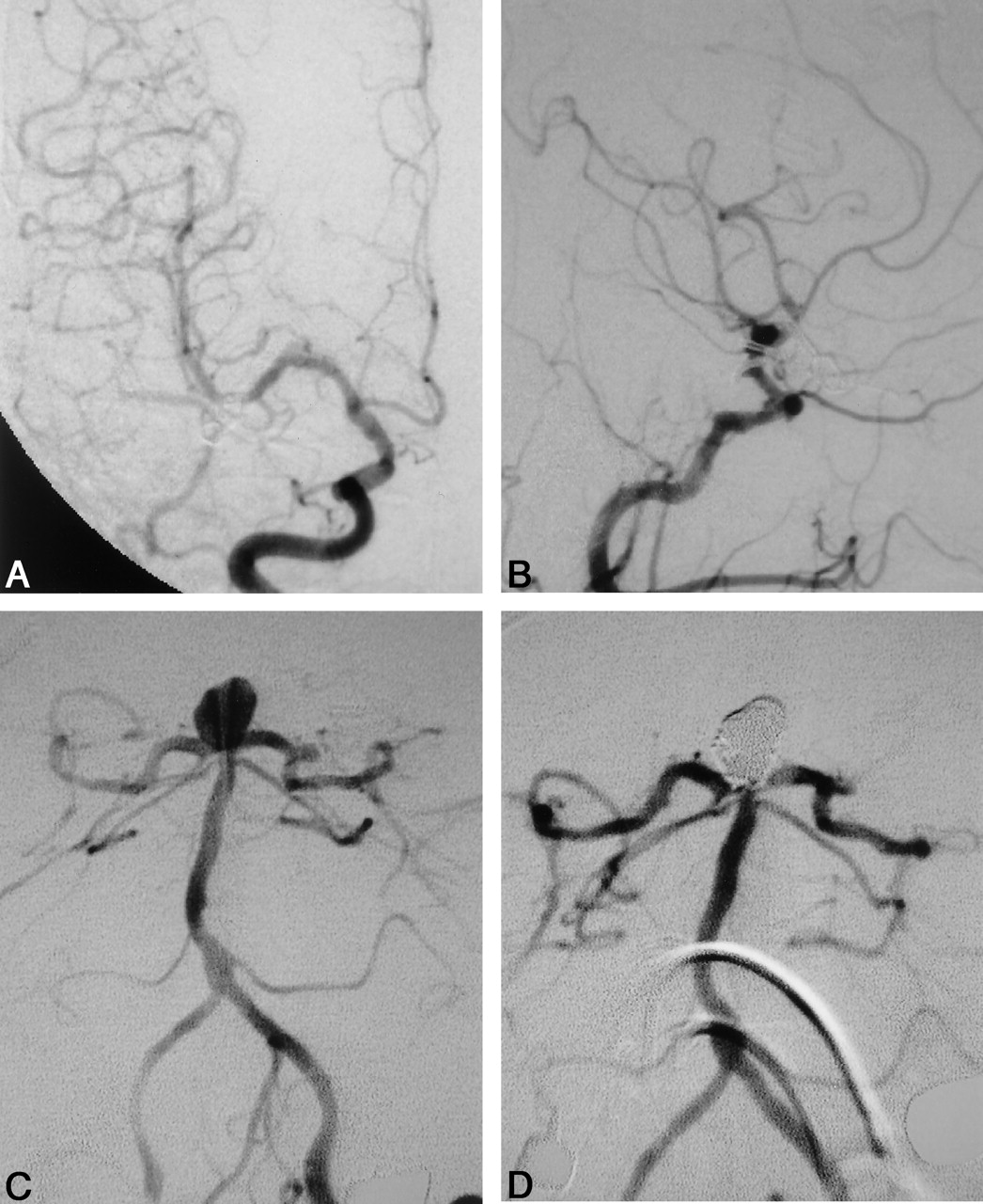
A 77-year-old woman presented with an acute headache followed by nausea and vomiting. At the age of 72, she underwent clipping of the incidental right middle cerebral artery aneurysm, left internal carotid-posterior communicating artery aneurysm and left carotid cave aneurysm (Fig 1A, B). She was nonsmoker, and hypertension was controlled with a calcium antagonist. Neurologic examination yielded normal findings. CT imaging showed an intraventricular hemorrhage (Fig 2), and cerebral angiography revealed a basilar top aneurysm (Fig 3C) that had not been observed in a previous angiography (Fig 1C). No regrowth of the previously treated aneurysms was observed (Fig 3A, B). She underwent successful embolization of the new aneurysm with Guglielmi detachable coils (Fig 3D). Her postoperative course was uneventful, and she was discharged 3 weeks after treatment.

Fig 1.
  • Download figure
  • Open in new tab
  • Download powerpoint
Fig 1.

Initial cerebral angiograms. Anteroposterior (AP) view of the right carotid angiogram displayed a middle cerebral artery aneurysm (A). The lateral view of the left carotid angiogram displayed an internal carotid-posterior communicating artery aneurysm and a carotid cave aneurysm (B). The vertebral angiogram showed no abnormal findings. (C)

Fig 2.
  • Download figure
  • Open in new tab
  • Download powerpoint
Fig 2.

CT scan showing an intraventricular hemorrhage.

Fig 3.
  • Download figure
  • Open in new tab
  • Download powerpoint
Fig 3.

Cerebral angiograms on admission. No regrowth of previously clipped aneurysms was observed. A, AP view of the right carotid angiogram. B, Lateral view of the left carotid angiogram. Vertebral angiography demonstrated a de novo aneurysm at the top of the basilar artery. C, Straight AP view of left vertebral angiogram. D, Successful coil embolization of the aneurysm.

Discussion

De novo formation of aneurysms is the result of the interplay between hemodynamic factors and structural weakness. Several authors have described the clinical characteristics of de novo aneurysms (1–4). The risk factors are considered to be carotid ligation, youth, female sex, a history of subarachnoid hemorrhage, smoking, and hypertension (3, 4). Tonn et al (4) reviewed 50 published cases and reported that the mean age of susceptibility to de novo aneurysms was 43.3 (range, 17–69 years). Patients in whom de novo aneurysms occur are commonly younger than those with the usual subarachnoid hemorrhage (1–4). The occurrence of de novo aneurysms in an elderly patient is extremely rare, and to the best of our knowledge, our patient is the oldest of all reported cases.

In addition, another unusual characteristic of our case lies in the multiplicity of the aneurysms. David et al (5) reported that five of their series of six de novo aneurysms had a history of multiple aneurysms. From reviews of the other previous reports, however, we were unable to uncover a relationship between the multiplicity of aneurysms at the time of the initial episode and formation of the de novo aneurysms. The etiology itself is obscure, but we speculate the possibility that multiple parts of the arterial wall may be “pathologic” in patients with multiple intracranial aneurysms. We maintain that strict follow-up is necessary for patients with known risk factors or multisegmental aneurysms, even if the patient is old, because de novo aneurysms may arise at any time, place, and age.

References

  1. Briganti F, Cirillo S, Caranci F, et al. Development of “de novo” aneurysms following endovascular procedures. Neuroradiology 2002;44:604–609
  2. Rinne JK, Hernesniemi JA. De novo aneurysms: special multiple intracranial aneurysms. Neurosurgery 1993;33:981–985
  3. Sakaki T, Tominaga M, Miyamoto K, et al. Clinical studies of de novo aneurysms. Neurosurgery 1993;32:512–517
  4. Tonn JC, Hoffmann O, Hoffmann E, et al. “De novo” formation of intracranial aneurysms: who is at risk? Neuroradiology 1999;41:674–679
  5. David CA, Vishteh AG, Spetzler RF, et al. Late angiographic follow-up review of surgically treated aneurysms. J Neurosurg 1999;91:396–401
  • Received May 25, 2004.
  • Accepted after revision July 11, 2004.
  • American Society of Neuroradiology
Advertisement

Indexed Content

  • Current Issue
  • Accepted Manuscripts
  • Article Preview
  • Past Issues
  • Editorials
  • Editors Choice
  • Fellow Journal Club
  • Letters to the Editor

Cases

  • Case Collection
  • Archive - Case of the Week
  • Archive - Case of the Month
  • Archive - Classic Case

Special Collections

  • Special Collections

Resources

  • News and Updates
  • Turn around Times
  • Submit a Manuscript
  • Author Policies
  • Manuscript Submission Guidelines
  • Evidence-Based Medicine Level Guide
  • Publishing Checklists
  • Graphical Abstract Preparation
  • Imaging Protocol Submission
  • Submit a Case
  • Become a Reviewer/Academy of Reviewers
  • Get Peer Review Credit from Publons

Multimedia

  • AJNR Podcast
  • AJNR SCANtastic
  • Video Articles

About Us

  • About AJNR
  • Editorial Board
  • Not an AJNR Subscriber? Join Now
  • Alerts
  • Feedback
  • Advertise with us
  • Librarian Resources
  • Permissions
  • Terms and Conditions

American Society of Neuroradiology

  • Not an ASNR Member? Join Now

© 2025 by the American Society of Neuroradiology All rights, including for text and data mining, AI training, and similar technologies, are reserved.
Print ISSN: 0195-6108 Online ISSN: 1936-959X

Powered by HighWire