Advertisement

Main menu

  • Home
  • Content
    • Current Issue
    • Accepted Manuscripts
    • Article Preview
    • Past Issue Archive
    • Video Articles
    • AJNR Case Collection
    • Case of the Week Archive
    • Case of the Month Archive
    • Classic Case Archive
  • Special Collections
    • AJNR Awards
    • Low-Field MRI
    • Alzheimer Disease
    • ASNR Foundation Special Collection
    • Photon-Counting CT
    • View All
  • Multimedia
    • AJNR Podcasts
    • AJNR SCANtastic
    • Trainee Corner
    • MRI Safety Corner
    • Imaging Protocols
  • For Authors
    • Submit a Manuscript
    • Submit a Video Article
    • Submit an eLetter to the Editor/Response
    • Manuscript Submission Guidelines
    • Statistical Tips
    • Fast Publishing of Accepted Manuscripts
    • Graphical Abstract Preparation
    • Imaging Protocol Submission
    • Author Policies
  • About Us
    • About AJNR
    • Editorial Board
    • Editorial Board Alumni
  • More
    • Become a Reviewer/Academy of Reviewers
    • Subscribers
    • Permissions
    • Alerts
    • Feedback
    • Advertisers
    • ASNR Home

User menu

  • Alerts
  • Log in

Search

  • Advanced search
American Journal of Neuroradiology

American Journal of Neuroradiology

ASHNR American Society of Functional Neuroradiology ASHNR American Society of Pediatric Neuroradiology ASSR
  • Alerts
  • Log in

Advanced Search

  • Home
  • Content
    • Current Issue
    • Accepted Manuscripts
    • Article Preview
    • Past Issue Archive
    • Video Articles
    • AJNR Case Collection
    • Case of the Week Archive
    • Case of the Month Archive
    • Classic Case Archive
  • Special Collections
    • AJNR Awards
    • Low-Field MRI
    • Alzheimer Disease
    • ASNR Foundation Special Collection
    • Photon-Counting CT
    • View All
  • Multimedia
    • AJNR Podcasts
    • AJNR SCANtastic
    • Trainee Corner
    • MRI Safety Corner
    • Imaging Protocols
  • For Authors
    • Submit a Manuscript
    • Submit a Video Article
    • Submit an eLetter to the Editor/Response
    • Manuscript Submission Guidelines
    • Statistical Tips
    • Fast Publishing of Accepted Manuscripts
    • Graphical Abstract Preparation
    • Imaging Protocol Submission
    • Author Policies
  • About Us
    • About AJNR
    • Editorial Board
    • Editorial Board Alumni
  • More
    • Become a Reviewer/Academy of Reviewers
    • Subscribers
    • Permissions
    • Alerts
    • Feedback
    • Advertisers
    • ASNR Home
  • Follow AJNR on Twitter
  • Visit AJNR on Facebook
  • Follow AJNR on Instagram
  • Join AJNR on LinkedIn
  • RSS Feeds

AJNR Awards, New Junior Editors, and more. Read the latest AJNR updates

LetterLetter

Is Lumbar Puncture Contraindicated in Patients with Chiari I Malformation?

Sami H. Erbay, Mark G. O’Callaghan and Rafeeque Bhadelia
American Journal of Neuroradiology April 2005, 26 (4) 985;
Sami H. Erbay
  • Find this author on Google Scholar
  • Find this author on PubMed
  • Search for this author on this site
Mark G. O’Callaghan
  • Find this author on Google Scholar
  • Find this author on PubMed
  • Search for this author on this site
Rafeeque Bhadelia
  • Find this author on Google Scholar
  • Find this author on PubMed
  • Search for this author on this site

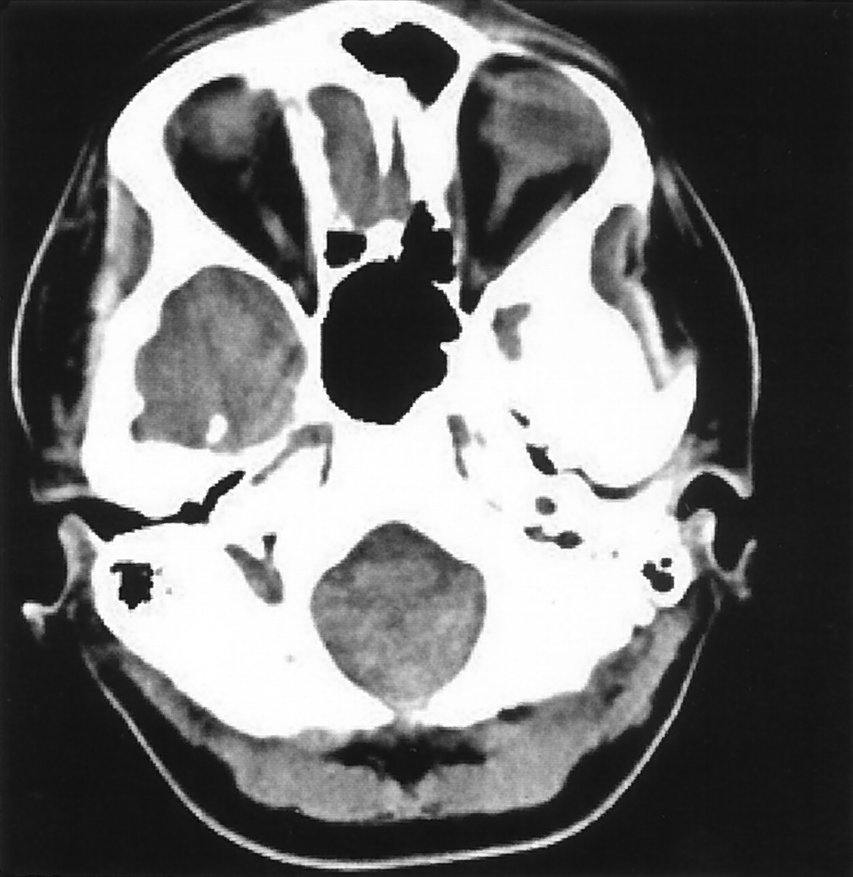
Dynamic brain stem compression due to unsuspected Chiari malformation (CM) may mimic a meningeal process and can pose a diagnostic challenge to both the neuroradiologist and the clinician. CM usually goes undetected on the axial CT examination, where severe beam hardening creates difficulty in evaluating the craniocervical junction (Fig 1). In the setting of CM, the craniospinal pressure dissociation can be worsened if CSF is removed from the spinal canal.

A 23-year-old woman with a clinical diagnosis of Ehlers-Danlos syndrome presented to our emergency room (ER) with increasing nausea, vomiting, and what she described as the worst headache of her life, which radiated to the skull base. There were no visual symptoms or cranial nerve dysfunction. A nonenhanced CT brain study was reported as normal. The patient underwent lumbar puncture (LP) in the ER to exclude subarachnoid hemorrhage. Approximately 14 mL of clear, colorless CSF was removed. The CSF pressures were 21 cm of water on opening and 13 on closing. There were no immediate complications from the LP. She was transferred to the medical ward in a stable condition. Ten hours after the LP, the patient experienced acute worsening of her initial symptoms and had a witnessed episode of apnea lasting 20 seconds. She fully recovered from the episode without any sequela. CM was diagnosed later that afternoon on an MR imaging study, which showed significant stenosis of the foramen magnum evidenced by complete obliteration of the CSF (Fig 2). No focal lesion or abnormal signal intensity was seen in the brain stem.

In a previously reported case of acquired CM, declining visual function was described after placement of a lumboperitoneal shunt. The condition improved after ventriculoperitoneal shunt surgery (1). Clinically, CM is not regarded as an absolute contraindication for an LP. In our patient, it was difficult to establish a definite correlation between the LP and the acute clinical episode described 10 hours later, although there was concern for a causal relationship between the two events. The response to an LP likely depends on the degree of tonsillar herniation and the amount of CSF removed. In our institution, opinion of whether to proceed with LP in the setting of known CM was divided among the clinical staff (neurologists and neurosurgeons). Life-threatening conditions such as subarachnoid hemorrhage or meningitis will always be strong justifications for the LP. In known cases of CM, better visualization of the craniocervical junction with MR imaging should probably be attempted to assess the severity of compromise at the foramen magnum. For undiagnosed cases, the radiologist needs to remain vigilant, because CT will likely remain the standard imaging technique before the LP. High index of clinical suspicion for dynamic brain stem compression remains the key factor for accurate detection of CM by imaging studies.

It is difficult to predict the true incidence of this presentation and its medicolegal implications. We would be interested in the experiences of others in this particular situation.

Fig 1.
  • Download figure
  • Open in new tab
  • Download powerpoint
Fig 1.

Axial CT image at the level of foramen magnum. It is difficult to visualize the true extent of foramen magnum crowding, because of beam-hardening artifact.

Fig 2.
  • Download figure
  • Open in new tab
  • Download powerpoint
Fig 2.

Sagittal T1-weighted image (TR/TE, 500/4 ms) reveals severe inferior herniation of cerebellar tonsils into the upper cervical canal effacing the CSF at the foramen magnum.

Reference

  1. Lancione RR Jr, Kosmorsky GS. When does low mean high? Isolated cerebral ventricular increased intracranial pressure in a patient with a Chiari I malformation. J Neuroophthalmol 2001;21:118–120.
  • American Society of Neuroradiology
Advertisement

Indexed Content

  • Current Issue
  • Accepted Manuscripts
  • Article Preview
  • Past Issues
  • Editorials
  • Editor's Choice
  • Fellows' Journal Club
  • Letters to the Editor
  • Video Articles

Cases

  • Case Collection
  • Archive - Case of the Week
  • Archive - Case of the Month
  • Archive - Classic Case

More from AJNR

  • Trainee Corner
  • Imaging Protocols
  • MRI Safety Corner
  • Book Reviews

Multimedia

  • AJNR Podcasts
  • AJNR Scantastics

Resources

  • Turnaround Time
  • Submit a Manuscript
  • Submit a Video Article
  • Submit an eLetter to the Editor/Response
  • Manuscript Submission Guidelines
  • Statistical Tips
  • Fast Publishing of Accepted Manuscripts
  • Graphical Abstract Preparation
  • Imaging Protocol Submission
  • Evidence-Based Medicine Level Guide
  • Publishing Checklists
  • Author Policies
  • Become a Reviewer/Academy of Reviewers
  • News and Updates

About Us

  • About AJNR
  • Editorial Board
  • Editorial Board Alumni
  • Alerts
  • Permissions
  • Not an AJNR Subscriber? Join Now
  • Advertise with Us
  • Librarian Resources
  • Feedback
  • Terms and Conditions
  • AJNR Editorial Board Alumni

American Society of Neuroradiology

  • Not an ASNR Member? Join Now

© 2025 by the American Society of Neuroradiology All rights, including for text and data mining, AI training, and similar technologies, are reserved.
Print ISSN: 0195-6108 Online ISSN: 1936-959X

Powered by HighWire