Advertisement

Main menu

  • Home
  • Content
    • Current Issue
    • Accepted Manuscripts
    • Article Preview
    • Past Issue Archive
    • AJNR Case Collection
    • Case of the Week Archive
    • Classic Case Archive
    • Case of the Month Archive
  • Special Collections
    • Spinal CSF Leak Articles (Jan 2020-June 2024)
    • 2024 AJNR Journal Awards
    • Most Impactful AJNR Articles
  • Multimedia
    • AJNR Podcast
    • AJNR Scantastics
    • Video Articles
  • For Authors
    • Submit a Manuscript
    • Author Policies
    • Fast publishing of Accepted Manuscripts
    • Graphical Abstract Preparation
    • Manuscript Submission Guidelines
    • Imaging Protocol Submission
    • Submit a Case for the Case Collection
  • About Us
    • About AJNR
    • Editorial Board
  • More
    • Become a Reviewer/Academy of Reviewers
    • Subscribers
    • Permissions
    • Alerts
    • Feedback
    • Advertisers
    • ASNR Home
  • Other Publications
    • ajnr

User menu

  • Alerts
  • Log in

Search

  • Advanced search
American Journal of Neuroradiology

American Journal of Neuroradiology

ASHNR American Society of Functional Neuroradiology ASHNR American Society of Pediatric Neuroradiology ASSR
  • Alerts
  • Log in

Advanced Search

  • Home
  • Content
    • Current Issue
    • Accepted Manuscripts
    • Article Preview
    • Past Issue Archive
    • AJNR Case Collection
    • Case of the Week Archive
    • Classic Case Archive
    • Case of the Month Archive
  • Special Collections
    • Spinal CSF Leak Articles (Jan 2020-June 2024)
    • 2024 AJNR Journal Awards
    • Most Impactful AJNR Articles
  • Multimedia
    • AJNR Podcast
    • AJNR Scantastics
    • Video Articles
  • For Authors
    • Submit a Manuscript
    • Author Policies
    • Fast publishing of Accepted Manuscripts
    • Graphical Abstract Preparation
    • Manuscript Submission Guidelines
    • Imaging Protocol Submission
    • Submit a Case for the Case Collection
  • About Us
    • About AJNR
    • Editorial Board
  • More
    • Become a Reviewer/Academy of Reviewers
    • Subscribers
    • Permissions
    • Alerts
    • Feedback
    • Advertisers
    • ASNR Home
  • Follow AJNR on Twitter
  • Visit AJNR on Facebook
  • Follow AJNR on Instagram
  • Join AJNR on LinkedIn
  • RSS Feeds

Welcome to the new AJNR, Updated Hall of Fame, and more. Read the full announcements.


AJNR is seeking candidates for the position of Associate Section Editor, AJNR Case Collection. Read the full announcement.


LetterLetter

Multiple Cranial Nerve Enhancement: A New MR Imaging Finding in Metachromatic Leukodystrophy

Antonio C.M. Maia, Antônio J. da Rocha, Carlos J. da Silva and Sérgio Rosemberg
American Journal of Neuroradiology June 2007, 28 (6) 999; DOI: https://doi.org/10.3174/ajnr.A0526
Antonio C.M. Maia Jr
  • Find this author on Google Scholar
  • Find this author on PubMed
  • Search for this author on this site
Antônio J. da Rocha
  • Find this author on Google Scholar
  • Find this author on PubMed
  • Search for this author on this site
Carlos J. da Silva
  • Find this author on Google Scholar
  • Find this author on PubMed
  • Search for this author on this site
Sérgio Rosemberg
  • Find this author on Google Scholar
  • Find this author on PubMed
  • Search for this author on this site

Metachromatic leukodystrophy (MLD) is a set of several disorders caused by deficient lysosomal activity. This deficiency results in accumulation of a metachromatic lipid material, galactosylceramide sulfatide, leading to the breakdown of the myelin sheath in both central and peripheral nervous systems, initially sparing the subcortical “U” fibers.1,2 Several MR imaging features of MLD have been described, but to the best of our knowledge, cranial nerve involvement demonstrated by MR imaging was never mentioned in this setting.

An undernourished 2-year-old girl was hospitalized as a result of uncontrollable vomiting and retarded neuropsychomotor development. Deep tendon reflexes were abolished in the 4 limbs, and the plantar reflex showed bilateral extension response. The routine laboratory tests, cranial nerve examination, and funduscopy were unremarkable. Electroneuromyography showed signs of predominantly sensory peripheral neuropathy in the lower limbs, with a demyelinating pattern that involved all 4 of the limbs, and an ultrastructural examination of the sural nerve was carried out, which showed abnormalities consistent with MLD. The disease was confirmed by a severe decrease of arylsulfatase A activity in leukocytes (1.0 nmol/h/mg of protein; reference range, 5–20 nmol/h/mg).

The baseline MR imaging scan showed bilateral and symmetrical involvement of the posterior periventricular white matter and splenium of the corpus callosum, sparing the U fibers. Gadolinium (Gd)-enhanced T1-weighted images showed bilateral and symmetrical abnormal enhancement of several cranial nerves, including the optic, oculomotor, trigeminal, abducens, facial, and vestibulocochlear nerves. The follow-up scan (Fig 1) demonstrated diffuse and symmetrical demyelination in the periventricular white matter and centrum semiovale, with numerous hypointense linear structures in a radiating (“tigroid”) pattern, with involvement of the internal capsules, corticospinal tracts, cerebral peduncles, and cerebellar white matter. There was no evidence of leptomeningeal or parenchymal enhancement. There was no evidence of nerve hypertrophy. There was a considerable worsening of the neurologic deficits characterized by spastic tetraplegia without any contact with the environment.

Cranial nerve Gd enhancement can be seen in a variety of entities, including neoplastic, infectious, demyelinating, and idiopathic diseases.3–5 Among leukodystrophies, cranial nerve involvement has been documented in infantile Krabbe disease.6,7 Proposed mechanisms for the abnormal nerve root enhancement have included altered vascular permeability with breakdown of the blood-nerve barrier as a result of perivascular inflammation or infiltration8 and enhancement in areas of active myelin breakdown.6

We presume that multiple cranial nerve enhancement in MLD is secondary to the accumulation of the metachromatic lipid material and to the disruption of the myelin sheath, similar to the process that affects peripheral nerves. A future investigation with a larger series is required to evaluate the usefulness of cranial nerve MR imaging analysis in suspected MLD patients.

  Fig 1.
  • Download figure
  • Open in new tab
  • Download powerpoint
Fig 1.

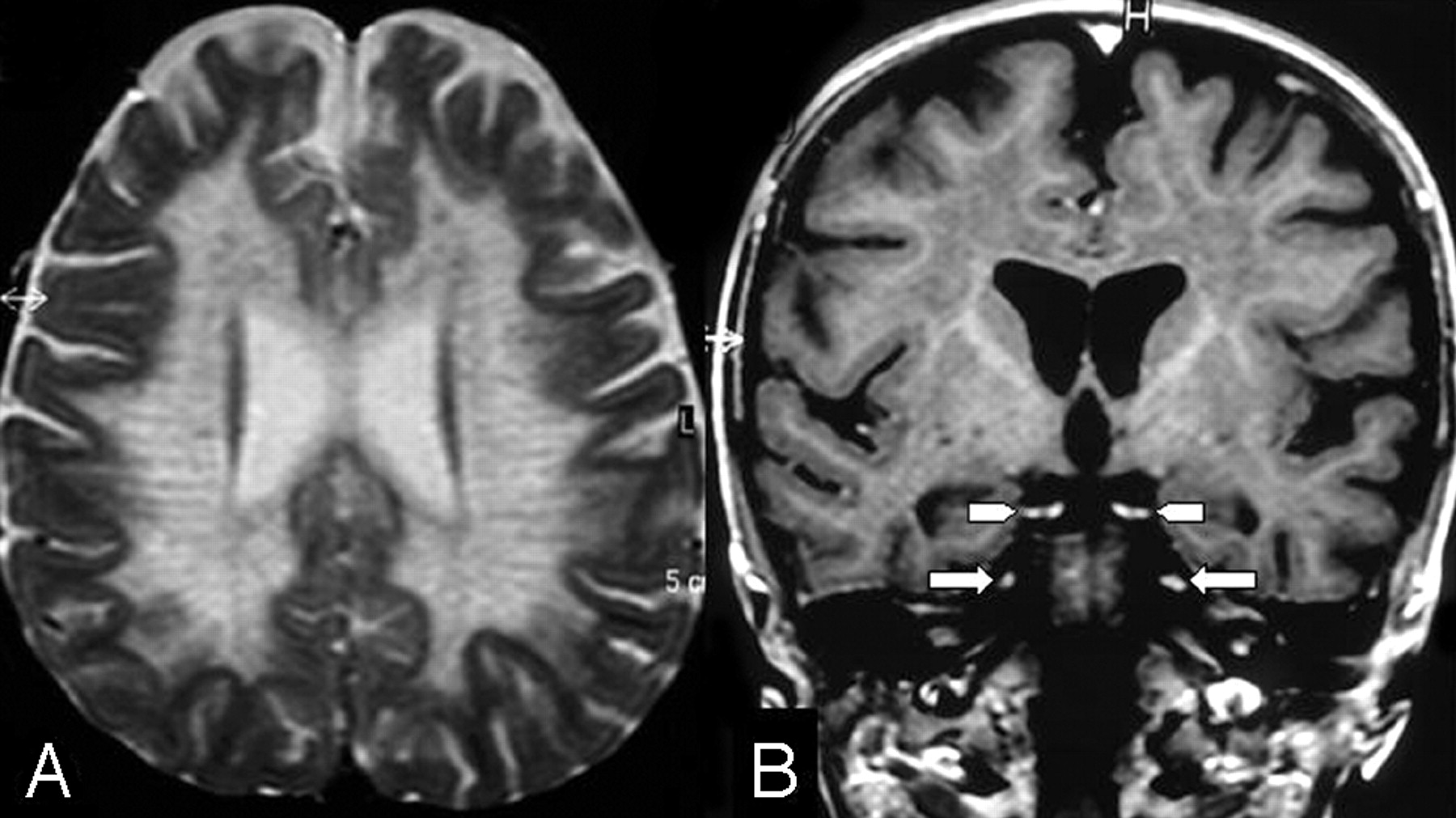
A, Axial T2-weighted image shows the white matter involvement with a tigroid pattern.

B, Coronal postcontrast T1-weighted image shows bilateral and symmetrical abnormal Gd enhancement of the oculomotor (arrowheads) and trigeminal nerves (arrows).

References

  1. Cheon JE, Kim IO, Hwang YS, et al. Leukodystrophy in children: a pictorial review of MR imaging features. Radiographics 2002;22:461–76
  2. Faerber EN, Melvin J, Smergel EM. MRI appearances of metachromatic leukodystrophy. Pediatr Radiol 1999;29:669–72
  3. Saremi F, Helmy M, Farzin S, et al. MRI of cranial nerve enhancement. AJR Am J Roentgenol 2005;185:1487–97
  4. da Silva CJ, da Rocha AJ, Mendes MF, et al. Trigeminal involvement in multiple sclerosis: magnetic resonance imaging findings with clinical correlation in a series of patients. Mult Scler 2005;11:282–85
  5. de Fátima Soares M, Braga FT, da Rocha AJ, et al. Optic nerve infiltration by acute lymphoblastic leukemia: MRI contribution. Pediatr Radiol 2005;35:799–802
  6. Bernal OG, Lenn N. Multiple cranial nerve enhancement in early infantile Krabbe's disease. Neurology 2000;54:2348–49
  7. Jones BV, Barron TF, Towfighi J. Optic nerve enlargement in Krabbe's disease. AJNR Am J Neuroradiol 1999;20:1228–31
  8. Vasconcellos E, Smith M. MRI nerve root enhancement in Krabbe disease. Pediatr Neurol 1998;19:151–52
  • Copyright © American Society of Neuroradiology
Advertisement

Indexed Content

  • Current Issue
  • Accepted Manuscripts
  • Article Preview
  • Past Issues
  • Editorials
  • Editors Choice
  • Fellow Journal Club
  • Letters to the Editor

Cases

  • Case Collection
  • Archive - Case of the Week
  • Archive - Case of the Month
  • Archive - Classic Case

Special Collections

  • Special Collections

Resources

  • News and Updates
  • Turn around Times
  • Submit a Manuscript
  • Author Policies
  • Manuscript Submission Guidelines
  • Evidence-Based Medicine Level Guide
  • Publishing Checklists
  • Graphical Abstract Preparation
  • Imaging Protocol Submission
  • Submit a Case
  • Become a Reviewer/Academy of Reviewers
  • Get Peer Review Credit from Publons

Multimedia

  • AJNR Podcast
  • AJNR SCANtastic
  • Video Articles

About Us

  • About AJNR
  • Editorial Board
  • Not an AJNR Subscriber? Join Now
  • Alerts
  • Feedback
  • Advertise with us
  • Librarian Resources
  • Permissions
  • Terms and Conditions

American Society of Neuroradiology

  • Not an ASNR Member? Join Now

© 2025 by the American Society of Neuroradiology All rights, including for text and data mining, AI training, and similar technologies, are reserved.
Print ISSN: 0195-6108 Online ISSN: 1936-959X

Powered by HighWire