Advertisement

Main menu

  • Home
  • Content
    • Current Issue
    • Accepted Manuscripts
    • Article Preview
    • Past Issue Archive
    • AJNR Case Collection
    • Case of the Week Archive
    • Classic Case Archive
    • Case of the Month Archive
  • Special Collections
    • Spinal CSF Leak Articles (Jan 2020-June 2024)
    • 2024 AJNR Journal Awards
    • Most Impactful AJNR Articles
  • Multimedia
    • AJNR Podcast
    • AJNR Scantastics
    • Video Articles
  • For Authors
    • Submit a Manuscript
    • Author Policies
    • Fast publishing of Accepted Manuscripts
    • Graphical Abstract Preparation
    • Manuscript Submission Guidelines
    • Imaging Protocol Submission
    • Submit a Case for the Case Collection
  • About Us
    • About AJNR
    • Editorial Board
  • More
    • Become a Reviewer/Academy of Reviewers
    • Subscribers
    • Permissions
    • Alerts
    • Feedback
    • Advertisers
    • ASNR Home
  • Other Publications
    • ajnr

User menu

  • Alerts
  • Log in

Search

  • Advanced search
American Journal of Neuroradiology

American Journal of Neuroradiology

ASHNR American Society of Functional Neuroradiology ASHNR American Society of Pediatric Neuroradiology ASSR
  • Alerts
  • Log in

Advanced Search

  • Home
  • Content
    • Current Issue
    • Accepted Manuscripts
    • Article Preview
    • Past Issue Archive
    • AJNR Case Collection
    • Case of the Week Archive
    • Classic Case Archive
    • Case of the Month Archive
  • Special Collections
    • Spinal CSF Leak Articles (Jan 2020-June 2024)
    • 2024 AJNR Journal Awards
    • Most Impactful AJNR Articles
  • Multimedia
    • AJNR Podcast
    • AJNR Scantastics
    • Video Articles
  • For Authors
    • Submit a Manuscript
    • Author Policies
    • Fast publishing of Accepted Manuscripts
    • Graphical Abstract Preparation
    • Manuscript Submission Guidelines
    • Imaging Protocol Submission
    • Submit a Case for the Case Collection
  • About Us
    • About AJNR
    • Editorial Board
  • More
    • Become a Reviewer/Academy of Reviewers
    • Subscribers
    • Permissions
    • Alerts
    • Feedback
    • Advertisers
    • ASNR Home
  • Follow AJNR on Twitter
  • Visit AJNR on Facebook
  • Follow AJNR on Instagram
  • Join AJNR on LinkedIn
  • RSS Feeds

Welcome to the new AJNR, Updated Hall of Fame, and more. Read the full announcements.


AJNR is seeking candidates for the position of Associate Section Editor, AJNR Case Collection. Read the full announcement.


Research ArticleAdult Brain

Blood Flow Mimicking Aneurysmal Wall Enhancement: A Diagnostic Pitfall of Vessel Wall MRI Using the Postcontrast 3D Turbo Spin-Echo MR Imaging Sequence

E. Kalsoum, A. Chabernaud Negrier, T. Tuilier, A. Benaïssa, R. Blanc, S. Gallas, J.-P. Lefaucheur, A. Gaston, R. Lopes, P. Brugières and J. Hodel
American Journal of Neuroradiology June 2018, 39 (6) 1065-1067; DOI: https://doi.org/10.3174/ajnr.A5616
E. Kalsoum
aFrom the Departments of Neuroradiology (E.K., A.C.N., T.T., A.B., S.G., A.G., P.B., J.H.)
  • Find this author on Google Scholar
  • Find this author on PubMed
  • Search for this author on this site
  • ORCID record for E. Kalsoum
A. Chabernaud Negrier
aFrom the Departments of Neuroradiology (E.K., A.C.N., T.T., A.B., S.G., A.G., P.B., J.H.)
  • Find this author on Google Scholar
  • Find this author on PubMed
  • Search for this author on this site
  • ORCID record for A. Chabernaud Negrier
T. Tuilier
aFrom the Departments of Neuroradiology (E.K., A.C.N., T.T., A.B., S.G., A.G., P.B., J.H.)
  • Find this author on Google Scholar
  • Find this author on PubMed
  • Search for this author on this site
  • ORCID record for T. Tuilier
A. Benaïssa
aFrom the Departments of Neuroradiology (E.K., A.C.N., T.T., A.B., S.G., A.G., P.B., J.H.)
  • Find this author on Google Scholar
  • Find this author on PubMed
  • Search for this author on this site
  • ORCID record for A. Benaïssa
R. Blanc
cDepartment of Interventional Neuroradiology (R.B.), Rothschild Foundation Hospital, Paris, France
  • Find this author on Google Scholar
  • Find this author on PubMed
  • Search for this author on this site
  • ORCID record for R. Blanc
S. Gallas
aFrom the Departments of Neuroradiology (E.K., A.C.N., T.T., A.B., S.G., A.G., P.B., J.H.)
  • Find this author on Google Scholar
  • Find this author on PubMed
  • Search for this author on this site
  • ORCID record for S. Gallas
J.-P. Lefaucheur
bNeurophysiology (J.-P.L.), Centre Hospitalier Universitaire Henri Mondor, Créteil, France
  • Find this author on Google Scholar
  • Find this author on PubMed
  • Search for this author on this site
  • ORCID record for J.-P. Lefaucheur
A. Gaston
aFrom the Departments of Neuroradiology (E.K., A.C.N., T.T., A.B., S.G., A.G., P.B., J.H.)
  • Find this author on Google Scholar
  • Find this author on PubMed
  • Search for this author on this site
  • ORCID record for A. Gaston
R. Lopes
dNeuroradiology Department (R.L.), Univ. Lille, INSERM, CHU Lille, U1171 - Degenerative and Vascular Cognitive Disorders, Lille, France.
  • Find this author on Google Scholar
  • Find this author on PubMed
  • Search for this author on this site
  • ORCID record for R. Lopes
P. Brugières
aFrom the Departments of Neuroradiology (E.K., A.C.N., T.T., A.B., S.G., A.G., P.B., J.H.)
  • Find this author on Google Scholar
  • Find this author on PubMed
  • Search for this author on this site
  • ORCID record for P. Brugières
J. Hodel
aFrom the Departments of Neuroradiology (E.K., A.C.N., T.T., A.B., S.G., A.G., P.B., J.H.)
  • Find this author on Google Scholar
  • Find this author on PubMed
  • Search for this author on this site
  • ORCID record for J. Hodel

Abstract

SUMMARY: Our aim was to compare the detectability of aneurysmal wall enhancement in unruptured intracranial aneurysms between conventional and motion-sensitized driven equilibrium–prepared postcontrast 3D T1-weighted TSE sequences (sampling perfection with applicationoptimized contrasts by using different flip angle evolution, SPACE). Twenty-two patients with 30 unruptured intracranial aneurysms were scanned at 3T. Aneurysmal wall enhancement was more significantly detected using conventional compared with motion-sensitized driven equilibrium–prepared SPACE sequences (10/30 versus 2/30, P < .0001). Contrast-to-noise ratio measurements did not differ between conventional and motion-sensitized driven equilibrium–prepared sequences (P = .51). Flowing blood can mimic aneurysmal wall enhancement using conventional SPACE sequences with potential implications for patient care.

ABBREVIATIONS:

AWE
aneurysmal wall enhancement
CNR
contrast-to-noise ratio
MSDE
motion-sensitized driven equilibrium
SPACE
sampling perfection with application-optimized contrasts by using different flip angle evolution
UIA
unruptured intracranial aneurysm

Vessel wall MR imaging is being increasingly used in patients with intracranial aneurysms,1 with aneurysmal wall enhancement (AWE) considered suggestive of instability/rupture.2⇓⇓⇓⇓⇓–8 The most commonly used sequence for vessel wall MR imaging is 3D TSE, including a black-blood effect9; however flowing blood can still lead to lumen enhancement.1,8

Motion-sensitized driven equilibrium (MSDE) uses flow-sensitive dephasing gradients to suppress residual blood flow.10 For the evaluation of AWE, only 1 recent study used MSDE,6 while all the other studies2⇓–4,7,11⇓⇓–14 used a postcontrast sequence without any additional blood-suppression technique (conventional sequence).

The influence of blood-suppression techniques on the detectability of AWE has not yet been evaluated, to our knowledge. We aimed to compare the detectability of AWE in consecutive patients with unruptured intracranial aneurysms (UIAs) using conventional and an MSDE-prepared postcontrast 3D TSE sequence (sampling perfection with application-optimized contrasts by using different flip angle evolution, SPACE).

Materials and Methods

Patients and Brain Imaging

This study was approved by the institutional review board of the Rothschild Foundation Hospital, Paris, France. From September 2016 to February 2017, twenty-two consecutive patients (17 women; 58.1 years of age; range, 25–85 years) with 30 asymptomatic UIAs were included. All patients underwent both DSA and 3T MR imaging within 1 week. Aneurysms were considered nonruptured on the basis of clinical (absence of recent or past acute headaches, no history of subarachnoid hemorrhage) and imaging (absence of recent or past SAH on FLAIR and SWI) data.

MR imaging was performed at 3T (Magnetom Skyra; Siemens, Erlangen, Germany) including DWI, FLAIR, SWI, TOF-MRA, contrast-enhanced MRA, and pre- and postcontrast 3D T1-weighted SPACE acquired with and without MSDE (4 acquisitions per patient). The order of the postcontrast SPACE sequence was randomized. SPACE parameters were similar to those in previous studies2,4,11,12: sagittal plane: TR/TE, 700/11 ms; slice thickness, 0.9 mm; FOV, 230 × 230 mm; matrix, 256 × 256 (interpolated to 512 × 512); voxel size, 0.45 mm; acquisition time, 4 minutes 33 seconds. MSDE preparation consisted of a first-order gradient moment of 500 mT × ms2/m and a b-value of 1.75 s/mm2, according to a previous study.10

Image Analysis

UIAs were defined in consensus using DSA images by 2 neuroradiologists (R.B. and E.K.). SPACE images were blindly and independently assessed in multiplanar mode (with image analysis in arbitrary planes) by 3 neuroradiologists: J.H., T.T., and A.C. Readers were blinded to clinical data and other sequences (except TOF-MRA) but were aware of patient aneurysm location.

Two independent blinded-reading sessions took place 2 weeks apart to avoid recall bias. Each UIA was analyzed only once per session, with pre- and postcontrast either conventional or MSDE-prepared SPACE sequences. For each of the 2 sets of SPACE acquisitions, the readers evaluated each UIA for AWE defined as a thick (>2 mm) area of aneurysmal wall with increased signal intensity between pre- and postcontrast images. Discrepancies were resolved by consensus.

Contrast-to-noise ratio (CNR) measurements were performed to ensure that the differences observed between SPACE sequences were not artifactual. Standardized ROIs (same size and location) were drawn within the following: 1) aneurysm lumen, 2) brain parenchyma, 3) anterior pituitary gland (ie, T1-enhancing tissue), and 4) background noise. CNR was measured in comparison with the parenchyma [CNR = (SI1 − SI2)/SDnoise] for aneurysm lumen and pituitary gland, with SI indicating signal intensity.

Statistical Analysis

Interobserver agreement was evaluated (Fleiss κ). Occurrence of AWE in the whole cohort and CNR values were compared between conventional and MSDE-prepared SPACE images (Fisher exact and paired t test, respectively).

Results

Thirty UIAs (mean long axis, 6.5 mm; range, 2.5–16 mm; mean short axis, 4.6 mm; range, 2–13 mm) were visible in the 22 patients (1.4 per patient; maximum, 4), affecting the basilar (n = 1), anterior cerebral (n = 1), middle cerebral (n = 10), anterior communicating (n = 8), or internal carotid artery (n = 10), including ophthalmic (n = 4) and intracavernous (n = 2) carotid segments.

Interrater agreement was almost perfect (κ = 0.87; 95% CI, 0.72–1.00; and κ = 0.81; 95% CI, 0.63–0.98) with conventional and MSDE-prepared SPACE images, respectively.

AWE was more frequently detected using a conventional (10/30 UIAs, 33.3%) than the MSDE-prepared (2/30, 6.7%) SPACE sequence (P < .0001). AWE was found using both sequences in 2 UIAs (Fig 1) and only with conventional SPACE in 8 with a circumferential appearance (Fig 2).

Fig 1.
  • Download figure
  • Open in new tab
  • Download powerpoint
Fig 1.

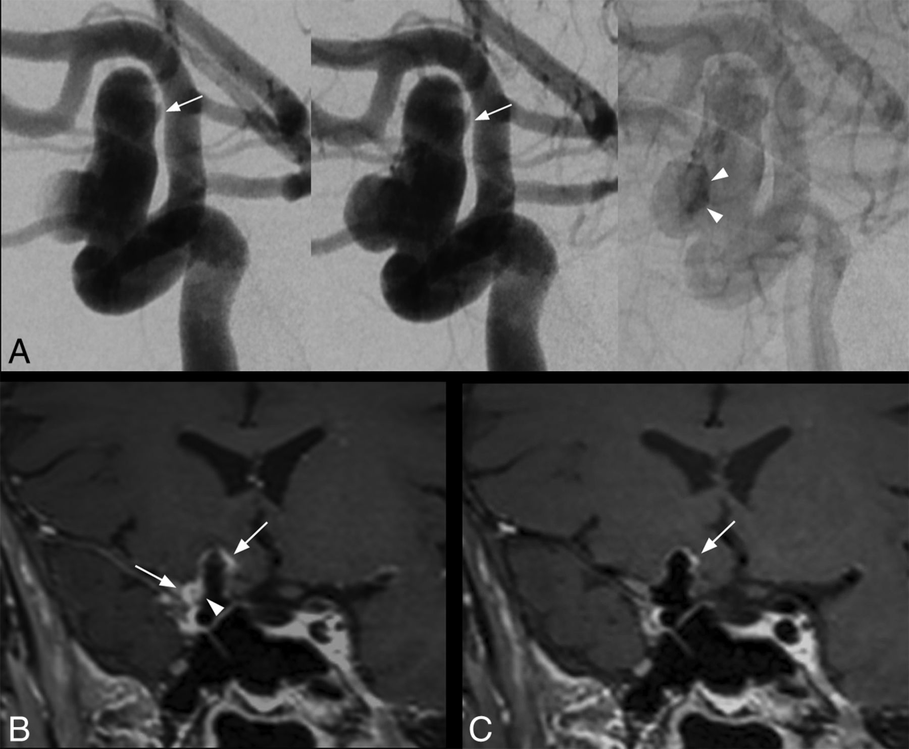
Saccular intracranial aneurysm in a 63-year-old patient. DSA angiogram (A, from left to right: early arterial phase, arterial phase, and venous phase) shows a 14-mm right internal carotid artery aneurysm with intrasaccular slow flowing/stagnant blood (arrowheads) and irregular shape (arrow). With conventional SPACE imaging (B), extensive AWE is visible, more prominent at the apex and at the lateral portion of the aneurysm (B, arrows). Note the enhancement within the aneurysm lumen visible only using conventional SPACE imaging (B, arrowhead), matching the stagnant blood on DSA (A, arrowheads). With MSDE SPACE imaging, AWE is only visible at the apex (C, arrow).

Fig 2.
  • Download figure
  • Open in new tab
  • Download powerpoint
Fig 2.

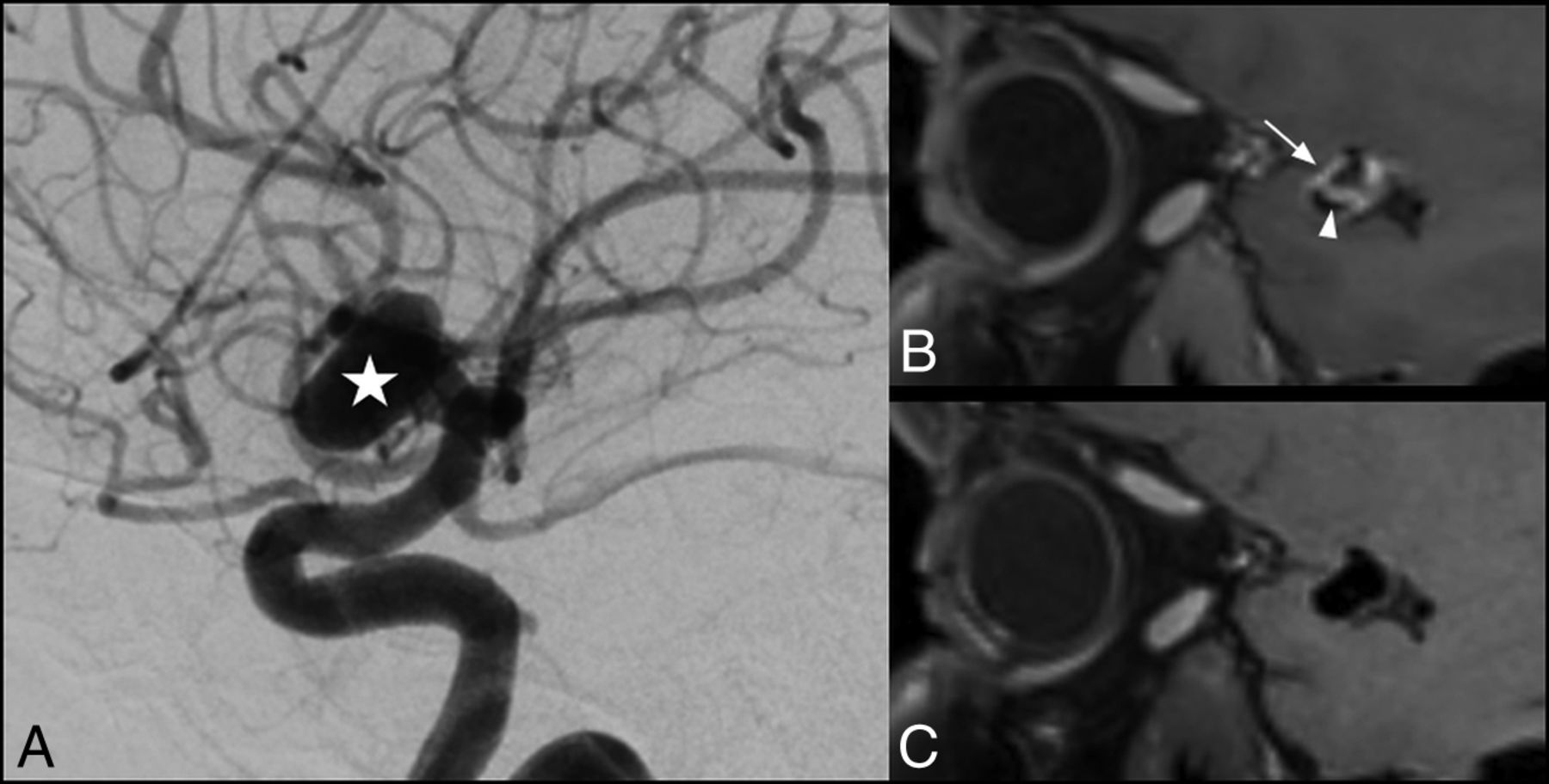
Saccular intracranial aneurysm in a 42-year-old patient. DSA angiogram shows an 11-mm aneurysm (A, star) arising from the M1 segment of the right middle cerebral artery. AWE is visible on the conventional SPACE image (B, arrow), while there is no enhancement visible on the corresponding MSDE SPACE image (C). Enhancement within the aneurysm lumen was also visible using a conventional SPACE image (B, arrowhead).

CNR did not differ between the 2 SPACE sequences for the pituitary gland (ie, T1-enhancing tissue) (321.9 ± 103.7 versus 304.8 ± 60.4 for conventional and MSDE-SPACE, respectively, P = .51) and was higher for the aneurysm lumen with MSDE-prepared SPACE sequences (102.4 ± 83.1 versus 180.2 ± 36.4, P = .008).

Discussion

MSDE preparation reduced the occurrence of AWE, suggesting flowing blood mimicking AWE on conventional images. Such pitfalls may have biased previous studies evaluating AWE and could also have implications for patient care.

Double inversion recovery, MSDE,10 or delay alternating with nutation for tailored excitation (DANTE) can be used for vessel wall MR imaging. We performed MSDE, considering its excellent blood suppression and adaptability to 3D imaging readout (while double inversion recovery is time-consuming and not easily adaptable for large-FOV 3D imaging). Low first-order gradient moment/b-values and composite pulses were used to avoid signal loss and eddy current effects, respectively.

With conventional SPACE sequences, our results (AWE in 33.3% of UIAs) were similar to those previously published.2 The rate of AWE was lower (6.7%) using MSDE, similar to that of Nagahata et al6 also using this technique (4.8% of UIAs). AWE detected using MSDE-prepared SPACE sequences can be related to wall inflammation and/or alternatively to an incomplete suppression of flowing blood not yet degraded by gradients. Such slow inflows may also be associated with degenerative remodeling and thus aneurysm growth.15

There is conflicting evidence on the relationship between AWE and aneurysm size. Some studies found no relationship between wall enhancement and aneurysm size,2,4 with others demonstrating a positive correlation.12 Considering our results, such discrepancy could be due to the relationship between intrasaccular flowing blood and aneurysm size.2,12

Limitations include a small number of patients and lack of histopathologic analysis (reference standard for AWE) because patients did not undergo an operation. However, our purpose was not to assess accuracy for the diagnosis of wall inflammation because we aimed to evaluate the influence of MSDE on AWE detectability. We focused only on UIA; further studies including patients with ruptured aneurysms with histologic analysis available may demonstrate the advantages of MSDE preparation.

ACKNOWLEDGMENTS

We thank Aurélien Monnet and Wadie Ben Hassen from Siemens Healthineers for their support.

REFERENCES

  1. 1.
    1. Mandell DM,
    2. Mossa-Basha M,
    3. Qiao Y, et al
    ; Vessel Wall Imaging Study Group of the American Society of Neuroradiology. Intracranial vessel wall MRI: Principles and Expert Consensus Recommendations of the American Society of Neuroradiology. AJNR Am J Neuroradiol 2017;38:218–29 doi:10.3174/ajnr.A4893 pmid:27469212
  2. 2.
    1. Edjlali M,
    2. Gentric JC,
    3. Régent-Rodriguez C, et al
    . Does aneurysmal wall enhancement on vessel wall MRI help to distinguish stable from unstable intracranial aneurysms? Stroke 2014;45:3704–06 doi:10.1161/STROKEAHA.114.006626 pmid:25325912
  3. 3.
    1. Endo H,
    2. Niizuma K,
    3. Fujimura M, et al
    . Ruptured cerebral microaneurysm diagnosed by 3-dimensional fast spin-echo T1 imaging with variable flip angles. J Stroke Cerebrovasc Dis 2015;24:e231–35 doi:10.1016/j.jstrokecerebrovasdis.2015.04.031 pmid:26009499
  4. 4.
    1. Hu P,
    2. Yang Q,
    3. Wang DD, et al
    . Wall enhancement on high-resolution magnetic resonance imaging may predict an unsteady state of an intracranial saccular aneurysm. Neuroradiology 2016;58:979–85 doi:10.1007/s00234-016-1729-3 pmid:27438805
  5. 5.
    1. Matouk CC,
    2. Mandell DM,
    3. Gunel M, et al
    . Vessel wall magnetic resonance imaging identifies the site of rupture in patients with multiple intracranial aneurysms: proof of principle. Neurosurgery 2013;72:492–96 doi:10.1227/NEU.0b013e31827d1012 pmid:23151622
  6. 6.
    1. Nagahata S,
    2. Nagahata M,
    3. Obara M, et al
    . Wall enhancement of the intracranial aneurysms revealed by magnetic resonance vessel wall imaging using three-dimensional turbo spin-echo sequence with motion-sensitized driven-equilibrium: a sign of ruptured aneurysm? Clin Neuroradiol 2016;26:277–83 doi:10.1007/s00062-014-0353-z pmid:25332151
  7. 7.
    1. Omodaka S,
    2. Endo H,
    3. Niizuma K, et al
    . Circumferential wall enhancement on magnetic resonance imaging is useful to identify rupture site in patients with multiple cerebral aneurysms. Neurosurgery 2017 Jun 6. [Epub ahead of print] doi:10.1093/neuros/nyx267 pmid:28586440
  8. 8.
    1. Omodaka S,
    2. Endo H,
    3. Niizuma K, et al
    . Quantitative assessment of circumferential enhancement along the wall of cerebral aneurysms using MR imaging. AJNR Am J Neuroradiol 2016;37:1262–66 doi:10.3174/ajnr.A4722 pmid:26939634
  9. 9.
    1. Busse RF,
    2. Hariharan H,
    3. Vu A, et al
    . Fast spin echo sequences with very long echo trains: design of variable refocusing flip angle schedules and generation of clinical T2 contrast. Magn Reson Med 2006;55:1030–37 doi:10.1002/mrm.20863 pmid:16598719
  10. 10.
    1. Wang J,
    2. Yarnykh VL,
    3. Hatsukami T, et al
    . Improved suppression of plaque-mimicking artifacts in black-blood carotid atherosclerosis imaging using a multislice motion-sensitized driven-equilibrium (MSDE) turbo spin-echo (TSE) sequence. Magn Reson Med 2007;58:973–81 doi:10.1002/mrm.21385 pmid:17969103
  11. 11.
    1. Fu Q,
    2. Guan S,
    3. Liu C, et al
    . Clinical significance of circumferential aneurysmal wall enhancement in symptomatic patients with unruptured intracranial aneurysms: a high-resolution MRI study. Clin Neuroradiol 2017 Jun 27. [Epub ahead of print] doi:10.1007/s00062-017-0598-4 pmid:28656370
  12. 12.
    1. Liu P,
    2. Qi H,
    3. Liu A, et al
    . Relationship between aneurysm wall enhancement and conventional risk factors in patients with unruptured intracranial aneurysms: a black-blood MRI study. Interv Neuroradiol 2016;22:501–05 doi:10.1177/1591019916653252 pmid:27341856
  13. 13.
    1. Qiao Y,
    2. Steinman DA,
    3. Qin Q, et al
    . Intracranial arterial wall imaging using three-dimensional high isotropic resolution black blood MRI at 3.0 Tesla. J Magn Reson Imaging 2011;34:22–30 doi:10.1002/jmri.22592 pmid:21698704
  14. 14.
    1. Zhang L,
    2. Zhang N,
    3. Wu J, et al
    . High resolution three dimensional intracranial arterial wall imaging at 3 T using T1 weighted SPACE. Magn Reson Imaging 2015;33:1026–34 doi:10.1016/j.mri.2015.06.006 pmid:26143482
  15. 15.
    1. Cebral J,
    2. Ollikainen E,
    3. Chung BJ, et al
    . Flow conditions in the intracranial aneurysm lumen are associated with inflammation and degenerative changes of the aneurysm wall. AJNR Am J Neuroradiol 2017;38:119–26 doi:10.3174/ajnr.A4951 pmid:27686488
  • Received January 29, 2018.
  • Accepted after revision February 5, 2018.
  • © 2018 by American Journal of Neuroradiology
Advertisement

Indexed Content

  • Current Issue
  • Accepted Manuscripts
  • Article Preview
  • Past Issues
  • Editorials
  • Editors Choice
  • Fellow Journal Club
  • Letters to the Editor

Cases

  • Case Collection
  • Archive - Case of the Week
  • Archive - Case of the Month
  • Archive - Classic Case

Special Collections

  • Special Collections

Resources

  • News and Updates
  • Turn around Times
  • Submit a Manuscript
  • Author Policies
  • Manuscript Submission Guidelines
  • Evidence-Based Medicine Level Guide
  • Publishing Checklists
  • Graphical Abstract Preparation
  • Imaging Protocol Submission
  • Submit a Case
  • Become a Reviewer/Academy of Reviewers
  • Get Peer Review Credit from Publons

Multimedia

  • AJNR Podcast
  • AJNR SCANtastic
  • Video Articles

About Us

  • About AJNR
  • Editorial Board
  • Not an AJNR Subscriber? Join Now
  • Alerts
  • Feedback
  • Advertise with us
  • Librarian Resources
  • Permissions
  • Terms and Conditions

American Society of Neuroradiology

  • Not an ASNR Member? Join Now

© 2025 by the American Society of Neuroradiology All rights, including for text and data mining, AI training, and similar technologies, are reserved.
Print ISSN: 0195-6108 Online ISSN: 1936-959X

Powered by HighWire