Advertisement

Main menu

  • Home
  • Content
    • Current Issue
    • Accepted Manuscripts
    • Article Preview
    • Past Issue Archive
    • Video Articles
    • AJNR Case Collection
    • Case of the Week Archive
    • Case of the Month Archive
    • Classic Case Archive
  • Special Collections
    • AJNR Awards
    • Low-Field MRI
    • Alzheimer Disease
    • ASNR Foundation Special Collection
    • Photon-Counting CT
    • View All
  • Multimedia
    • AJNR Podcasts
    • AJNR SCANtastic
    • Trainee Corner
    • MRI Safety Corner
    • Imaging Protocols
  • For Authors
    • Submit a Manuscript
    • Submit a Video Article
    • Submit an eLetter to the Editor/Response
    • Manuscript Submission Guidelines
    • Statistical Tips
    • Fast Publishing of Accepted Manuscripts
    • Graphical Abstract Preparation
    • Imaging Protocol Submission
    • Author Policies
  • About Us
    • About AJNR
    • Editorial Board
    • Editorial Board Alumni
  • More
    • Become a Reviewer/Academy of Reviewers
    • Subscribers
    • Permissions
    • Alerts
    • Feedback
    • Advertisers
    • ASNR Home

User menu

  • Alerts
  • Log in

Search

  • Advanced search
American Journal of Neuroradiology

American Journal of Neuroradiology

ASHNR American Society of Functional Neuroradiology ASHNR American Society of Pediatric Neuroradiology ASSR
  • Alerts
  • Log in

Advanced Search

  • Home
  • Content
    • Current Issue
    • Accepted Manuscripts
    • Article Preview
    • Past Issue Archive
    • Video Articles
    • AJNR Case Collection
    • Case of the Week Archive
    • Case of the Month Archive
    • Classic Case Archive
  • Special Collections
    • AJNR Awards
    • Low-Field MRI
    • Alzheimer Disease
    • ASNR Foundation Special Collection
    • Photon-Counting CT
    • View All
  • Multimedia
    • AJNR Podcasts
    • AJNR SCANtastic
    • Trainee Corner
    • MRI Safety Corner
    • Imaging Protocols
  • For Authors
    • Submit a Manuscript
    • Submit a Video Article
    • Submit an eLetter to the Editor/Response
    • Manuscript Submission Guidelines
    • Statistical Tips
    • Fast Publishing of Accepted Manuscripts
    • Graphical Abstract Preparation
    • Imaging Protocol Submission
    • Author Policies
  • About Us
    • About AJNR
    • Editorial Board
    • Editorial Board Alumni
  • More
    • Become a Reviewer/Academy of Reviewers
    • Subscribers
    • Permissions
    • Alerts
    • Feedback
    • Advertisers
    • ASNR Home
  • Follow AJNR on Twitter
  • Visit AJNR on Facebook
  • Follow AJNR on Instagram
  • Join AJNR on LinkedIn
  • RSS Feeds

AJNR Awards, New Junior Editors, and more. Read the latest AJNR updates

Research ArticleHEAD AND NECK IMAGING
Open Access

High-Resolution MR Imaging of the Parasellar Ligaments

Ian T. Mark, Myung-Ho In, Daehun Kang, Eric Stinson, John Huston, Yunhong Shu, Luciano Leonel and Maria Peris Celda
American Journal of Neuroradiology June 2025, DOI: https://doi.org/10.3174/ajnr.A8658
Ian T. Mark
aFrom the Department of Radiology (I.T.M., M.I., D.K., E.S., J.H., Y.S.), Mayo Clinic, Rochester, Minnesota
  • Find this author on Google Scholar
  • Find this author on PubMed
  • Search for this author on this site
  • ORCID record for Ian T. Mark
Myung-Ho In
aFrom the Department of Radiology (I.T.M., M.I., D.K., E.S., J.H., Y.S.), Mayo Clinic, Rochester, Minnesota
  • Find this author on Google Scholar
  • Find this author on PubMed
  • Search for this author on this site
  • ORCID record for Myung-Ho In
Daehun Kang
aFrom the Department of Radiology (I.T.M., M.I., D.K., E.S., J.H., Y.S.), Mayo Clinic, Rochester, Minnesota
  • Find this author on Google Scholar
  • Find this author on PubMed
  • Search for this author on this site
  • ORCID record for Daehun Kang
Eric Stinson
aFrom the Department of Radiology (I.T.M., M.I., D.K., E.S., J.H., Y.S.), Mayo Clinic, Rochester, Minnesota
  • Find this author on Google Scholar
  • Find this author on PubMed
  • Search for this author on this site
John Huston
aFrom the Department of Radiology (I.T.M., M.I., D.K., E.S., J.H., Y.S.), Mayo Clinic, Rochester, Minnesota
  • Find this author on Google Scholar
  • Find this author on PubMed
  • Search for this author on this site
  • ORCID record for John Huston
Yunhong Shu
aFrom the Department of Radiology (I.T.M., M.I., D.K., E.S., J.H., Y.S.), Mayo Clinic, Rochester, Minnesota
  • Find this author on Google Scholar
  • Find this author on PubMed
  • Search for this author on this site
  • ORCID record for Yunhong Shu
Luciano Leonel
bDepartment of Neurosurgery (L.L., M.P.C.), Mayo Clinic, Rochester, Minnesota
cDepartment of Anatomy (L.L., M.P.C.), Mayo Clinic, Rochester, Minnesota
  • Find this author on Google Scholar
  • Find this author on PubMed
  • Search for this author on this site
Maria Peris Celda
bDepartment of Neurosurgery (L.L., M.P.C.), Mayo Clinic, Rochester, Minnesota
cDepartment of Anatomy (L.L., M.P.C.), Mayo Clinic, Rochester, Minnesota
  • Find this author on Google Scholar
  • Find this author on PubMed
  • Search for this author on this site

SUMMARY:

The parasellar ligaments have been previously described in cadaver specimens and intraoperatively, but identification on MR imaging has eluded radiologists. Using high-resolution T2-weighted MR imaging, we identified the parasellar ligaments as T2-hypointense, bandlike structures that emanate from the medial wall of the cavernous sinus. Subsequent dissection of the same specimen provided matching anatomic images of the parasellar ligaments identified on MRI. This imaging finding is important because resection of the medial wall of the cavernous sinus has been tied to improved outcomes for gross total resection and endocrinologic remission of functioning pituitary adenomas.

ABBREVIATIONS:

MWCS
medial wall of the cavernous sinus
NEX
number of excitations

The cavernous sinus is a complex structure that houses critical anatomy such as the ICAs and cranial nerves.1 Recent studies have underscored the importance of the medial wall of the cavernous sinus (MWCS) in improving outcomes for functional pituitary adenoma surgery.2 Within the cavernous sinus, the parasellar ligaments are important structures that extend from the MWCS to the ICAs and cranial nerves.3 Despite their anatomic significance, the parasellar ligaments have remained elusive on MR imaging. The purpose of this report is to describe the imaging appearance of the parasellar ligaments on MR imaging.

Imaging

All imaging was performed on a research-only compact 3T scanner (GE Healthcare).4 A standard coronal T2-weighted sequence that mimics our clinically used protocol was initially run (slice thickness, 3 mm; TR, 3025 ms; TE, 101 ms; 160-mm field of view; number of excitations (NEX), 1; acquisition time 55 seconds). This clinical sequence demonstrated hazy T2-isointense signal in the cavernous sinus, immediately adjacent to the pituitary gland (Fig 1A). In our experience, ill-defined signal in the cavernous sinuses on the coronal T2-weighted sequence is a typical finding, without having sufficient spatial resolution to identify the parasellar ligaments.

FIG 1.
  • Download figure
  • Open in new tab
  • Download powerpoint
FIG 1.

A, A standard 3-mm-thick coronal T2-weighted sequence shows hazy signal (arrows) in the cavernous sinus. B, High-resolution 0.5-mm slice thickness imaging resolves these hazy structures as distinct T2-hypointense bands that represent the parasellar ligaments (upper arrow indicates the anterior horizontal ligament; lower arrow, the periosteal ligament).

An additional high-resolution T2-weighted sequence was then performed to further resolve the structures within the cavernous sinus (slice thickness. 0.5 mm; TR, 4372 ms; TE, 100 ms; 160-mm field of view; NEX, 6; acquisition time, 21 minutes 13 seconds).

The higher-resolution imaging further resolved the ill-defined signal in the cavernous sinus as distinct T2-hypointense bands (Fig 1B). The bands run from the MWCS that directly abuts the lateral aspect of the pituitary gland and fan outward as they extend laterally toward the ICA and the lateral wall of the cavernous sinus. These distinct bands correlate with the parasellar ligaments (Fig 2), specifically the anterior horizontal ligament and periosteal ligament.

FIG 2.
  • Download figure
  • Open in new tab
  • Download powerpoint
FIG 2.

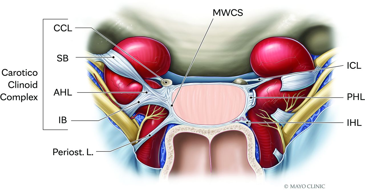
Illustration of the parasellar ligaments demonstrate the “candy wrapper” configuration that originates on the MWCS and fans out laterally as the ligaments extend to the cavernous segment of the ICA and the lateral wall of the cavernous sinus. CCL indicates carotid-clinoid ligament; AHL, anterior horizontal ligament and the associated SB, superior branch; IB, inferior branch; Periost. L., periosteal ligament; ICL, interclinoid ligament; PHL, posterior horizontal ligament; IHL, interclinoid ligament. This illustration has been modified from the original version.3

Anatomy Report

For this study, we used 1 embalmed cadaveric specimen prepared as previously described.5 Briefly, for the latex injection both the common carotid and vertebral arteries as well as internal jugular veins were isolated and cannulated with stainless steel cannulas and perfused with red and blue latex, respectively.

To expose and describe the pituitary region and ligaments, we performed a regular transsphenoidal approach. With a zero-degree endoscope, both middle and superior turbinates on both sides of the nasal cavity were lateralized, revealing the ostium of the sphenoid sinus. A Kerrison rongeur was used to enlarge the ostium and enter the sphenoid sinus cavity. Within the sphenoid sinus, the septum was drilled, and the bony landmarks, recesses, and prominences were exposed. The dissection proceeded by drilling the sellar prominence and moving laterally to the carotid prominence. Once the dura covering the pituitary gland and anterior bend of the ICA were exposed, a retractable knife was used to incise and expose the dura. The incision was performed at the level of the anterior intercavernous sinus, moving laterally to open the anterior wall of the cavernous sinus and its contents. Last, using 0° and 30° scopes, we dissected the pituitary region and cavernous sinus and photo documented them, preserving the pituitary ligaments and their landmarks, in particular their origins and insertions in the surrounding structures (Fig 3, Supplemental Data).

FIG 3.
  • Download figure
  • Open in new tab
  • Download powerpoint
FIG 3.

Endoscopic image from the same cadaver specimen from Fig 1. The distinct T2-hypointense bands in the right cavernous sinus seen on MRI correspond to the right parasellar ligaments. Specifically. the anterior horizontal ligament (upper arrow) and the periosteal ligament (lower arrow).

DISCUSSION

The cavernous sinus is an important anatomic region to understand during resection of functioning pituitary adenomas. Resection of the MWCS, even in the absence of macroscopic invasion, has been shown to be associated with improved outcomes including gross total resection and endocrinologic remission.2 Surgical resection of the MWCS is not without risk, with many important structures including the ICA and cranial nerves residing in the cavernous sinus. The parasellar ligaments have been described as key anatomic structures that connect the MWCS to the ICA and are therefore required to be transected during MWCS resection. Preoperative identification of the parasellar ligaments could therefore help with surgical planning to avoid carotid artery injury. While these structures have been described in cadaver specimens, we present a case of MR imaging of the parasellar ligaments with anatomic correlation.

The MWCS is a thin single-layer structure comprising the meningeal dura that is difficult to see on MRI.3 The resection of the MWCS has been shown to be of increasing importance in achieving biochemical cure of functioning pituitary adenomas and to prevent recurrence in nonfunctioning adenomas.6⇓-8 Parasellar ligament transection is important in the resection of the MWCS from the surrounding neurovascular structures.3

Kehrli et al9 theorized that the fetal pituitary gland is wrapped in a mesenchymal mass, of which the parasellar ligaments are remnants, a process that leads to variability in the ligaments. The parasellar ligaments are fan-shaped structures originating from the MWCS and extending to the tunica adventitia of the ICAs and laterally to the cranial nerves in what has been described as a “candy-wrapper” configuration.3 The parasellar ligaments, when studied in cadaver models, have a variable configuration but are grossly organized into anterior and posterior groups. The anterior horizontal ligament and posterior horizontal ligament, identified on our specimen, reside in the anterior and posterior groups, respectively.

Case Summary

We present high-resolution MR imaging of the parasellar ligaments. These ligaments are important structures that connect the MWCS to the ICAs and cranial nerves and have previously been described with gross visualization in cadaver models and intraoperatively, but not previously, with MR imaging. The MWCS is an increasingly important structure in achieving endocrinologic remission following the resection of functioning pituitary adenomas; therefore, the parasellar ligaments have newly recognized relevance for surgical planning.

Footnotes

  • Disclosure forms provided by the authors are available with the full text and PDF of this article at www.ajnr.org.

Indicates open access to non-subscribers at www.ajnr.org

References

  1. 1.
    1. Truong HQ,
    2. Lieber S,
    3. Najera E, et al
    . The medial wall of the cavernous sinus, Part 1: surgical anatomy, ligaments, and surgical technique for its mobilization and/or resection. J Neurosurg 2019;131:122–30 doi:10.3171/2018.3.JNS18596 pmid:30192192
  2. 2.
    1. Pontes JP,
    2. Udoma-Udofa OC,
    3. de Oliveira JS, et al
    . Efficacy and safety of cavernous sinus medial wall resection in pituitary adenoma surgery: a systematic review and a single-arm meta-analysis. Pituitary 2023;26:340–51 doi:10.1007/s11102-023-01332-5 pmid:37382779
  3. 3.
    1. Serioli S,
    2. Plou P,
    3. Leonel LC, et al
    . The “candy wrapper” of the pituitary gland: a road map to the parasellar ligaments and the medial wall of the cavernous sinus. Acta Neurochir (Wien) 2023;165:3431–44 doi:10.1007/s00701-023-05736-x pmid:37594638
  4. 4.
    1. Foo TK,
    2. Laskaris E,
    3. Vermilyea M, et al
    . Lightweight, compact, and high-performance 3T MR system for imaging the brain and extremities. Magn Reson Med 2018;80:2232–45 doi:10.1002/mrm.27175 pmid:29536587
  5. 5.
    1. Leonel LC,
    2. Carlstrom LP,
    3. Graffeo CS, et al
    . Foundations of advanced neuroanatomy: technical guidelines for specimen preparation, dissection, and 3D-photodocumentation in a surgical anatomy laboratory. J Neurol Surg B Skull Base 2021;82:e248–58 doi:10.1055/s-0039-3399590 pmid:34306946
  6. 6.
    1. de Macêdo Filho LJ,
    2. Diógenes AV,
    3. Barreto EG, et al
    . Endoscopic endonasal resection of the medial wall of the cavernous sinus and its impact on outcomes of pituitary surgery: a systematic review and meta-analysis. Brain Sci 2022;12:1354 doi:10.3390/brainsci12101354 pmid:36291288
  7. 7.
    1. Cohen-Cohen S,
    2. Gardner PA,
    3. Alves-Belo JT, et al
    . The medial wall of the cavernous sinus, Part 2: selective medial wall resection in 50 pituitary adenoma patients. J Neurosurg 2019;131:131–40 doi:10.3171/2018.5.JNS18595 pmid:30192191
  8. 8.
    1. Borghei-Razavi H,
    2. Muhsen BA,
    3. Joshi K, et al
    . Endoscopic extracapsular resection of an adrenocorticotropic hormone-secreting macroadenoma with selective resection of the medial cavernous sinus wall. World Neurosurg 2020;144:199 doi:10.1016/j.wneu.2020.09.087 pmid:32971280
  9. 9.
    1. Kehrli P,
    2. Ali M,
    3. Reis M, et al
    . Anatomy and embryology of the lateral sellar compartment (cavernous sinus) medial wall. Neurol Res 1998;20:585–92 doi:10.1080/01616412.1998.11740568 pmid:9785585
  • Received November 19, 2024.
  • Accepted after revision January 11, 2025.
  • © 2025 by American Journal of Neuroradiology
Advertisement

Indexed Content

  • Current Issue
  • Accepted Manuscripts
  • Article Preview
  • Past Issues
  • Editorials
  • Editor's Choice
  • Fellows' Journal Club
  • Letters to the Editor
  • Video Articles

Cases

  • Case Collection
  • Archive - Case of the Week
  • Archive - Case of the Month
  • Archive - Classic Case

More from AJNR

  • Trainee Corner
  • Imaging Protocols
  • MRI Safety Corner
  • Book Reviews

Multimedia

  • AJNR Podcasts
  • AJNR Scantastics

Resources

  • Turnaround Time
  • Submit a Manuscript
  • Submit a Video Article
  • Submit an eLetter to the Editor/Response
  • Manuscript Submission Guidelines
  • Statistical Tips
  • Fast Publishing of Accepted Manuscripts
  • Graphical Abstract Preparation
  • Imaging Protocol Submission
  • Evidence-Based Medicine Level Guide
  • Publishing Checklists
  • Author Policies
  • Become a Reviewer/Academy of Reviewers
  • News and Updates

About Us

  • About AJNR
  • Editorial Board
  • Editorial Board Alumni
  • Alerts
  • Permissions
  • Not an AJNR Subscriber? Join Now
  • Advertise with Us
  • Librarian Resources
  • Feedback
  • Terms and Conditions
  • AJNR Editorial Board Alumni

American Society of Neuroradiology

  • Not an ASNR Member? Join Now

© 2025 by the American Society of Neuroradiology All rights, including for text and data mining, AI training, and similar technologies, are reserved.
Print ISSN: 0195-6108 Online ISSN: 1936-959X

Powered by HighWire