Advertisement

Main menu

  • Home
  • Content
    • Current Issue
    • Accepted Manuscripts
    • Article Preview
    • Past Issue Archive
    • Video Articles
    • AJNR Case Collection
    • Case of the Week Archive
    • Case of the Month Archive
    • Classic Case Archive
  • Special Collections
    • AJNR Awards
    • Low-Field MRI
    • Alzheimer Disease
    • ASNR Foundation Special Collection
    • Photon-Counting CT
    • View All
  • Multimedia
    • AJNR Podcasts
    • AJNR SCANtastic
    • Trainee Corner
    • MRI Safety Corner
    • Imaging Protocols
  • For Authors
    • Submit a Manuscript
    • Submit a Video Article
    • Submit an eLetter to the Editor/Response
    • Manuscript Submission Guidelines
    • Statistical Tips
    • Fast Publishing of Accepted Manuscripts
    • Graphical Abstract Preparation
    • Imaging Protocol Submission
    • Author Policies
  • About Us
    • About AJNR
    • Editorial Board
    • Editorial Board Alumni
  • More
    • Become a Reviewer/Academy of Reviewers
    • Subscribers
    • Permissions
    • Alerts
    • Feedback
    • Advertisers
    • ASNR Home

User menu

  • Alerts
  • Log in

Search

  • Advanced search
American Journal of Neuroradiology

American Journal of Neuroradiology

ASHNR American Society of Functional Neuroradiology ASHNR American Society of Pediatric Neuroradiology ASSR
  • Alerts
  • Log in

Advanced Search

  • Home
  • Content
    • Current Issue
    • Accepted Manuscripts
    • Article Preview
    • Past Issue Archive
    • Video Articles
    • AJNR Case Collection
    • Case of the Week Archive
    • Case of the Month Archive
    • Classic Case Archive
  • Special Collections
    • AJNR Awards
    • Low-Field MRI
    • Alzheimer Disease
    • ASNR Foundation Special Collection
    • Photon-Counting CT
    • View All
  • Multimedia
    • AJNR Podcasts
    • AJNR SCANtastic
    • Trainee Corner
    • MRI Safety Corner
    • Imaging Protocols
  • For Authors
    • Submit a Manuscript
    • Submit a Video Article
    • Submit an eLetter to the Editor/Response
    • Manuscript Submission Guidelines
    • Statistical Tips
    • Fast Publishing of Accepted Manuscripts
    • Graphical Abstract Preparation
    • Imaging Protocol Submission
    • Author Policies
  • About Us
    • About AJNR
    • Editorial Board
    • Editorial Board Alumni
  • More
    • Become a Reviewer/Academy of Reviewers
    • Subscribers
    • Permissions
    • Alerts
    • Feedback
    • Advertisers
    • ASNR Home
  • Follow AJNR on Twitter
  • Visit AJNR on Facebook
  • Follow AJNR on Instagram
  • Join AJNR on LinkedIn
  • RSS Feeds

AJNR Awards, New Junior Editors, and more. Read the latest AJNR updates

Research ArticleSpine Imaging and Spine Image-Guided Interventions

Armed Kyphoplasty: An Indirect Central Canal Decompression Technique in Burst Fractures

A. Venier, L. Roccatagliata, M. Isalberti, P. Scarone, D.E. Kuhlen, M. Reinert, G. Bonaldi, J.A. Hirsch and A. Cianfoni
American Journal of Neuroradiology October 2019, DOI: https://doi.org/10.3174/ajnr.A6285
A. Venier
aFrom the Departments of Neurosurgery (A.V., P.S., D.E.K., M.R.)
  • Find this author on Google Scholar
  • Find this author on PubMed
  • Search for this author on this site
  • ORCID record for A. Venier
L. Roccatagliata
bNeuroradiology (L.R., M.I., A.C.), Neurocenter of Southern Switzerland, Lugano, Switzerland
  • Find this author on Google Scholar
  • Find this author on PubMed
  • Search for this author on this site
  • ORCID record for L. Roccatagliata
M. Isalberti
bNeuroradiology (L.R., M.I., A.C.), Neurocenter of Southern Switzerland, Lugano, Switzerland
  • Find this author on Google Scholar
  • Find this author on PubMed
  • Search for this author on this site
  • ORCID record for M. Isalberti
P. Scarone
aFrom the Departments of Neurosurgery (A.V., P.S., D.E.K., M.R.)
  • Find this author on Google Scholar
  • Find this author on PubMed
  • Search for this author on this site
  • ORCID record for P. Scarone
D.E. Kuhlen
aFrom the Departments of Neurosurgery (A.V., P.S., D.E.K., M.R.)
  • Find this author on Google Scholar
  • Find this author on PubMed
  • Search for this author on this site
  • ORCID record for D.E. Kuhlen
M. Reinert
aFrom the Departments of Neurosurgery (A.V., P.S., D.E.K., M.R.)
  • Find this author on Google Scholar
  • Find this author on PubMed
  • Search for this author on this site
  • ORCID record for M. Reinert
G. Bonaldi
cDepartment of Neuroradiology (G.B.), Papa Giovanni XXIII Hospital, Bergamo, Italy
dDepartment of Neurosurgery (G.B.), Clinica Igea, Milan, Italy
  • Find this author on Google Scholar
  • Find this author on PubMed
  • Search for this author on this site
  • ORCID record for G. Bonaldi
J.A. Hirsch
eDepartment of Neuroradiology (J.A.H.), Massachusetts General Hospital, Harvard Medical School, Boston, Massachusetts
  • Find this author on Google Scholar
  • Find this author on PubMed
  • Search for this author on this site
  • ORCID record for J.A. Hirsch
A. Cianfoni
bNeuroradiology (L.R., M.I., A.C.), Neurocenter of Southern Switzerland, Lugano, Switzerland
fDepartment of Neuroradiology (A.C.), Inselspital, University Hospital of Bern, Bern, Switzerland.
  • Find this author on Google Scholar
  • Find this author on PubMed
  • Search for this author on this site
  • ORCID record for A. Cianfoni
  • Article
  • Figures & Data
  • Info & Metrics
  • Responses
  • References
  • PDF
Loading

Article Figures & Data

Figures

  • Fig 1.
    • Download figure
    • Open in new tab
    • Download powerpoint
    Fig 1.

    Severe L3 vertebral body collapse (A) in a 74-year-old woman with osteopenia following minor trauma. Intraoperative myelogram through intradural injection of contrast agent at L1–L2 (arrow, A) shows an opacification defect of the dural sac dorsal to the retropusled fragment at L3 (arrowhead, A). On fracture reduction through balloon-expanded vertebral body stents (B), the myelogram shows greater opacification of the dural sac at L3 (arrowhead, B), a real-time indirect sign of ligamentotaxis and partial central canal clearance. C and D, Preoperative and postoperative midsagittal CT images used for measurement of vertebral body height at the maximum point of collapse (red arrows) and of posterior wall retropulsion (white arrows) perpendicular to the dashed white line connecting the postero-inferior corner of the cranial vertebral body and the postero-superior corner of the caudal one, representing the expected original posterior wall, now intersecting the PWR.

  • Fig 2.
    • Download figure
    • Open in new tab
    • Download powerpoint
    Fig 2.

    Lung cancer metastatic T4 fracture in a 67-year-old man, with disabling back pain. MR imaging (A) and CT (B) show a lytic lesion, with vertebral body collapse and retropulsion of an osseous fragment (arrowhead, B), causing spinal cord compression, but the patient was neurologically intact. The patient underwent armed kyphoplasty with the SAIF technique (C–E) with bilateral stent and screw implant, with a decompressive surgery backup plan on standby. The procedure was uneventful, and the patient showed no neurologic worsening. Postoperative CT (F–H) shows a 3D view of the stent-screw-cement complex (F) and, most notably, the vertebral body height restoration and correction of posterior wall retropulsion (arrow, H) through ligamentotaxis. The patient was ambulating the same day and could undergo radiation treatment during the following days.
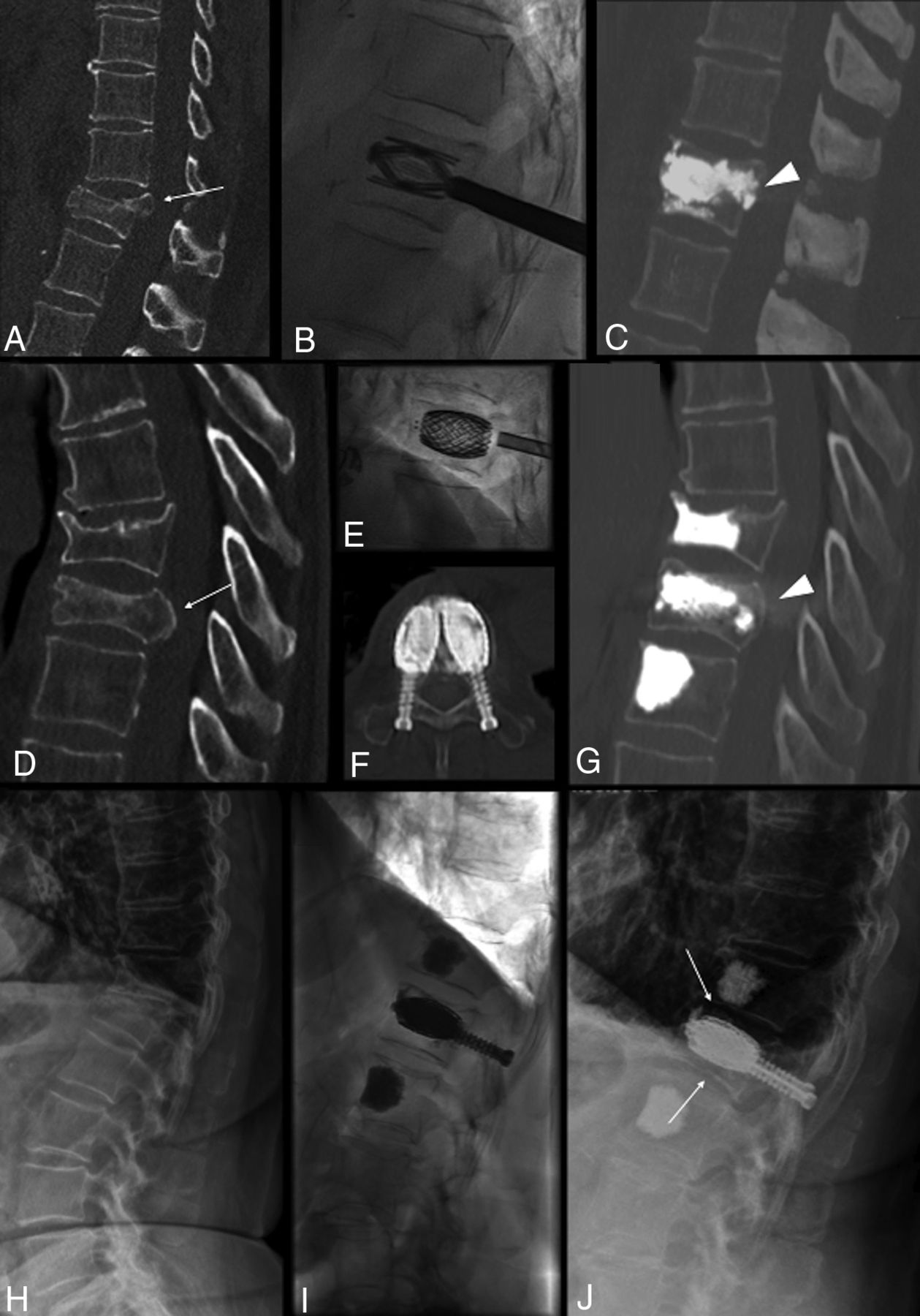
  • Fig 3.
    • Download figure
    • Open in new tab
    • Download powerpoint
    Fig 3.

    Three different cases (A–C), (D–G), and (H–J). A–C, Treatment with the SpineJack of a traumatic incomplete burst fracture of T12 in a 55-year-old man with posterior wall retropulsion (arrow, A) and junctional kyphosis. Postoperative CT shows vertebral height restoration, central canal clearance through retropulsed fragment correction (arrowhead, C), and kyphosis correction. D–G, Treatment with the SAIF technique of a traumatic T10 fracture in a 78-year-old man with osteoporosis with >50% height loss and posterior wall retropulsion (arrow, D), with effective height restoration and posterior wall reposition (arrowhead, G). H–J, Treatment of an L1 osteoporotic fracture with bone subsidence (arrows, J) around the cement cast at 1-month follow-up, not compromising alignment and curvature. The patient was asymptomatic.

  • Fig 4.
    • Download figure
    • Open in new tab
    • Download powerpoint
    Fig 4.

    Complete burst fracture of L1 following high-energy trauma in a 40-year-old man with marked vertebral body fragmentation (A–C) and posterior wall retropulsion (arrowhead, A). Sagittal fat-saturated proton density MR image (D) shows an epidural hematoma and compression of the conus medullaris, but the patient was neurologically intact. The patient underwent surgical treatment in a hybrid operation room, including, in sequence, L1 decompressive laminectomy, pedicular screw placement, fracture reduction and vertebral body augmentation with percutaneous bilateral SpineJack, and posterior stabilization with spinal rods (E and F). Postprocedural CT (G and H) shows fracture and kyphosis reduction and, most important, central canal clearance (arrow, H) through ligamentotaxis. Notably, no maneuvers of direct fragment impaction or of posterior fracture distraction were performed. Follow-up imaging with standing plain films at 6 months (I and J) and with CT at 12 months (K) shows preserved vertebral body height and alignment and osseous healing around the cement cast. In this case, a more invasive procedure of corpectomy and anterior column stabilization could be successfully avoided by armed kyphoplasty.

PreviousNext
Back to top
Advertisement
Print
Download PDF
Email Article

Thank you for your interest in spreading the word on American Journal of Neuroradiology.

NOTE: We only request your email address so that the person you are recommending the page to knows that you wanted them to see it, and that it is not junk mail. We do not capture any email address.

Enter multiple addresses on separate lines or separate them with commas.
Armed Kyphoplasty: An Indirect Central Canal Decompression Technique in Burst Fractures
(Your Name) has sent you a message from American Journal of Neuroradiology
(Your Name) thought you would like to see the American Journal of Neuroradiology web site.
CAPTCHA
This question is for testing whether or not you are a human visitor and to prevent automated spam submissions.
Cite this article
A. Venier, L. Roccatagliata, M. Isalberti, P. Scarone, D.E. Kuhlen, M. Reinert, G. Bonaldi, J.A. Hirsch, A. Cianfoni
Armed Kyphoplasty: An Indirect Central Canal Decompression Technique in Burst Fractures
American Journal of Neuroradiology Oct 2019, DOI: 10.3174/ajnr.A6285

Citation Manager Formats

  • BibTeX
  • Bookends
  • EasyBib
  • EndNote (tagged)
  • EndNote 8 (xml)
  • Medlars
  • Mendeley
  • Papers
  • RefWorks Tagged
  • Ref Manager
  • RIS
  • Zotero
0 Responses
Respond to this article
Share
Bookmark this article
Armed Kyphoplasty: An Indirect Central Canal Decompression Technique in Burst Fractures
A. Venier, L. Roccatagliata, M. Isalberti, P. Scarone, D.E. Kuhlen, M. Reinert, G. Bonaldi, J.A. Hirsch, A. Cianfoni
American Journal of Neuroradiology Oct 2019, DOI: 10.3174/ajnr.A6285
del.icio.us logo Twitter logo Facebook logo Mendeley logo
  • Tweet Widget
  • Facebook Like
  • Google Plus One
Purchase

Jump to section

  • Article
    • Abstract
    • ABBREVIATIONS:
    • MATERIALS AND METHODS
    • RESULTS
    • DISCUSSION
    • CONCLUSIONS
    • Footnotes
    • References
  • Figures & Data
  • Info & Metrics
  • Responses
  • References
  • PDF

Related Articles

  • No related articles found.
  • PubMed
  • Google Scholar

Cited By...

  • Armed kyphoplasty-the future?
  • 'Armed kyphoplasty with posterior stabilization avoids corpectomy in complex thoracolumbar spine fractures: a case series
  • Middle column Stent-screw Assisted Internal Fixation (SAIF): a modified minimally-invasive approach to rescue vertebral middle column re-fractures
  • The 'armed concrete' approach: stent-screw-assisted internal fixation (SAIF) reconstructs and internally fixates the most severe osteoporotic vertebral fractures
  • Crossref
  • Google Scholar

This article has not yet been cited by articles in journals that are participating in Crossref Cited-by Linking.

More in this TOC Section

Spine Imaging and Spine Image-Guided Interventions

  • Management Outcomes For VO Spine Biopsy
  • Characteristics of SIH Type I Culprit Lesions
  • Advanced Imaging of Type 2 Spinal CSF Leaks
Show more Spine Imaging and Spine Image-Guided Interventions

Neurointervention

  • A Retrospective Study in Tentorial DAVFs
  • Proximal Protection Devices for Carotid Stenting
  • Rescue Reentry in Carotid Near-Occlusion
Show more Neurointervention

Similar Articles

Advertisement

Indexed Content

  • Current Issue
  • Accepted Manuscripts
  • Article Preview
  • Past Issues
  • Editorials
  • Editor's Choice
  • Fellows' Journal Club
  • Letters to the Editor
  • Video Articles

Cases

  • Case Collection
  • Archive - Case of the Week
  • Archive - Case of the Month
  • Archive - Classic Case

More from AJNR

  • Trainee Corner
  • Imaging Protocols
  • MRI Safety Corner
  • Book Reviews

Multimedia

  • AJNR Podcasts
  • AJNR Scantastics

Resources

  • Turnaround Time
  • Submit a Manuscript
  • Submit a Video Article
  • Submit an eLetter to the Editor/Response
  • Manuscript Submission Guidelines
  • Statistical Tips
  • Fast Publishing of Accepted Manuscripts
  • Graphical Abstract Preparation
  • Imaging Protocol Submission
  • Evidence-Based Medicine Level Guide
  • Publishing Checklists
  • Author Policies
  • Become a Reviewer/Academy of Reviewers
  • News and Updates

About Us

  • About AJNR
  • Editorial Board
  • Editorial Board Alumni
  • Alerts
  • Permissions
  • Not an AJNR Subscriber? Join Now
  • Advertise with Us
  • Librarian Resources
  • Feedback
  • Terms and Conditions
  • AJNR Editorial Board Alumni

American Society of Neuroradiology

  • Not an ASNR Member? Join Now

© 2025 by the American Society of Neuroradiology All rights, including for text and data mining, AI training, and similar technologies, are reserved.
Print ISSN: 0195-6108 Online ISSN: 1936-959X

Powered by HighWire