Advertisement

Main menu

  • Home
  • Content
    • Current Issue
    • Accepted Manuscripts
    • Article Preview
    • Past Issue Archive
    • Video Articles
    • AJNR Case Collection
    • Case of the Week Archive
    • Case of the Month Archive
    • Classic Case Archive
  • Special Collections
    • AJNR Awards
    • Low-Field MRI
    • Alzheimer Disease
    • ASNR Foundation Special Collection
    • Photon-Counting CT
    • View All
  • Multimedia
    • AJNR Podcasts
    • AJNR SCANtastic
    • Trainee Corner
    • MRI Safety Corner
    • Imaging Protocols
  • For Authors
    • Submit a Manuscript
    • Submit a Video Article
    • Submit an eLetter to the Editor/Response
    • Manuscript Submission Guidelines
    • Statistical Tips
    • Fast Publishing of Accepted Manuscripts
    • Graphical Abstract Preparation
    • Imaging Protocol Submission
    • Author Policies
  • About Us
    • About AJNR
    • Editorial Board
    • Editorial Board Alumni
  • More
    • Become a Reviewer/Academy of Reviewers
    • Subscribers
    • Permissions
    • Alerts
    • Feedback
    • Advertisers
    • ASNR Home

User menu

  • Alerts
  • Log in

Search

  • Advanced search
American Journal of Neuroradiology

American Journal of Neuroradiology

ASHNR American Society of Functional Neuroradiology ASHNR American Society of Pediatric Neuroradiology ASSR
  • Alerts
  • Log in

Advanced Search

  • Home
  • Content
    • Current Issue
    • Accepted Manuscripts
    • Article Preview
    • Past Issue Archive
    • Video Articles
    • AJNR Case Collection
    • Case of the Week Archive
    • Case of the Month Archive
    • Classic Case Archive
  • Special Collections
    • AJNR Awards
    • Low-Field MRI
    • Alzheimer Disease
    • ASNR Foundation Special Collection
    • Photon-Counting CT
    • View All
  • Multimedia
    • AJNR Podcasts
    • AJNR SCANtastic
    • Trainee Corner
    • MRI Safety Corner
    • Imaging Protocols
  • For Authors
    • Submit a Manuscript
    • Submit a Video Article
    • Submit an eLetter to the Editor/Response
    • Manuscript Submission Guidelines
    • Statistical Tips
    • Fast Publishing of Accepted Manuscripts
    • Graphical Abstract Preparation
    • Imaging Protocol Submission
    • Author Policies
  • About Us
    • About AJNR
    • Editorial Board
    • Editorial Board Alumni
  • More
    • Become a Reviewer/Academy of Reviewers
    • Subscribers
    • Permissions
    • Alerts
    • Feedback
    • Advertisers
    • ASNR Home
  • Follow AJNR on Twitter
  • Visit AJNR on Facebook
  • Follow AJNR on Instagram
  • Join AJNR on LinkedIn
  • RSS Feeds

AJNR Awards, New Junior Editors, and more. Read the latest AJNR updates

LetterOTHER CONTENT

Reply

Zhong Qian and Wilfrido R. Castañeda
American Journal of Neuroradiology May 2000, 21 (5) 988-990;
Zhong Qian
M.D
  • Find this author on Google Scholar
  • Find this author on PubMed
  • Search for this author on this site
Wilfrido R. Castañeda
M.D
  • Find this author on Google Scholar
  • Find this author on PubMed
  • Search for this author on this site
  • Article
  • Figures & Data
  • Info & Metrics
  • Responses
  • References
  • PDF
Loading

We would like to thank Dr. Massoud for his interest in our recently published arteriovenous malformation (AVM) model in sheep. As it was stated in our article, the purpose of the study was to evaluate the feasibility of creating an animal AVM model without the need for complex endovascular procedures. In his commentary of our article, Dr. Massoud made a strong defense of the swine model described by his group and criticized the sheep model as overly complicated and unrepresentative of a cerebral AVM. We will not even attempt to respond to all of his criticisms, because the response would need to be even longer than the original manuscript. We will simply try to delineate the rationale we followed in developing our experimental model and clarify some of his misunderstanding.

Despite some similarities to human AVMs, animal models are unable to reproduce the complex angioarchitectural features of human AVMs, such as arteriovenous shunts, multiple compartments in the nidus, tortuous feeders, intranidal vascular aneurysms, propensity to hemorrhage, variable hemodynamic resistance, abnormal venous drainage, “steal effect,” as well as the “perfusion pressure breakthrough” after treatment. Contrary to what Massoud likes to believe, this great variability in the morphologic and pathophysiologic features in AVMs makes it impossible to classify any one animal model as a “typical” AVM or to claim that it embodies most of the desired features. The fact is that, to date, none of the described animal models, including the swine, are truly representative of a human AVM.

Dr. Massoud emphasized in his original publication that endovascular occlusion of the right occipital, ascending pharyngeal, and the external carotid artery in the swine model was necessary to maximize blood shunting across the rete to the fistula. Their pilot study revealed that if these vessels were left unoccluded, they became major collateral pathways, diverting most of the blood flow away from the rete itself (1). Although Massoud argued in his letter that he eliminated the need for endovascular procedures in a subsequent study (2, 3), there was still the need to place a large 8F guiding catheter into the right carotid artery adjacent to the fistula in order to decrease the flow through this vessel and “thus allow for a greater contribution to the fistula from the cephalad aspect of the artery and thus increase the flow reversal down the drainage portion of the AVM model.” This was done in order to optimize the inadequate flow obtained through the rete in the swine.

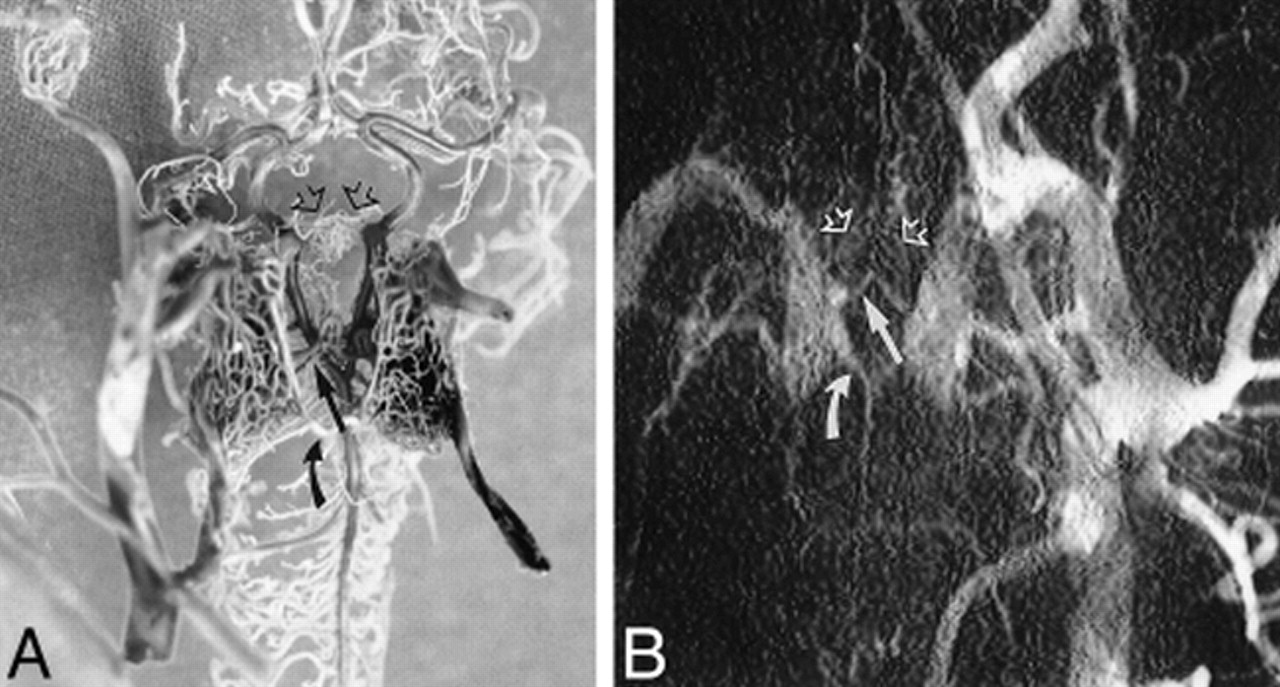
Another “deficiency” of the sheep animal model is, according to Dr. Massoud, the existence of a single vessel connecting the two retia. We disagree with his interpretation of the images presented, because multiple vascular connections were clearly shown in the cast specimens as well as angiographically in our study (Fig 1A and B).

fig 1.
  • Download figure
  • Open in new tab
  • Download powerpoint
fig 1.

A, Photograph of the corrosion cast of the cerebrovascular anatomy on ventral view with 20° cephalad angulation shows two vascular connections in the posterior (curved arrow) and middle (straight arrow) portions as well as a dense vascular network connecting the anterior portion of the carotid retia. B, Close-up view of the retial structure on a left common carotid arteriogram after surgery confirms the vascular connections between both retia demonstrated on the corrosion cast

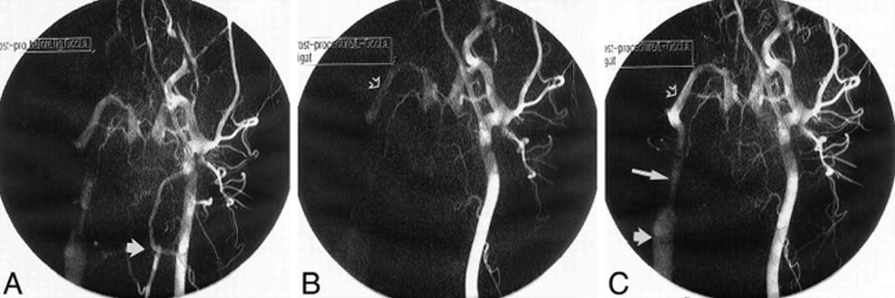
Dr. Massoud also pointed out in his letter that, in the sheep model, most of the flow diversion from the contralateral side to the fistula occurred at the level of the occipital arteries and not through the retia. We disagree with his interpretation and, in order to prove our point, we recreated the model in an additional animal and repeated the angiographic study after ligation of the occipital artery contralateral to the fistula. As expected, angiograms performed before and after ligation demonstrated, as expected, the same differences in density between the distal and proximal carotid artery (Fig 2A and B). Additionally, it was apparent in the postligation angiogram that the internal maxillary artery on the side of the fistula was densely opacified during the early phase, even before the carotid artery was opacified (Fig 2C), discounting Dr. Massoud's misconception that this segment had filled in a retrograde manner through the occipital artery. The relatively low density in the segment above the junction with the right occipital artery is easily explained, not as a result of diverted blood flow at the level of the occipital arteries, but instead as the final effect of the contrast column on the proximal and mid segments that are curved, as seen on the lateral projection, giving them a relatively higher density. This observation was demonstrated on the anteroposterior and lateral angiograms performed before and after surgery (4).

fig 2.
  • Download figure
  • Open in new tab
  • Download powerpoint
fig 2.

A, Postoperative left carotid arteriogram taken before occipital artery ligation shows the opacified left occipital artery (arrow). B, Left carotid arteriogram after occipital artery ligation. The early phase shows both retial structures and opacification of the internal maxillary artery (open arrow) on the anastomotic side. C, Late phase of carotid arteriogram shows opacification of the external carotid artery (long arrow) and jugular vein (short arrow) on the anastomotic side, confirming the direction of flow diversion through the retia toward the arteriovenous fistula

Dr. Massoud stated that the lack of validation of the morphologic and hemodynamic behavior of the sheep model made this model inadequate for scientific purposes. We would like to comment that, although hemodynamic assessments of the swine model, during AVM embolization by Massoud et al (2, 3), provided some interesting data (2, 3), the validity of these studies was questioned by several investigators (5, 6). Cockroff and Steinberg (5) expressed in their commentary that “the validity of the conclusions drawn depends on the model relevance to human AVMs.” They further expressed that “it remained to be determined whether these techniques will provide useful information on humans with complex AVMs.” Wakhloo et al (6), among others, also stated that the “use of blood flow velocity measurements in an AVM draining vein to quantify the extent of an embolization may have limited application.”

Rather than respond to all of Dr. Massoud's criticisms, which are mostly an ardent defense of “his model,” we think it is more important to keep in perspective that the importance of these models is their value for research and training in neurovascular interventions, because they do have arterial feeder(s), a nidal structure, draining vein(s), and high flow through the “AVM,” regardless of their morphologic characteristics.

References

  1. 12.↵
    Massoud TF, Ji C, Vinuela F, Guglielmi G, Robert J, Duckwiler GR, Gobin YP. An experimental arteriovenous malformation model in swine:anatomic basis and construction technique. AJNR Am J Neuroradiol 1994;15:1537-1545
    Abstract/FREE Full Text
  2. 13.↵
    Murayama Y, Massoud TF, Vinuela F. Transvenous hemodynamic assessment of experimental arteriovenous malformations: Doppler guidewire monitoring of embolotherapy in a swine model. Stroke 1996;27:1365-1372
    Abstract/FREE Full Text
  3. 14.
    Murayama Y, Massoud TF, Vinuela F. Hemodynamic changes in arterial feeders and draining veins during embolotherapy of arteriovenous malformations: an experimental study in a swine model. Neurosurgery 1998;43:96-104
    CrossRefPubMed
  4. 15.↵
    Qian Z, Climent S, Maynar M, Uson-Garallo J, Lima-Rodrigues MA, Calles C, Robertson H, Castaneda-Zuniga WR. A simplified arteriovenous malformation model in sheep: feasibility study. AJNR Am J Neuroradiol 1999;20:765-770
    Abstract/FREE Full Text
  5. 16.↵
    Cockroft KM, Steinberg GK. Comments on Murayama Y, Massoud TF, Vinuela F. Hemodynamic changes in arterial feeders and draining veins during embolotherapy of arteriovenous malformations: an experimental study in a swine model. Neurosurgery 1998;43:104
  6. 17.↵
    Wakhloo AK, Lieber BB, Hopkins LN. Comments on Murayama Y, Massoud TF, Vinuela F Hemodynamic changes in arterial feeders and draining veins during embolotherapy of arteriovenous malformations: an experimental study in a swine model. Neurosurgery 1998;43:105-106
  • Copyright © American Society of Neuroradiology
PreviousNext
Back to top

In this issue

American Journal of Neuroradiology
Vol. 21, Issue 5
1 May 2000
  • Table of Contents
  • Index by author
Advertisement
Print
Download PDF
Email Article

Thank you for your interest in spreading the word on American Journal of Neuroradiology.

NOTE: We only request your email address so that the person you are recommending the page to knows that you wanted them to see it, and that it is not junk mail. We do not capture any email address.

Enter multiple addresses on separate lines or separate them with commas.
Reply
(Your Name) has sent you a message from American Journal of Neuroradiology
(Your Name) thought you would like to see the American Journal of Neuroradiology web site.
CAPTCHA
This question is for testing whether or not you are a human visitor and to prevent automated spam submissions.
Cite this article
Zhong Qian, Wilfrido R. Castañeda
Reply
American Journal of Neuroradiology May 2000, 21 (5) 988-990;

Citation Manager Formats

  • BibTeX
  • Bookends
  • EasyBib
  • EndNote (tagged)
  • EndNote 8 (xml)
  • Medlars
  • Mendeley
  • Papers
  • RefWorks Tagged
  • Ref Manager
  • RIS
  • Zotero
0 Responses
Respond to this article
Share
Bookmark this article
Reply
Zhong Qian, Wilfrido R. Castañeda
American Journal of Neuroradiology May 2000, 21 (5) 988-990;
del.icio.us logo Twitter logo Facebook logo Mendeley logo
  • Tweet Widget
  • Facebook Like
  • Google Plus One

Jump to section

  • Article
    • References
  • Figures & Data
  • Info & Metrics
  • Responses
  • References
  • PDF

Related Articles

  • No related articles found.
  • Google Scholar

Cited By...

  • No citing articles found.
  • Crossref
  • Google Scholar

This article has not yet been cited by articles in journals that are participating in Crossref Cited-by Linking.

More in this TOC Section

  • Dr Doppman: The Lone Radiologist
  • Emergency Interventional Stroke Therapy: A Statement from the American Society of Interventional and Therapeutic Neuroradiology  Stroke Task Force of the American Society of Neuroradiology and The Society of Cardiovascular and Interventional Radiology
  • Biography: Wade Wong, Sixth President of the American Society of Spine Radiology
Show more OTHER CONTENT

Similar Articles

Advertisement

Indexed Content

  • Current Issue
  • Accepted Manuscripts
  • Article Preview
  • Past Issues
  • Editorials
  • Editor's Choice
  • Fellows' Journal Club
  • Letters to the Editor
  • Video Articles

Cases

  • Case Collection
  • Archive - Case of the Week
  • Archive - Case of the Month
  • Archive - Classic Case

More from AJNR

  • Trainee Corner
  • Imaging Protocols
  • MRI Safety Corner
  • Book Reviews

Multimedia

  • AJNR Podcasts
  • AJNR Scantastics

Resources

  • Turnaround Time
  • Submit a Manuscript
  • Submit a Video Article
  • Submit an eLetter to the Editor/Response
  • Manuscript Submission Guidelines
  • Statistical Tips
  • Fast Publishing of Accepted Manuscripts
  • Graphical Abstract Preparation
  • Imaging Protocol Submission
  • Evidence-Based Medicine Level Guide
  • Publishing Checklists
  • Author Policies
  • Become a Reviewer/Academy of Reviewers
  • News and Updates

About Us

  • About AJNR
  • Editorial Board
  • Editorial Board Alumni
  • Alerts
  • Permissions
  • Not an AJNR Subscriber? Join Now
  • Advertise with Us
  • Librarian Resources
  • Feedback
  • Terms and Conditions
  • AJNR Editorial Board Alumni

American Society of Neuroradiology

  • Not an ASNR Member? Join Now

© 2025 by the American Society of Neuroradiology All rights, including for text and data mining, AI training, and similar technologies, are reserved.
Print ISSN: 0195-6108 Online ISSN: 1936-959X

Powered by HighWire