
Abstract
SUMMARY: The term “bovine arch” is widely used to describe a common anatomic variant of the human aortic arch branching. This so-called bovine aortic arch has no resemblance to the bovine aortic arch. We describe the most common human aortic arch branching patterns and compare these with the bovine aortic arch.
One of the most widely used misnomers in the medical literature is that of the “bovine aortic arch” in humans.1,2 This term refers to a common anatomic configuration of the aortic arch. By its name, the bovine aortic arch in humans would presumably resemble the aortic arch branching pattern found in the family of ruminant animals, including cattle and buffalo. However, the bovine aortic arch configuration ascribed to the most common human aortic arch variants bears no resemblance to the aortic arch branching pattern found in cattle. We describe the specific anatomic appearance of human and bovine aortic arch branching patterns and propose a simple nomenclature scheme that should supplant the use of the term “bovine aortic arch” in humans.
Human Aortic Arch
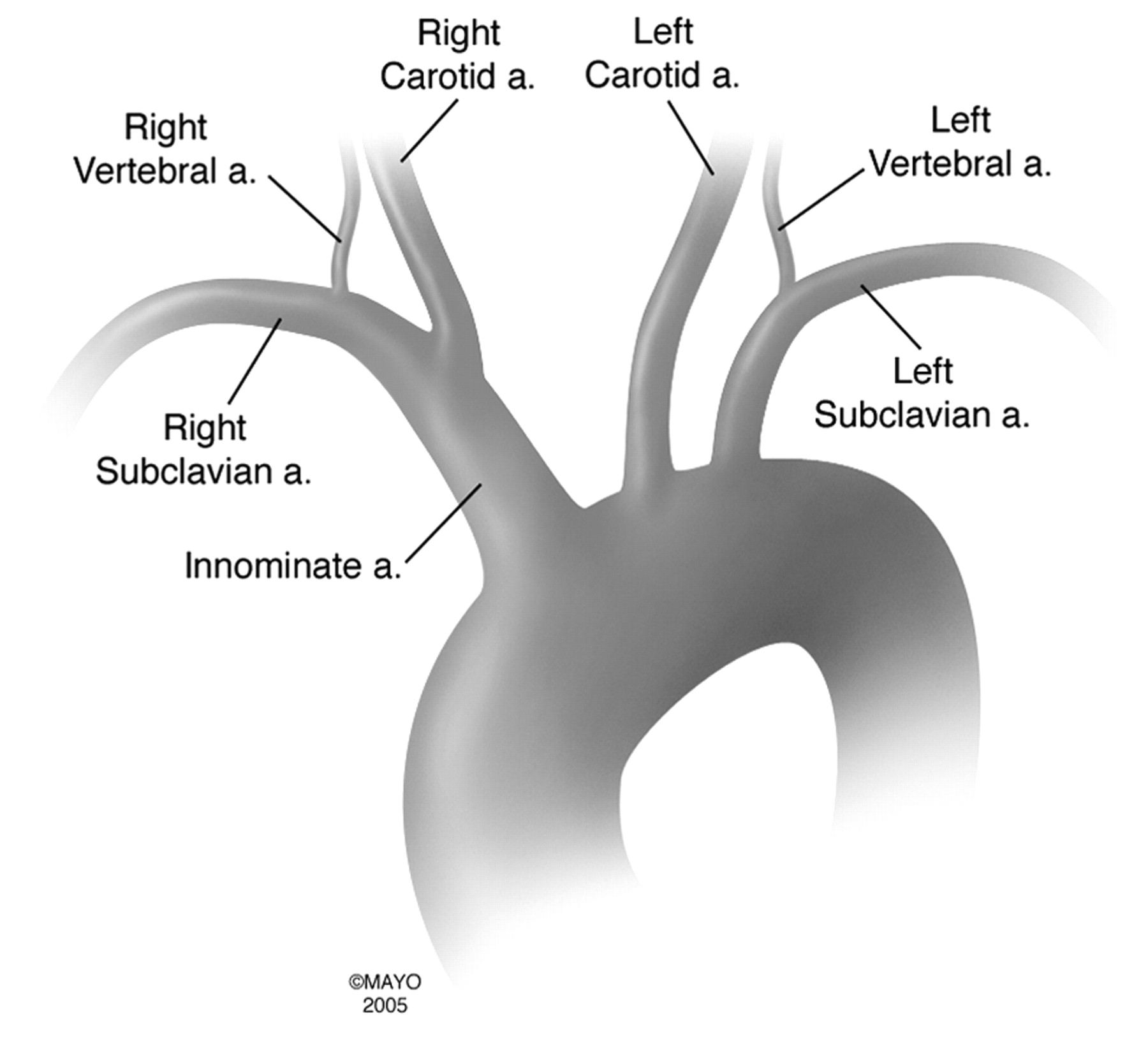
The most common aortic arch branching pattern in humans consists of 3 great vessels originating from the arch of the aorta (Fig 1). The first branch is the innominate artery, which branches into the right subclavian artery and the right common carotid artery. The second branch in the most common pattern is the left common carotid artery, and the last branch is the left subclavian artery. The final configuration of the aortic arch and its branches is probably related to different growth rates in the various arteries and the associated “migration” and “merging” of the branches.3 The second most common variant of aortic arch branching occurs when the left common carotid artery has a common origin with the innominate artery. Rather than arising directly from the aortic arch as a separate branch, the left common carotid artery origin is moved to the right and merges with the origin of the innominate artery. This variant is most often termed a “bovine aortic arch” (Fig 2).1-4 A similar but less common variant occurs when the left common carotid artery originates directly from the innominate artery rather than as a common trunk (Fig 3). Both variants of left common carotid artery origin have been called in various textbooks and medical articles a “bovine-type arch,” though this term is most commonly ascribed to the common trunk variety.1,2,4 More than 20 different aortic arch configurations have been described, but those specifically described previously are by far the most commonly encountered.3-6

The most common aortic arch branching pattern found in humans has separate origins for the innominate, left common carotid, and left subclavian arteries.

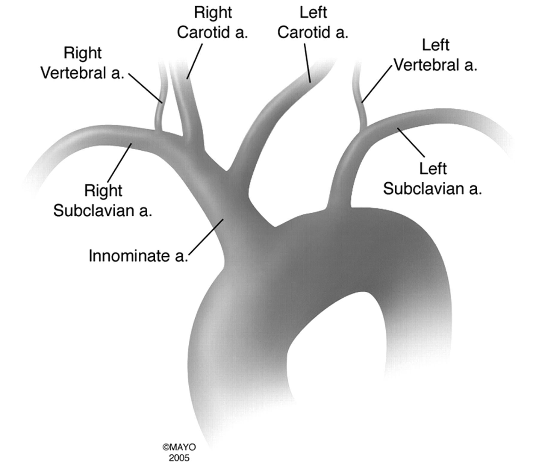
The second most common pattern of human aortic arch branching has a common origin for the innominate and left common carotid arteries. This pattern has erroneously been referred to as a “bovine arch.”

In this variant of aortic arch branching, the left common carotid artery originates separately from the innominate artery. This pattern has also been erroneously referred to as a “bovine arch.”
True Bovine Arch
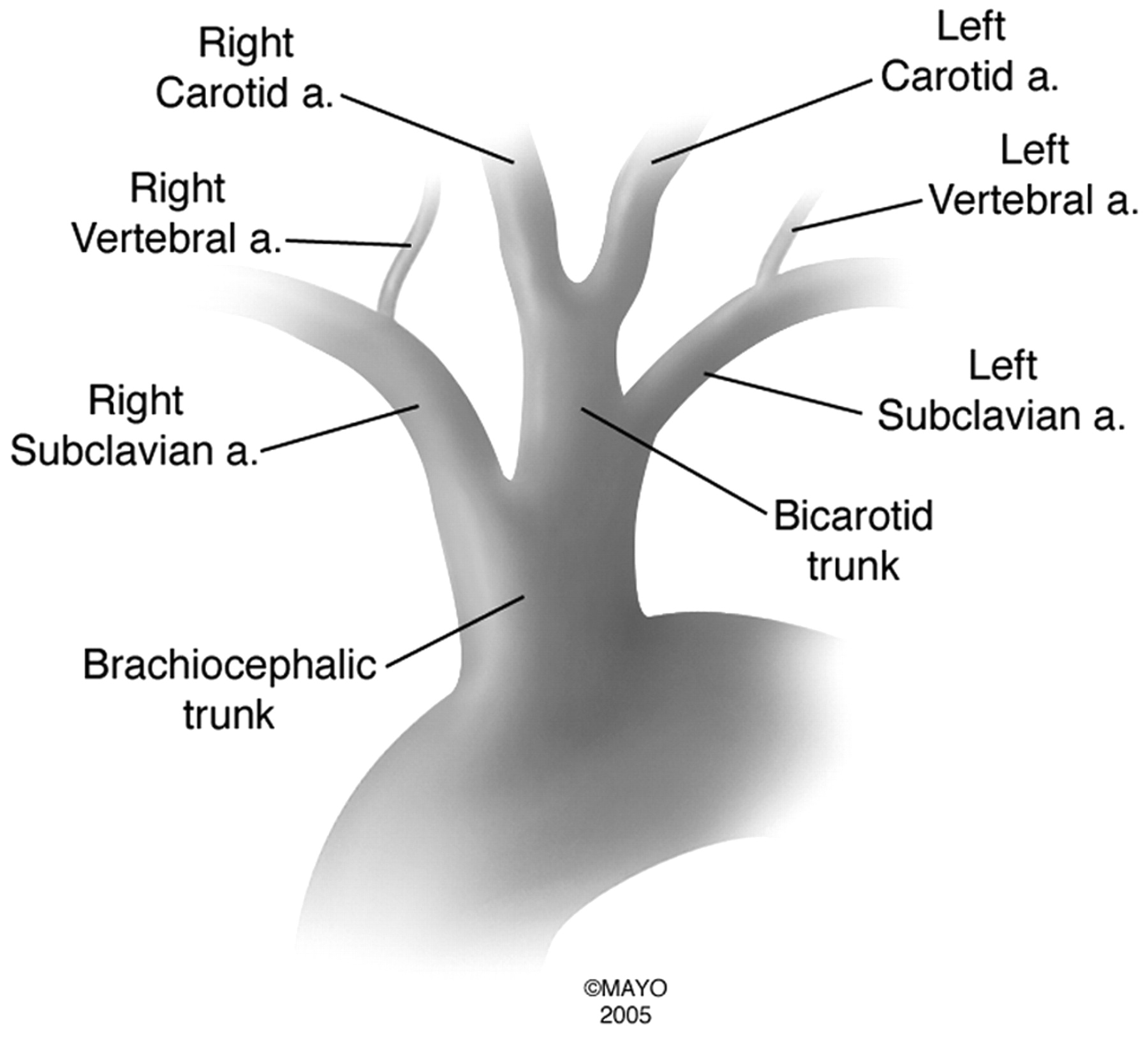
A true bovine aortic arch bears no resemblance to any of the common human aortic arch variations. In cattle, a single great vessel originates from the aortic arch.7 This large brachiocephalic trunk gives rise to both subclavian arteries and a bicarotid trunk. The bicarotid trunk then bifurcates into the left common carotid artery and right common carotid artery (Fig 4). The presence of a single long vessel originating from the aortic arch is a common occurrence in animals with deep chests. The general thinking among veterinary anatomists is that the long distance from the aortic arch to the thoracic inlet is the reason that all the great vessels come off the arch as a single vessel, the brachiocephalic trunk.

The aortic arch branching pattern found in cattle has a single brachiocephalic trunk originating from the aortic arch and eventually splits into the bilateral subclavian arteries and a bicarotid trunk.
Discussion
Standard Aortic Arch.
This is the most common anatomic appearance in humans (Fig 1). There are 3 great vessels that originate from the arch: the innominate artery, left common carotid artery, and left subclavian artery. The innominate artery subsequently branches into the right common carotid artery and the right subclavian artery. The incidence of this pattern is reported to be between 48% and 84%, with the variation largely attributable to differences between white and black individuals.5 It is generally accepted that this “normal” configuration occurs in approximately 70% of patients.3
Common Origin of the Innominate Artery and Left Common Carotid Artery.
This configuration replaces the misnomer of a “bovine” arch. The innominate artery and the left common carotid artery have a common origin. Therefore, only 2 great vessels originate from the aortic arch (Fig 2). This aortic arch branching pattern is found more often in blacks, and previous cadaveric reports have documented occurrences in 25% of blacks and in only 8% of whites.5 Overall, this pattern of branching is seen in approximately 13% of patients.3
Origin of the Left Common Carotid Artery from the Innominate Artery.
When the left common carotid artery originates from the innominate artery directly, there are 2 great vessels arising from the arch. This variant is similar to the common origin variety, except that the left common carotid artery originates from the innominate artery more distally, rather than as a common trunk (Fig 3). The left common carotid artery originates off the innominate artery at an average distance of less than 1 cm from the aortic arch with the maximal distance being 2.5 cm.5 This variant also occurs more commonly in blacks (10%) compared with whites (5%), with an overall rate of 9% in the general population.3,5
Conclusion
The term “bovine aortic arch” is a common misnomer in the medical literature, and the human aortic arch branching patterns to which it is ascribed are not commonly found in cattle. We suggest a descriptive aortic arch naming scheme that facilitates communication between practitioners and excludes the use of misnomers such as “bovine arch.”
- Received January 30, 2006.
- Accepted after revision March 10, 2006.
- Copyright © American Society of Neuroradiology
In this issue


Jump to section
Related Articles
Cited By...
- Comment on "Common Origin of Brachiocephalic and Left Common Carotid Arteries: Proposal of New Terminology"
- Common Origin of Brachiocephalic and Left Common Carotid Arteries: Proposal of New Terminology
- Republished: A rare case of absent left common carotid artery with bovine origin of the left external carotid artery
- A rare case of absent left common carotid artery with bovine origin of the left external carotid artery
- Initial experience of a novel sheath guide for transbrachial carotid artery stenting: technical note
- 2011 ASA/ACCF/AHA/AANN/AANS/ACR/ASNR/CNS/SAIP/SCAI/SIR/SNIS/SVM/SVS Guideline on the Management of Patients With Extracranial Carotid and Vertebral Artery Disease: Executive Summary: A Report of the American College of Cardiology Foundation/American Heart Association Task Force on Practice Guidelines, and the American Stroke Association, American Association of Neuroscience Nurses, American Association of Neurological Surgeons, American College of Radiology, American Society of Neuroradiology, Congress of Neurological Surgeons, Society of Atherosclerosis Imaging and Prevention, Society for Cardiovascular Angiography and Interventions, Society of Interventional Radiology, Society of NeuroInterventional Surgery, Society for Vascular Medicine, and Society for Vascular Surgery
- 2011 ASA/ACCF/AHA/AANN/AANS/ACR/ASNR/CNS/SAIP/SCAI/SIR/SNIS/SVM/SVS Guideline on the Management of Patients With Extracranial Carotid and Vertebral Artery Disease: A Report of the American College of Cardiology Foundation/American Heart Association Task Force on Practice Guidelines, and the American Stroke Association, American Association of Neuroscience Nurses, American Association of Neurological Surgeons, American College of Radiology, American Society of Neuroradiology, Congress of Neurological Surgeons, Society of Atherosclerosis Imaging and Prevention, Society for Cardiovascular Angiography and Interventions, Society of Interventional Radiology, Society of NeuroInterventional Surgery, Society for Vascular Medicine, and Society for Vascular Surgery
- 2011 ASA/ACCF/AHA/AANN/AANS/ACR/ASNR/CNS/SAIP/SCAI/SIR/SNIS/SVM/SVS guideline on the management of patients with extracranial carotid and vertebral artery disease: executive summary: A Report of the American College of Cardiology Foundation/American Heart Association Task Force on Practice Guidelines and the American Stroke Association, American Association of Neuroscience Nurses, American Association of Neurological Surgeons, American College of Radiology, American Society of Neuroradiology, Congress of Neurological Surgeons, Society of Atherosclerosis Imaging and Prevention, Society for Cardiovascular Angiography and Interventions, Society of Interventional Radiology, Society of NeuroInterventional Surgery, Society for Vascular Medicine and Society for Vascular Surgery Developed in collaboration with the American Academy of Neurology and Society of Cardiovascular Computed Tomography
- 2011 ASA/ACCF/AHA/AANN/AANS/ACR/ASNR/CNS/SAIP/SCAI/SIR/SNIS/SVM/SVS Guideline on the Management of Patients With Extracranial Carotid and Vertebral Artery Disease: A Report of the American College of Cardiology Foundation/American Heart Association Task Force on Practice Guidelines, and the American Stroke Association, American Association of Neuroscience Nurses, American Association of Neurological Surgeons, American College of Radiology, American Society of Neuroradiology, Congress of Neurological Surgeons, Society of Atherosclerosis Imaging and Prevention, Society for Cardiovascular Angiography and Interventions, Society of Interventional Radiology, Society of NeuroInterventional Surgery, Society for Vascular Medicine, and Society for Vascular Surgery Developed in Collaboration With the American Academy of Neurology and Society of Cardiovascular Computed Tomography
- 2011 ASA/ACCF/AHA/AANN/AANS/ACR/ASNR/CNS/SAIP/SCAI/SIR/SNIS/SVM/SVS Guideline on the Management of Patients With Extracranial Carotid and Vertebral Artery Disease: Executive Summary: A Report of the American College of Cardiology Foundation/American Heart Association Task Force on Practice Guidelines, and the American Stroke Association, American Association of Neuroscience Nurses, American Association of Neurological Surgeons, American College of Radiology, American Society of Neuroradiology, Congress of Neurological Surgeons, Society of Atherosclerosis Imaging and Prevention, Society for Cardiovascular Angiography and Interventions, Society of Interventional Radiology, Society of NeuroInterventional Surgery, Society for Vascular Medicine, and Society for Vascular Surgery Developed in Collaboration With the American Academy of Neurology and Society of Cardiovascular Computed Tomography