Advertisement

Main menu

  • Home
  • Content
    • Current Issue
    • Accepted Manuscripts
    • Article Preview
    • Past Issue Archive
    • AJNR Case Collection
    • Case of the Week Archive
    • Classic Case Archive
    • Case of the Month Archive
  • Special Collections
    • Spinal CSF Leak Articles (Jan 2020-June 2024)
    • 2024 AJNR Journal Awards
    • Most Impactful AJNR Articles
  • Multimedia
    • AJNR Podcast
    • AJNR Scantastics
    • Video Articles
  • For Authors
    • Submit a Manuscript
    • Author Policies
    • Fast publishing of Accepted Manuscripts
    • Graphical Abstract Preparation
    • Manuscript Submission Guidelines
    • Imaging Protocol Submission
    • Submit a Case for the Case Collection
  • About Us
    • About AJNR
    • Editorial Board
  • More
    • Become a Reviewer/Academy of Reviewers
    • Subscribers
    • Permissions
    • Alerts
    • Feedback
    • Advertisers
    • ASNR Home
  • Other Publications
    • ajnr

User menu

  • Alerts
  • Log in

Search

  • Advanced search
American Journal of Neuroradiology

American Journal of Neuroradiology

ASHNR American Society of Functional Neuroradiology ASHNR American Society of Pediatric Neuroradiology ASSR
  • Alerts
  • Log in

Advanced Search

  • Home
  • Content
    • Current Issue
    • Accepted Manuscripts
    • Article Preview
    • Past Issue Archive
    • AJNR Case Collection
    • Case of the Week Archive
    • Classic Case Archive
    • Case of the Month Archive
  • Special Collections
    • Spinal CSF Leak Articles (Jan 2020-June 2024)
    • 2024 AJNR Journal Awards
    • Most Impactful AJNR Articles
  • Multimedia
    • AJNR Podcast
    • AJNR Scantastics
    • Video Articles
  • For Authors
    • Submit a Manuscript
    • Author Policies
    • Fast publishing of Accepted Manuscripts
    • Graphical Abstract Preparation
    • Manuscript Submission Guidelines
    • Imaging Protocol Submission
    • Submit a Case for the Case Collection
  • About Us
    • About AJNR
    • Editorial Board
  • More
    • Become a Reviewer/Academy of Reviewers
    • Subscribers
    • Permissions
    • Alerts
    • Feedback
    • Advertisers
    • ASNR Home
  • Follow AJNR on Twitter
  • Visit AJNR on Facebook
  • Follow AJNR on Instagram
  • Join AJNR on LinkedIn
  • RSS Feeds

Welcome to the new AJNR, Updated Hall of Fame, and more. Read the full announcements.


AJNR is seeking candidates for the position of Associate Section Editor, AJNR Case Collection. Read the full announcement.


LetterLetter

T2 Hypointense Signal of Rathke Cleft Cyst

Fabrice Bonneville, Jacques Chiras, Françoise Cattin and Jean-François Bonneville
American Journal of Neuroradiology March 2007, 28 (3) 397;
Fabrice Bonneville
  • Find this author on Google Scholar
  • Find this author on PubMed
  • Search for this author on this site
Jacques Chiras
  • Find this author on Google Scholar
  • Find this author on PubMed
  • Search for this author on this site
Françoise Cattin
  • Find this author on Google Scholar
  • Find this author on PubMed
  • Search for this author on this site
Jean-François Bonneville
  • Find this author on Google Scholar
  • Find this author on PubMed
  • Search for this author on this site
  • Article
  • Figures & Data
  • Info & Metrics
  • Responses
  • References
  • PDF
Loading

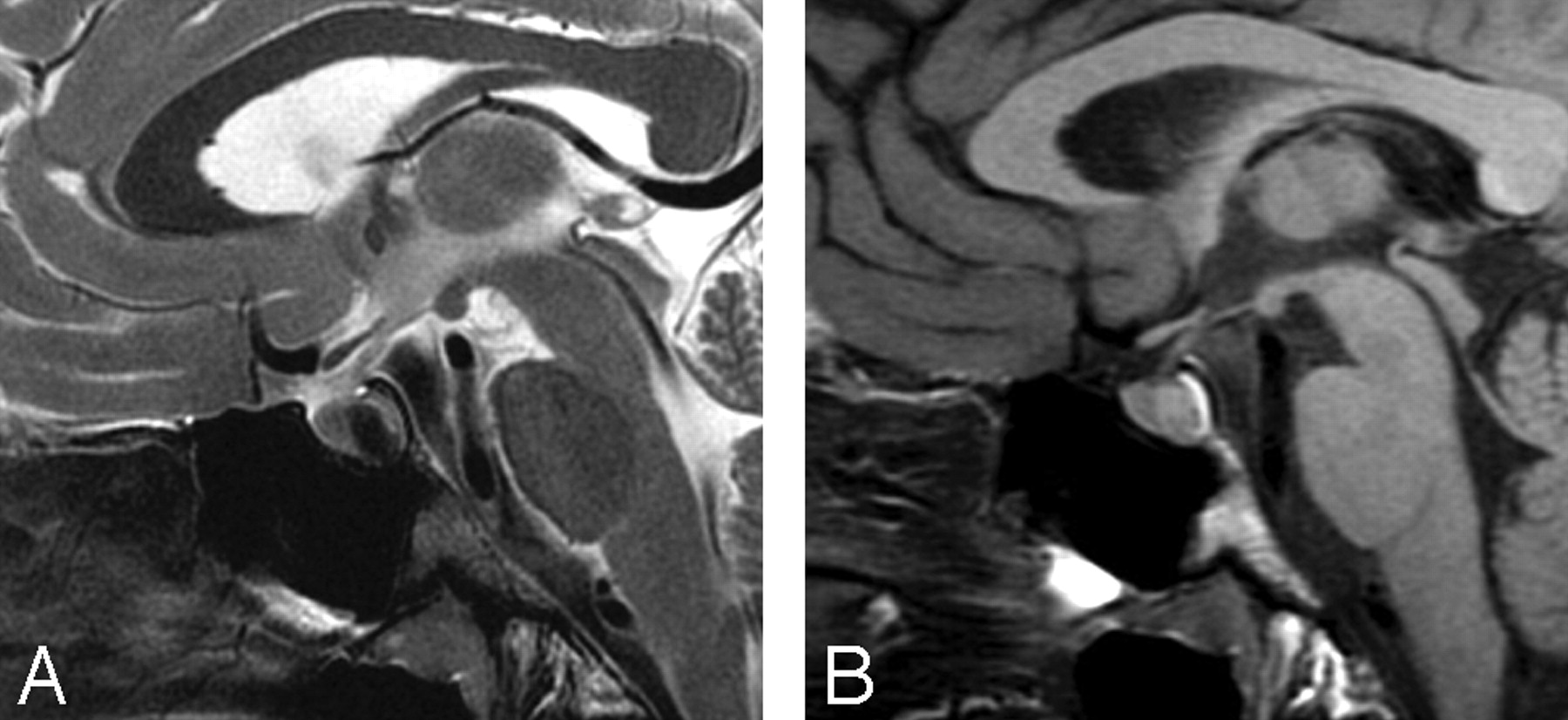
We read with interest the article by Megdiche-Bazarbacha et al,1 in which the authors report a case of a giant intrasphenoidal Rathke cleft cyst (RCC). In our recent review of RCCs,2 we mentioned a comparable case reported by Meyer et al3 in the AJNR of such a giant RCC centered on the sphenoid bone, mimicking a chordoma. A search of PubMed with the keywords “Rathke cyst sphenoid” revealed at least another very similar case recently published in the Journal of Neurosurgery.4 The MR signal intensity characteristics of all these lesions were identical, with T1 and T2 signal intensities slightly greater than that of CSF. It is well known that the signal intensities of RCCs are variable and directly depend on their biochemical content, with most of the reported RCCs demonstrating T2 high signal intensity. It is, however, crucial to point out for practicing radiologists that a homogeneous T2 hypointense signal intensity within a nonenhancing midline sellar cyst is highly suggestive of a RCC.2 Figure 1 illustrates this feature and also demonstrates how T2-weighted images are more sensitive than T1-weighted images in depicting small cysts located between the anterior and the posterior pituitary lobes.

Finally, the postoperative RCC recurrence rate of only 5% of Megdiche-Bazarbacha et al1 seems quite low. Although considered as benign lesions, RCCs commonly recur, with reported rates as high as 33%.5 Independent of the neurosurgeons skill, this recurrence is due to the fact that the most common and safest surgical technique consists of draining the contents of the cyst with only a partial excision or fenestration of the paper-thin wall cyst, leaving in place the source of the secretions that may re-accumulate.

  Fig 1.
  • Download figure
  • Open in new tab
  • Download powerpoint
Fig 1.

A Rathke cleft cyst in a 25-year-old woman with headaches. A, Sagittal T2-weighted image easily demonstrates a homogeneous hypointense-signal-intensity mass perfectly located on the midline between both pituitary lobes. B, Sagittal T1-weighted image barely shows the isointense-to-slightly-hyperintense signal intensity of the lesion.

References

  1. ↵
    Megdiche-Bazarbacha H, Ben Hammouda K, Aicha AB, et al. Intrasphenoidal rathke cleft cyst. AJNR Am J Neuroradiol 2006;27:1098–100
    Abstract/FREE Full Text
  2. ↵
    Bonneville F, Cattin F, Bonneville JF, et al. Rathke's cleft cyst [in French]. J Neuroradiol 2003;30:238–48
    PubMed
  3. ↵
    Meyer JR, Quint DJ, McKeever PE, et al. Giant Rathke cleft cyst. AJNR Am J Neuroradiol 1994;15:533–36
    Abstract/FREE Full Text
  4. ↵
    Ben Hamouda K, Khaldi M, Jemel H, et al. Rathke cleft cyst of the sphenoid bone and nasopharynx: case illustration. J Neurosurg 2005;103:1095
    PubMed
  5. ↵
    Mukherjee JJ, Islam N, Kaltsas G, et al. Clinical, radiological and pathological features of patients with Rathke's cleft cysts: tumors that may recur. J Clin Endocrinol Metab 1997;82:2357–62
    CrossRefPubMed
  • Copyright © American Society of Neuroradiology
PreviousNext
Back to top

In this issue

American Journal of Neuroradiology: 28 (3)
American Journal of Neuroradiology
Vol. 28, Issue 3
March 2007
  • Table of Contents
  • Index by author
Advertisement
Print
Download PDF
Email Article

Thank you for your interest in spreading the word on American Journal of Neuroradiology.

NOTE: We only request your email address so that the person you are recommending the page to knows that you wanted them to see it, and that it is not junk mail. We do not capture any email address.

Enter multiple addresses on separate lines or separate them with commas.
T2 Hypointense Signal of Rathke Cleft Cyst
(Your Name) has sent you a message from American Journal of Neuroradiology
(Your Name) thought you would like to see the American Journal of Neuroradiology web site.
CAPTCHA
This question is for testing whether or not you are a human visitor and to prevent automated spam submissions.
Cite this article
Fabrice Bonneville, Jacques Chiras, Françoise Cattin, Jean-François Bonneville
T2 Hypointense Signal of Rathke Cleft Cyst
American Journal of Neuroradiology Mar 2007, 28 (3) 397;

Citation Manager Formats

  • BibTeX
  • Bookends
  • EasyBib
  • EndNote (tagged)
  • EndNote 8 (xml)
  • Medlars
  • Mendeley
  • Papers
  • RefWorks Tagged
  • Ref Manager
  • RIS
  • Zotero
0 Responses
Respond to this article
Share
Bookmark this article
T2 Hypointense Signal of Rathke Cleft Cyst
Fabrice Bonneville, Jacques Chiras, Françoise Cattin, Jean-François Bonneville
American Journal of Neuroradiology Mar 2007, 28 (3) 397;
del.icio.us logo Twitter logo Facebook logo Mendeley logo
  • Tweet Widget
  • Facebook Like
  • Google Plus One

Jump to section

  • Article
    • References
  • Figures & Data
  • Info & Metrics
  • Responses
  • References
  • PDF

Related Articles

  • No related articles found.
  • PubMed
  • Google Scholar

Cited By...

  • Assessing Disease Experience across the Life Span for Individuals with Osteogenesis Imperfecta: Challenges and Opportunities for Patient-Reported Outcomes (PROs) Measurement
  • Crossref
  • Google Scholar

This article has not yet been cited by articles in journals that are participating in Crossref Cited-by Linking.

More in this TOC Section

  • Brain AVM’s Nidus: What if We Hadn’t Understood Anything?
  • Letter to the Editor regarding “Automated Volumetric Software in Dementia: Help or Hindrance to the Neuroradiologist?”
  • Reply:
Show more Letters

Similar Articles

Advertisement

Indexed Content

  • Current Issue
  • Accepted Manuscripts
  • Article Preview
  • Past Issues
  • Editorials
  • Editors Choice
  • Fellow Journal Club
  • Letters to the Editor

Cases

  • Case Collection
  • Archive - Case of the Week
  • Archive - Case of the Month
  • Archive - Classic Case

Special Collections

  • Special Collections

Resources

  • News and Updates
  • Turn around Times
  • Submit a Manuscript
  • Author Policies
  • Manuscript Submission Guidelines
  • Evidence-Based Medicine Level Guide
  • Publishing Checklists
  • Graphical Abstract Preparation
  • Imaging Protocol Submission
  • Submit a Case
  • Become a Reviewer/Academy of Reviewers
  • Get Peer Review Credit from Publons

Multimedia

  • AJNR Podcast
  • AJNR SCANtastic
  • Video Articles

About Us

  • About AJNR
  • Editorial Board
  • Not an AJNR Subscriber? Join Now
  • Alerts
  • Feedback
  • Advertise with us
  • Librarian Resources
  • Permissions
  • Terms and Conditions

American Society of Neuroradiology

  • Not an ASNR Member? Join Now

© 2025 by the American Society of Neuroradiology All rights, including for text and data mining, AI training, and similar technologies, are reserved.
Print ISSN: 0195-6108 Online ISSN: 1936-959X

Powered by HighWire