
Abstract
BACKGROUND AND PURPOSE: Peripheral aneurysms of the posterior inferior cerebellar artery (PICA) are rare, and pre-existing literature concerning their endovascular treatment is limited. The purpose of this study was to assess the etiology and clinical characteristics of peripheral PICA aneurysms and to evaluate the angiographic and clinical results of the patients who underwent endovascular treatment for a peripheral PICA aneurysm in a single center.
MATERIALS AND METHODS: Twelve consecutive patients with 12 peripheral PICA aneurysms (10 ruptured) included in an internal data base were retrospectively reviewed. Posttreatment and follow-up angiograms were analyzed, and the clinical outcome was recorded.
RESULTS: The etiology was dissection in 7 (58%) and unknown in 5 cases (42%). Three dissecting aneurysms reruptured before endovascular treatment, and another 3 demonstrated angiographic progress. Four aneurysms were treated by endosaccular coiling, 6 (all dissecting) by parent artery occlusion, and in 2 cases endovascular treatment failed. Angiographic outcome was complete aneurysm and/or parent artery occlusion in 9 cases and neck remnant in 1 case. One aneurysm needed retreatment at follow-up. One lethal procedural complication occurred, and transient ischemic symptoms appeared in 2 patients. The clinical outcome was good in 7 patients, whereas 3 patients, all poor clinical grade, died (1 for unrelated reasons). No rebleedings have occurred during the follow-up.
CONCLUSION: In this series, most peripheral PICA aneurysms were secondary to arterial dissection. They were unstable with a high risk of rebleeding and a high mortality if not treated without delay. Endovascular treatment was effective in preventing rehemorrhage.
Posterior inferior cerebellar artery (PICA) aneurysms account for approximately 0.5% to 3.0% of all intracranial aneurysms,1,2 and most are located right at the origin or in the first anteromedullary segment of the vessel.2-4 Roughly, only a fifth of the PICA aneurysms are thought to arise from more distal segments of the PICA.2-4 Approximately 28% of all aneurysms located in the vertebrobasilar arteries, including PICA, have been estimated to be of dissecting origin.5 Among peripheral PICA aneurysms, dissecting etiology has been found in 0%-80% of the cases.6-9
Due to anatomic location of the PICAs (proximity to brain stem and lower cranial nerves), their surgical treatment is associated with significant risk of neurologic complications.10 Endovascular technique enables treating these lesions without craniotomy and the risks related to surgical manipulation. Despite the recently increased number of publications, the pre-existing literature concerning endovascular treatment of peripheral PICA aneurysms is limited and controversial.6-9,11-21 In the present study, a consecutive series of 12 patients harboring a peripheral PICA aneurysm allocated for endovascular treatment in a single center was retrospectively analyzed.
Materials and Methods
The cases were collected from the data base containing all of the patients who had undergone endovascular treatment for their intracranial aneurysm since the start of the endovascular coiling activity in our hospital on December 1993 until March 2008. From the 25 angiographically verified PICA aneurysms, 12 were located distally to the anteromedullary segment of the PICA and composed the case material of this study.
The mean age of the patients (7 women, 5 men) was 53.5 years (range, 34.0–80.0 years). Ten of the aneurysms presented with an acute intracranial hemorrhage, and 2 were incidental imaging findings. The indication for digital subtraction angiography (DSA) in these latter 2 cases was assessment of cervicocerebral vessels after intravenous thrombolysis of an acute stroke in one case and headaches of uncertain etiology in the other case (initially examined at 1.5T by MR imaging and MR angiography). All of the patients underwent a 4-vessel cerebral angiography, disclosing a peripheral PICA aneurysm (in one case the primary DSA was negative). The location of the aneurysm was recorded according to the classification described by Lister et al22 as anterior medullary, lateral medullary, tonsillomedullary, telovelotonsillar, or cortical. Proximal PICA aneurysms located at the vertebral artery-PICA junction or at anteromedullary segment and flow-related aneurysms associated with brain arteriovenous malformations were excluded. The etiology of the aneurysms was classified on the basis of angiographic findings as dissecting or unknown. Dissecting etiology was diagnosed if the lesion initially demonstrated a solitary or multiple irregular fusiform dilations with or without associated narrowing of the PICA. Other angiographic abnormalities revealed by DSA were also recorded. Patient and aneurysm characteristics are summarized in the Table.
Patient and aneurysm characteristics
Endovascular Treatment
During the study period, endovascular treatment was the primary treatment option in all of the cases of PICA aneurysms in our hospital. Embolization was performed with the patients awake under intravenous sedation in 2 patients (patients 1 and 2; Table) and under general anesthesia in 10 patients (patients 3–12; Table). All of the procedures were performed via the transfemoral route in an angiographic suite with biplane imaging capabilities (Neurostar; Siemens, Erlangen, Germany in cases 1–4 and Integris Allura; Philips, Best, the Netherlands, in cases 5–12). Three interventional radiologists were involved in the treatments. The embolization method was selected on the basis of the location and morphology and presumed etiology of the aneurysm. Selective embolization of the aneurysmal sac with parent artery preservation was preferred if an acutely ruptured dissecting aneurysm was excluded and the aneurysm had a reasonable neck to allow endosaccular packing with coils. Otherwise the parent artery was considered to be sacrificed. In each case, the capability of the patient to tolerate permanent PICA occlusion was assessed on the angiographic criteria based on the presence and size of potential collateral vessels (contralateral PICA, ipsilateral anterior inferior cerebellar artery, and superior cerebellar artery). Test occlusion before permanent occlusion was performed by temporary inflation of a balloon in the vertebral artery over the origin of the PICA in one case with simultaneous contrast injection into the contralateral vertebral artery (patient 4; Table). Detachable platinum microcoils were used in endosaccular packing of the aneurysmal sac. Parent artery occlusions were performed with coils whenever technically feasible or with glue, mixed with iodized oil. According to our routine practice, the patient was heparinized during the procedure with activated clotting time targeted from 200 to 300 seconds. Procedural complications or any adverse events were recorded. After the procedure, the patient was closely monitored in an intensive care unit or in a postoperative recovery unit for at least one day. All of the postprocedural CT and MR images were analyzed with the intention to detect new lesions.
Angiographic Result
After endosaccular coiling, the angiographic occlusion grade was measured according to the modified Raymond scale23 as complete occlusion, neck remnant, or incomplete occlusion. In cases of parent artery occlusion, the occlusion grade of the abnormal segment of PICA was graded as complete or incomplete. The initial angiographic result was assessed from the final postembolization angiograms. Follow-up DSA was performed on 6 patients, and the same grading system was used to determine the occlusion grade.
Clinical Evaluation
Pretreatment clinical status was recorded from the patient files according to the Hunt & Hess classification.24 Any new neurologic symptoms after the treatment were recorded. Clinical outcome of the patients was established from the patient files and by telephone call by using the Glasgow outcome scale.25 New bleeding episodes either before the treatment or after the treatment were recorded. Patients were excluded from follow-up if endovascular treatment was abandoned.
Results
The patient and the aneurysm characteristics are presented in the Table. Three of the patients (25%) were in poor clinical state before endovascular treatment (Hunt and Hess 4 or 5). In addition to subarachnoid and intraventricular hemorrhage, intracerebellar hematoma was evident in 4 patients, and 1 patient had also hemorrhage in the subdural space. Hydrocephalus was present on 8 patients.
The location of the aneurysm was lateromedullary in 1 (8%), tonsillomedullary in 4 (33%), telovelotonsillar in 3 (25%), and cortical in 4 cases (33%). Seven of the aneurysms (58%) were dissecting, and 5 (42%) were of unknown etiology. All 5 of the aneurysms with an unknown etiology had a relatively wide neck with a mean dome-to-neck ratio of 1.6; 4 of them were sidewall aneurysms and 1 was locating at bifurcation. The maximal diameter of the aneurysms ranged from 2 to 15 mm (mean, 7 mm). There was second incidental aneurysm at the tip of basilar artery in 1 patient (patient 10; Table). The peripheral dissecting PICA aneurysm was considered to be the ruptured one in this case based on the location of hemorrhage on CT. The basilar tip aneurysm was embolized with coils in the same session in which the peripheral PICA aneurysm was treated. In one case (patient 5; Table) there were luminal narrowings of the proximal PICA possibly due to another dissection or, alternatively, secondary to vasospasm.
Among the 10 ruptured aneurysms, 3 (30%; all dissecting) reruptured early before the endovascular treatment (Table). The interval between the initial subarachnoid hemorrhage and rehemorrhage was 18 days in patient 3, 6 hours in patient 7, and 10 days in patient 8. Among the ruptured 10 aneurysms, embolization was performed in the same session after the diagnostic DSA in 3 cases and in another session in 7 cases. All 5 of the dissecting aneurysms that underwent the second DSA had progressed.
Endovascular Treatment
The delay from the initial bleeding to the endovascular treatment ranged from 1 to 42 days (mean, 9.8 days). The endovascular treatment method was parent artery occlusion in 7 patients and endosaccular coiling with parent artery preservation in 5 patients. Parent artery occlusion was performed with coils in 2 cases, with glue in 4 cases, and was interrupted due to technical failure in 1 case. In this case (patient 5; Table) tight proximal narrowings of the PICA made selective catheterization impossible even with the smallest available microcatheter (Magic 1.2; Balt, Montmorency, France). This patient was subsequently operated on, and the aneurysm was successfully trapped. In another patient (patient 1; Table), the aneurysm ruptured after selective catheterization of the aneurysm before deploying any coils. According to the ongoing treatment policy in our hospital at that time (1994), the patient was immediately intubated and transferred to an operation room. Unfortunately, aneurysm exposure failed and the patient subsequently died 2 days later. In 10 (83%) of 12 cases, the endovascular treatment was technically successfully completed. One of these patients underwent endovascular retreatment of the neck remnant 2 months after the initial embolization (patient 4; Table).
There were 4 procedural complications among 13 treatment sessions, including 1 retreatment (31%). In addition to the previously described lethal aneurysm rupture, there were 2 cases of clinically asymptomatic transient unintended parent artery occlusions and 1 case where significant glue migration distal to the planned occlusion site occurred without any clinical sequelae. One of those unintended parent artery occlusions recanalized spontaneously and the other was successfully treated with intra-arterial injection of abciximab. The latter one was, however, reoccluded in follow-up, but the patient remained asymptomatic. Two patients had transient neurologic symptoms after the endovascular procedure. Procedural mortality, transient morbidity, and permanent morbidity were thus 1 in 13 (7.7%), 2 in 13 (15%), and 0 in 13 (0%), respectively. Nine patients were eligible for postprocedural imaging, which revealed new ischemic lesions in 7 patients (78%). In 3 cases, a small lacunar cerebellar infarct was detected (all asymptomatic), and in 4 cases, a cortical cerebellar infarct in PICA territory was evident (transient worsening of vertigo in one case, and in other cases the poor neurologic status remained unchanged). All of the cortical infarcts were related to parent artery occlusions with glue. In patient 8, with a history of cerebellar infarct 11 years earlier, there was a proximal occlusion of the contralateral PICA in initial angiography, and in postprocedural CT there were signs of bilateral infarction.
Angiographic Results
Among the 4 cases where the aneurysm was treated by endosaccular coiling with parent artery preservation, the initial angiographic result was complete occlusion in 2 and incomplete occlusion in 2 cases. Patient 4 (initially incomplete occlusion) was retreated 2 months after the initial treatment, and the occlusion grade improved to complete occlusion and the result has remained stable during 48 months of follow-up. Patient 9 (initially complete occlusion) underwent a follow-up angiography 12 months after the treatment, and a small neck remnant was detected due to coil compaction. Retreatment was not indicated, but further follow-up was scheduled. In patient 11, the initial angiographic result was incomplete occlusion, and in follow-up angiography 2 months later the aneurysm, as well as the parent artery at the aneurysm site, was completely occluded. With patient 12 (initially complete occlusion), the scheduled follow-up angiography has not yet been performed.
In cases of parent artery occlusion, the initial angiographic result was complete occlusion in all but one case. In patient 3, at the end of the procedure there still was a slow flow in the parent artery and the aneurysm. Angiography was repeated 2 days and one month after the treatment confirming a complete occlusion of the aneurysm and the parent vessel. In all 4 of the cases where the aneurysm was treated by parent artery occlusion with glue, the initial angiographic result was a complete occlusion, and follow-up angiography was considered unnecessary. However, in patient 10, one follow-up angiography was performed after 6 months due to a coincidental basilar tip aneurysm, which was embolized with coils in the same session. The follow-up angiography showed complete occlusion of both aneurysms.
Clinical Outcome
Ten patients were eligible for clinical follow-up with a mean length of 22.0 months (range, 0.5-63.0 months). The clinical outcome was good (Glasgow Outcome Scale [GOS] 5) in 7 cases, whereas 3 patients died. Two patients with poor preprocedural clinical condition died during their initial hospital stay. Patient 7 recovered to GOS 3 but died 24 months afterward due to pulmonary cancer. There have been no rebleedings after the endovascular treatment during the follow-up.
Discussion
In the present study, 12 consecutive patients harboring a peripheral PICA aneurysm allocated for endovascular treatment as a primary treatment method in a single center are presented. Most (58%) of the aneurysms were classified as dissecting aneurysms based on the angiographic findings and in one case also on the direct observation during surgery. The etiology of dissection was presumed to be spontaneous in all (no history of trauma and no known comorbidities associated with dissection). Concerning the endovascular treatment of dissecting PICA aneurysms, to our knowledge, the present series composes the largest consecutive series of patients (7 patients) published in its context so far.19 Four of the 5 aneurysms, which could not be classified as dissecting ones, were morphologically different from the typical intracranial aneurysms located at the circle of Willis. They were not arising from any vessel branching points but were more like wide-neck sidewall aneurysms located at the turning point of the artery. The curves and hairpin turns are typical in the course of PICA, and excessive hemodynamic stress has been suspected to be the etiological factor in these types of peripheral aneurysms, contributing to elastic lamina injury and subsequent aneurysm formation.26 It is possible that some of the aneurysms that we classified as “unknown etiology” may have resulted from acute or chronic dissection but did not fulfill the strict criteria of dissection in the present study. Patient 5 (Table and Fig 1) had an acute onset of headaches typical for dissection, and there were irregularities in the PICA both proximal and distal to aneurysm. Peripheral aneurysms may be rarely infectious or traumatic, but there were no such cases in the present study.27,28

A 46-year old woman (patient 4; Table) experienced acute onset of headache. MR imaging and MR angiography showed a right-sided PICA aneurysm without any evidence of intracranial bleeding. In initial DSA (A), a 9- × 5.5-mm sidewall aneurysm was visualized in a tonsillomedullary segment of the right PICA. The artery is narrowed proximally and dilated distally to the aneurysm neck. A balloon occlusion test was performed with indeterminate findings. The aneurysm was treated by endosaccular coiling with parent artery preservation. The initial occlusion grade was incomplete, but after retreatment a complete occlusion was achieved, and it has remained stable during follow-up (Fig 2B).

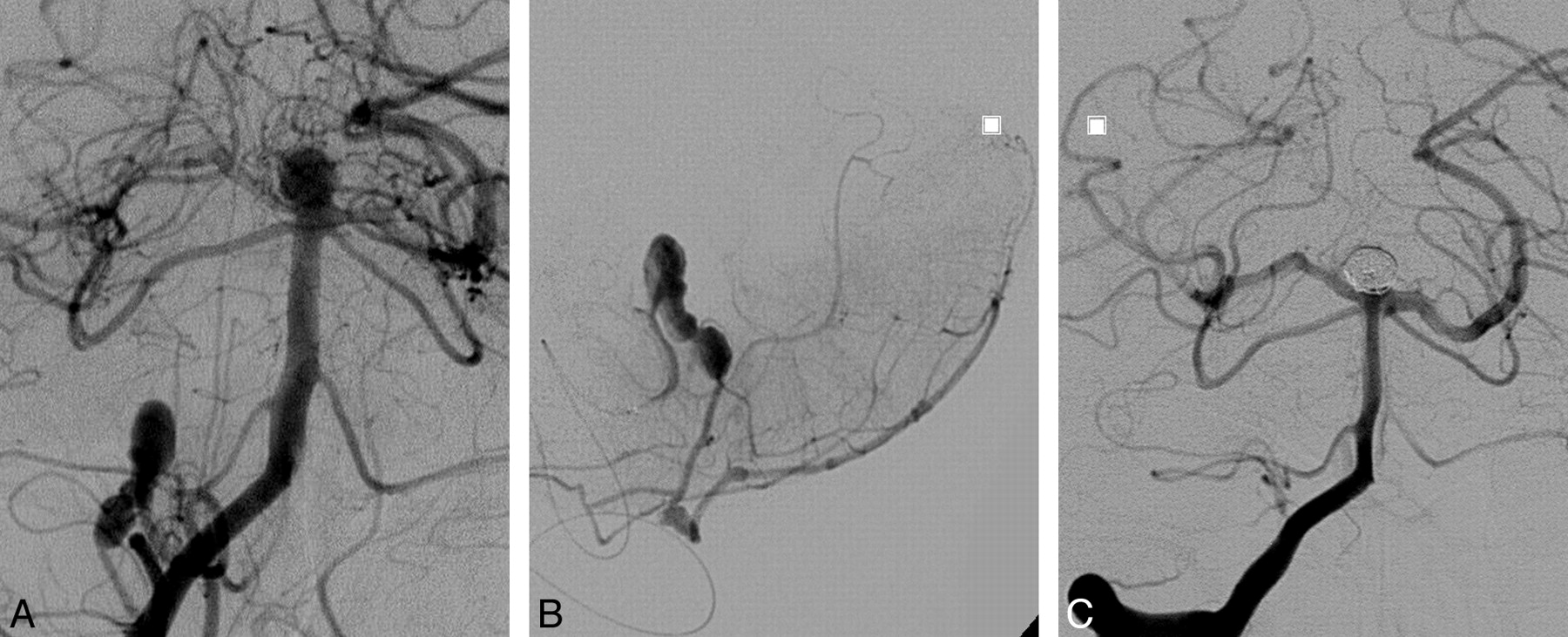
An 80-year-old woman (patient 10; Table) experienced severe vertigo with sudden onset. CT examination revealed a small right-sided intracerebral hematoma in cerebellar vermis, hemorrhage in the fourth ventricle, and prepontine cistern. Subsequent DSA revealed a dissecting aneurysm of the right PICA and a saccular aneurysm of the basilar tip (A). A superselective right PICA injection demonstrated an irregular, fusiform PICA aneurysm located in the cortical segment (B). Parent artery occlusion of the dissected cortical segment of PICA and coil embolization of the basilar tip aneurysm were performed in the same session. In control angiogram after the treatment, both aneurysms are occluded (C). Posttreatment CT showed PICA infarct, and the patient had temporary worsening of vertigo, but in 6 months of follow-up she recovered without any symptoms.
The most typical presentation of a dissecting PICA aneurysm is hemorrhage, which has been reported to account for two thirds of the cases.15 In the present series, all 7 of the dissecting aneurysms were ruptured, and 3 (43%) of them rebled early before endovascular treatment. Angiographic progression was detected in 5 cases of the ruptured dissecting aneurysms eligible for repeat angiography before the treatment. These findings are in concordance with earlier observations and support the necessity of early treatment to prevent rebleedings.5,29 During the present series, the mean delay from initial bleeding to endovascular treatment was 12 days, which is significantly longer than in cases of ruptured saccular aneurysms in our hospital.30 The prolonged delay is partly due to the poor clinical condition of the patient in many cases. On the other hand, limited knowledge regarding the optimal treatment method may also have prolonged the delay, especially in the first cases of our experience.
Endovascular treatment of a peripheral PICA aneurysm can technically be performed either by endosaccular coiling with parent artery preservation or by the parent artery occlusion method. Contrary to typical saccular aneurysms, peripheral PICA aneurysms usually have a fusiform shape or a very wide neck, making endosaccular coiling with parent artery preservation difficult or impossible. In addition, the small caliber of the parent artery does not usually allow the use of remodeling techniques, such as temporary balloon inflation or stent deployment.31,32 Rarely, the small caliber of the vessel may not even allow catheterization of the vessel, as in one of the cases in the present series. The advantage of the endosaccular coiling technique is the preservation of the parent artery, which could minimize the risk of ischemic complications. The disadvantage of this technique is the uncertain stability of the aneurysm occlusion and the risk of aneurysm recurrence or reopening,18,33 which may predispose the aneurysm to rerupture. On the other hand, parent artery occlusion of the PICA is potentially associated with 2 types of ischemic complication. First, from the 3 most proximal segments of the PICA (anterior medullary, lateral medullary, and tonsillomedullary), as well as from vertebral artery at the origin of PICA, perforating arteries supplying the brain stem may originate.6,21,34 It has been reported that, in 50% of the cases, no supply to the medulla exists from PICA, and the more dorsally (or laterally) the PICA originates, the smaller are the chances of its having medullary branches.35 The risk of brain stem ischemia in cases of permanent occlusion at the first 3 segments is also limited due to the numerous anastomoses of the perforating arteries forming a plexiform network on the medullary surface.36 Furthermore, aneurysmal dilations of the dissected arterial segments may be associated with absence of normal perforating branches, and occlusion of these segments can, thus, be performed without further damage to perforating territories.15 Although potentially carrying a high risk for morbidity, according to the literature and our own experience (2 patients), the risk for brain stem ischemia seems to be relatively low.8,12,15,37 Another potential risk of permanent PICA occlusion is cerebellar ischemia distal to the occlusion site. Collateral supply from the contralateral PICA or ipsilateral anterior cerebellar artery and/or superior cerebellar artery is reported to be sufficient to prevent ischemia in most cases.19,21 Basically, a test occlusion can be performed to investigate the patient's capability to tolerate permanent occlusion. In cases where collateral supply is insufficient, potentially there is the option of surgical bypass from the occipital artery to PICA. On the other hand, if permanent PICA occlusion is followed by cerebellar infarct, it is usually of limited size, and patients usually tolerate it well if it is unilateral.21 According to the present study, cerebellar infarct is common after permanent PICA occlusion, but the patients who survived the initial bleeding tolerated it well.
Due to the challenging anatomy of PICA, technical complications associated with endovascular treatment of peripheral PICA aneurysms seem to be more common than among intracranial aneurysms in general. During the present series, we experienced one lethal iatrogenic aneurysm rupture. This was the first patient in the series and one of the very first patients treated in our hospital for intracranial aneurysm with the endovascular technique. The patient was not under general anesthesia, and initially it was impossible to continue the procedure due to seizures associated with the rupture. This case demonstrates the steep learning curve associated with endovascular treatment of intracranial aneurysms, and, after our initial experience, we have done all of the intracranial procedures under general anesthesia and treat iatrogenic ruptures by endovascular techniques with a significantly better outcome.30 In addition to this lethal complication in our very early experience, we did not have any other cases of procedural mortality or permanent morbidity in this series, and endovascular treatment of peripheral PICA aneurysms can, thus, be considered relatively safe.
Final angiographic outcomes in the present series were good: complete aneurysm occlusion was achieved in 9 aneurysms (90%), and a residual neck was found in 1 aneurysm (10%). The results of the present study confirm that complete aneurysm occlusion can be reliably achieved by parent artery occlusion.19,21 Reopening of an occluded vessel harboring a distal aneurysm is unlikely,21 and we did not routinely perform follow-up angiography in cases where the parent artery was occluded with glue. With endosaccular coiling with parent artery preservation, complete aneurysm occlusion cannot be achieved in every case, and aneurysm recurrences may appear. Angiographic follow-up is, thus, mandatory in cases of endosaccular coiling, and retreatment may be indicated if the occlusion grade is insufficient or decreases in the follow-up. Fujimura et al18 have reported a case of fatal rebleeding 19 days after selective embolization of a dissecting PICA aneurysm, which had an angiographically stable neck remnant. The authors concluded that endosaccular coiling with parent artery preservation should be avoided when treating such lesions. In the present study, there were no rebleedings during follow-up. We agree with Fujimura et al18 that parent artery occlusion is a more reliable technique in preventing further bleedings. However, on the basis of other reports7-9,20 and our experience of a single case, endosaccular coiling with parent artery preservation might prevent the aneurysm from rerupture as well.
Among the 10 patients eligible for follow-up, the clinical outcome was good in 7 patients (70%) and poor in 3 patients (30%). All of the patients with poor clinical outcome were in poor (Hunt and Hess grade 4 or 5) clinical condition. As a limitation of this study, the number of patients is too few for relevant correlative statistical analysis. However, the clinical outcome seems to be strongly associated with the pretreatment clinical condition similar to large clinical series concerning endovascularly treated intracranial aneurysms in general.30,38,39 The retrospective nature and lack of a systematic postprocedural MR imaging protocol including diffusion-weighted imaging can also be considered as limitations of this study.
Conclusions
In conclusion, most peripheral PICA aneurysms in this series were secondary to arterial dissection. Ruptured dissecting PICA aneurysms were vulnerable lesions with a high risk of recurrent bleeding and mortality. Therefore, they should be treated without any inadvertent delay. Endovascular treatment was effective in preventing rehemorrhage. Endosaccular coiling with parent artery preservation was performed whenever the anatomy allowed the coils to be retained in the aneurysm sac but was impossible or more risky in case of an acute hemorrhage. Long-term occlusion grade needs to be followed if endosaccular coiling has been done. Parent artery occlusion resulted in permanent occlusion of the lesion. PICA territory infarcts were frequently found after permanent PICA occlusions. However, the patients tolerated them well, and the clinical outcome was mainly affected by the preprocedural clinical condition.
References
- Received April 2, 2008.
- Accepted after revision May 27, 2008.
- Copyright © American Society of Neuroradiology
In this issue


Jump to section
Related Articles
Cited By...
- Endovascular rescue treatment through stent positioning after surgical clipping of intracranial aneurysms complicated by parent artery obstruction
- Endovascular treatment of posterior inferior cerebellar artery aneurysms: a 7-year single-center experience
- Endovascular parent vessel sacrifice in ruptured dissecting vertebral and posterior inferior cerebellar artery aneurysms: clinical outcomes and review of the literature
- Onyx embolization in distal dissecting posterior inferior cerebellar artery aneurysms
- Elderly patients with intracranial aneurysms have higher quality of life after coil embolization: a decision analysis
- Off-Label Use of Drugs and Devices in the Neuroendovascular Suite
- Endovascular treatment of ruptured dissecting aneurysms of the posterior inferior cerebellar artery
- Dissecting Aneurysms of the Distal Segment of the Posterior Inferior Cerebellar Arteries: Clinical Presentation and Management
- Balloon test occlusion and endosurgical parent artery sacrifice for the evaluation and management of complex intracranial aneurysmal disease
- Reply:
- Onyx Embolization of Distal Aneurysms of the Posterior Circulation