Advertisement

Main menu

  • Home
  • Content
    • Current Issue
    • Accepted Manuscripts
    • Article Preview
    • Past Issue Archive
    • AJNR Case Collection
    • Case of the Week Archive
    • Classic Case Archive
    • Case of the Month Archive
  • Special Collections
    • Spinal CSF Leak Articles (Jan 2020-June 2024)
    • 2024 AJNR Journal Awards
    • Most Impactful AJNR Articles
  • Multimedia
    • AJNR Podcast
    • AJNR Scantastics
    • Video Articles
  • For Authors
    • Submit a Manuscript
    • Author Policies
    • Fast publishing of Accepted Manuscripts
    • Graphical Abstract Preparation
    • Manuscript Submission Guidelines
    • Imaging Protocol Submission
    • Submit a Case for the Case Collection
  • About Us
    • About AJNR
    • Editorial Board
  • More
    • Become a Reviewer/Academy of Reviewers
    • Subscribers
    • Permissions
    • Alerts
    • Feedback
    • Advertisers
    • ASNR Home
  • Other Publications
    • ajnr

User menu

  • Alerts
  • Log in

Search

  • Advanced search
American Journal of Neuroradiology

American Journal of Neuroradiology

ASHNR American Society of Functional Neuroradiology ASHNR American Society of Pediatric Neuroradiology ASSR
  • Alerts
  • Log in

Advanced Search

  • Home
  • Content
    • Current Issue
    • Accepted Manuscripts
    • Article Preview
    • Past Issue Archive
    • AJNR Case Collection
    • Case of the Week Archive
    • Classic Case Archive
    • Case of the Month Archive
  • Special Collections
    • Spinal CSF Leak Articles (Jan 2020-June 2024)
    • 2024 AJNR Journal Awards
    • Most Impactful AJNR Articles
  • Multimedia
    • AJNR Podcast
    • AJNR Scantastics
    • Video Articles
  • For Authors
    • Submit a Manuscript
    • Author Policies
    • Fast publishing of Accepted Manuscripts
    • Graphical Abstract Preparation
    • Manuscript Submission Guidelines
    • Imaging Protocol Submission
    • Submit a Case for the Case Collection
  • About Us
    • About AJNR
    • Editorial Board
  • More
    • Become a Reviewer/Academy of Reviewers
    • Subscribers
    • Permissions
    • Alerts
    • Feedback
    • Advertisers
    • ASNR Home
  • Follow AJNR on Twitter
  • Visit AJNR on Facebook
  • Follow AJNR on Instagram
  • Join AJNR on LinkedIn
  • RSS Feeds

Welcome to the new AJNR, Updated Hall of Fame, and more. Read the full announcements.


AJNR is seeking candidates for the position of Associate Section Editor, AJNR Case Collection. Read the full announcement.


Research ArticleInterventional

Surpass Streamline Flow-Diverter Embolization Device for Treatment of Iatrogenic and Traumatic Internal Carotid Artery Injuries

M. Ghorbani, H. Shojaei, K. Bavand and M. Azar
American Journal of Neuroradiology June 2018, 39 (6) 1107-1111; DOI: https://doi.org/10.3174/ajnr.A5607
M. Ghorbani
aFrom the Division of Vascular and Endovascular Neurosurgery (M.G., H.S., K.B.), Firoozgar Hospital, Iran University of Medical Sciences, Tehran, Iran
  • Find this author on Google Scholar
  • Find this author on PubMed
  • Search for this author on this site
  • ORCID record for M. Ghorbani
H. Shojaei
aFrom the Division of Vascular and Endovascular Neurosurgery (M.G., H.S., K.B.), Firoozgar Hospital, Iran University of Medical Sciences, Tehran, Iran
  • Find this author on Google Scholar
  • Find this author on PubMed
  • Search for this author on this site
  • ORCID record for H. Shojaei
K. Bavand
aFrom the Division of Vascular and Endovascular Neurosurgery (M.G., H.S., K.B.), Firoozgar Hospital, Iran University of Medical Sciences, Tehran, Iran
  • Find this author on Google Scholar
  • Find this author on PubMed
  • Search for this author on this site
  • ORCID record for K. Bavand
M. Azar
bDepartment of Neurosurgery (M.A.), School of Medicine, Rasoul-e-Akram Hospital, Iran University of Medical Sciences, Tehran, Iran.
  • Find this author on Google Scholar
  • Find this author on PubMed
  • Search for this author on this site
  • ORCID record for M. Azar
  • Article
  • Figures & Data
  • Info & Metrics
  • Responses
  • References
  • PDF
Loading

Abstract

SUMMARY: Iatrogenic and traumatic cerebral internal carotid artery injuries are uncommon but potentially lethal complications. Direct surgical repair of ICA injuries may be difficult in an acute setting. However, endovascular treatment with a flow-diverter embolization device is a feasible alternative technique that we experienced. In this clinical report, we describe demographic data, radiographic images, lesion characteristics, endovascular procedure notes, postprocedural hospital course, and follow-up digital subtraction angiography of 5 patients. At least 6-month follow-up was available in all patients without occurrence of rebleeding and other complications.

ABBREVIATIONS:

FDD
flow-diverter embolization device
FESS
functional endoscopic sinus surgery

Iatrogenic and traumatic cerebral internal carotid artery injuries are uncommon but potentially lethal complications. Immediate diagnosis and management of these injuries may be lifesaving. The iatrogenic vascular injuries were categorized according to each diagnostic or therapeutic procedure responsible for the injury such as transsphenoidal surgery, functional endoscopic sinus surgery (FESS), skull base surgery, tumor surgery, central venous catheterization, and the others.1 Traumatic vascular injuries can occur due to any penetrating or blunt trauma to the craniocervical region during gunshot, impulsion, and road traffic injuries.

Iatrogenic ICA injury during transsphenoidal surgery is a rare complication occurring in approximately in 0.20–1% of cases.2 The presentation of this potentially fatal complication includes severe perioperative or postoperative bleeding, a false aneurysm of the ICA, or a carotid cavernous fistula.3

Traditionally, surgical ligation or endovascular occlusion of the ICA has been used to treat ICA injuries; however, surgical ligation is associated with a high incidence of major complications.4 Lately, stent grafts have become a more usable option for the treatment of ICA injuries. Their complications are rare but include dissection, distal embolus, perforation, and occlusion. The low flexibility of stent-graft devices is another problem of deployment in vascular bends and curvature.

The Surpass Streamline flow-diverter device (FDD) (Stryker Neurovascular, Fremont, California) is part of a new generation of endoluminal devices for treating large or giant wide-neck aneurysms. The stent is a self-expanding tubular mesh made of cobalt-chromium with 30% metal coverage. In a series published by De Vries et al,5 the investigators used Surpass to treat 37 patients, with an occlusion rate of 94% at nonbifurcation sites and 50% at bifurcation locations. One patient had a stroke following treatment, and no mortality was reported.

In this article, we present our experience with the management of iatrogenic and traumatic ICA injuries and their outcomes after endovascular treatment with Surpass Streamline FDD placement.

Case Series

A retrospective study was conducted, and all patients with an ICA injury related to iatrogenic or traumatic causes who underwent endovascular treatment with the Surpass Streamline FDD in our institution (Firoozgar Hospital) were included from 2015 to 2017. Patient demographic data, clinical charts, indications for treatment, radiographic images, lesion characteristics, operative notes, endovascular procedural notes, postprocedural hospital course, and follow-up DSAs were reviewed.

The etiologies of the ICA injuries were as follows: transsphenoidal surgery (n = 2), FESS (n = 1), car collision (n = 1), and gunshot (n = 1). Patient information, mechanism of injury, angiographic findings, treatment, and outcome are summarized in the Table.

View this table:
  • View inline
  • View popup

Demographic data and clinical and angiographic findings of patients with ICA injury

Procedure Note.

Informed consent was obtained from the patients or legal representative for the use of the Surpass Streamline FDD, including its off-label use. All procedures were performed with the patient under general anesthesia and in the angiographic suite. In iatrogenic ICA injuries, the operative site was packed before transfer to the angiographic suite to maintain homeostasis.

First, through a right-sided femoral access, selective DSA identified or confirmed the site of extravasations and ICA injuries. In 2 patients, the area of extravasation was the cavernous segment, and in 3 patients, it was the paraclival segment of the ICA.

For endovascular treatment, the existing sheath was exchanged for a 90-cm-long sheath with a 260-cm guiding wire, and then a 6F Envoy (Codman & Shurtleff, Raynham, Massachusetts) guiding catheter was introduced to the right or left ICA by a 0.035-inch hydrophilic guiding wire. The Surpass embolization device was passed over a 0.014-inch microwire and was placed across the extravasated area. Devices were deployed slowly and precisely without complications, and control DSA revealed good position of devices.

After FDD positioning and just before deployment, heparin was administered with an initial IV bolus of 60–70 IU/kg (maximum, 5000 U), followed by an IV infusion of 12–15 IU/kg/h (maximum, 1000 U/h). Then the FDD was deployed, and it covered the site of injury. After deployment, all patients received 650 mg (2 × 325 mg) of aspirin and 600 mg (4 × 75 mg) of clopidogrel as loading doses through an orogastric tube and continued with 75 mg of clopidogrel and 325 mg of aspirin daily. Systemic heparin infusion was maintained for 48 hours. Dual-antiplatelet therapy (aspirin, 325 mg, and clopidogrel, 75 mg daily) was continued for 6 months in all patients. At follow-up visits, neurologic examinations were performed, and follow-up DSA was performed for all patients after 6 months and confirmed the patency of the stents.

On the third day after discontinuation of heparin, the nasal tampon was removed in the operating room by an endoscope with the patient under general anesthesia.

Case 1.

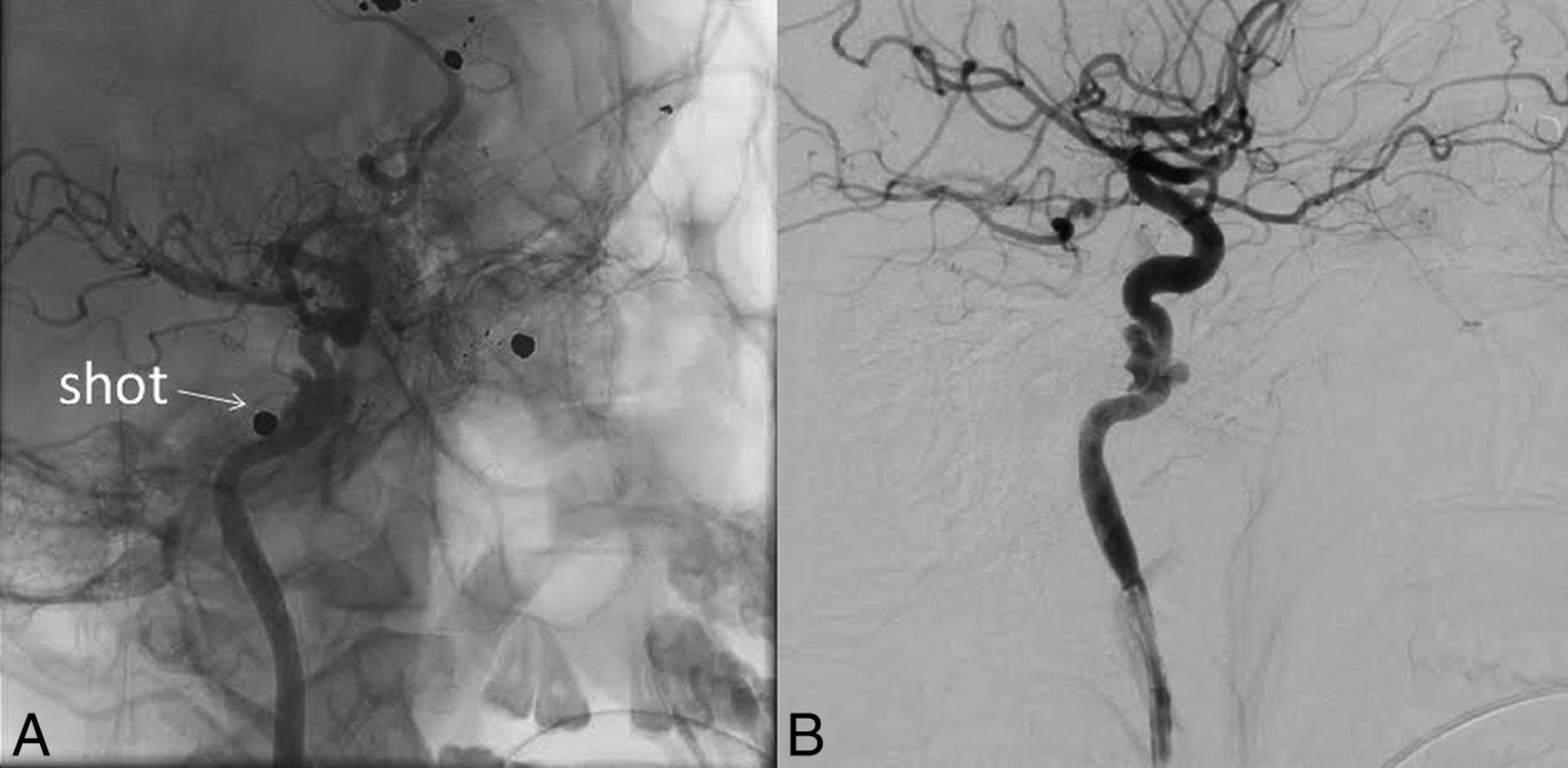
A 21-year-old man was shot and admitted to another hospital about 20 days prior. He underwent an operation to treat intracerebral hemorrhage and then was referred to our institution for more diagnostic and therapeutic interventions. His Glasgow Coma Scale score at this time was 15. His blood pressure and heart rate were stable. On neurologic examination, he had hemiparesis of the left-sided limbs. Noncontrast head CT revealed multiple shots in the cranial and cervical regions (Fig 1). Selective DSA revealed a vascular injury and dissecting aneurysm due to the gun injury in the paraclival segment of right ICA (Fig 2).

Fig 1.
  • Download figure
  • Open in new tab
  • Download powerpoint
Fig 1.

Axial spiral brain CT scans.

Fig 2.
  • Download figure
  • Open in new tab
  • Download powerpoint
Fig 2.

Lateral view of right ICA selective angiography.

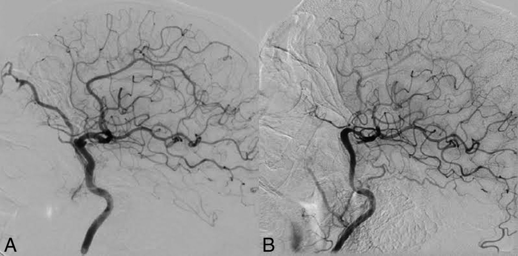
At this time, we decided to manage this injury with an endovascular technique. With the patient under general anesthesia, a Surpass Streamline FDD (4.0 × 20 mm) was deployed covering the site of injury, with heparin and dual-antiplatelet administration as mentioned before. Follow-up DSA was performed after 6 months and confirmed the patency of the stent with complete recovery of the dissecting aneurysm (Fig 3).

Fig 3.
  • Download figure
  • Open in new tab
  • Download powerpoint
Fig 3.

Posttreatment DSA. A, Lateral view of the right ICA, B, Anteroposterior view of the right ICA.

Case 2.

A 40-year-old female patient had massive arterial bleeding during an endoscopic transsphenoidal operation to treat clival chondrosarcoma. The site was packed and the patient was transferred to the angiographic suite under general anesthesia. Selective DSA via right femoral access showed a small iatrogenic injury at the medial wall of the paraclival segment of the left ICA with pseudoaneurysm formation (Fig 4A). A Surpass Streamline FDD (4.0 × 20 mm) was deployed covering the site of injury, with heparin and dual-antiplatelet administration as mentioned before. Follow-up DSA was performed after 6 months, confirming the patency of the stent with complete recovery of the injury (Fig 4B).

Fig 4.
  • Download figure
  • Open in new tab
  • Download powerpoint
Fig 4.

A, Anteroposterior view of left ICA selective angiography. B, Posttreatment anteroposterior view of left ICA angiography.

Case 3.

A 52-year-old man underwent functional endoscopic sinus surgery for a nasal polyp at another hospital and then was referred to our institution with massive epistaxis occurring after 2 days. Selective DSA detected an iatrogenic left ICA laceration at the cavernous segment (Fig 5). A Surpass Streamline FDD (4.0 × 20 mm) was deployed covering the site of injury, with heparin and dual-antiplatelet administration as mentioned before. After 6 months, follow-up DSA was performed and revealed patency of the stent with complete recovery of the injury (Fig 6).

Fig 5.
  • Download figure
  • Open in new tab
  • Download powerpoint
Fig 5.

A, Lateral view of left ICA selective angiography. B, Oblique view of left ICA angiography.

Fig 6.
  • Download figure
  • Open in new tab
  • Download powerpoint
Fig 6.

Posttreatment DSA. A, Lateral view of the left ICA. B, Anteroposterior view of the left ICA.

Case 4.

A 20-year-old man with a history of a car collision about 1 month before was admitted to another hospital with intermittent epistaxis; an endoscopic nasal examination was performed in the operating room. Then, the patient was referred to our institution with suspicion of an ICA injury. Selective DSA revealed a traumatic pseudoaneurysm formation at the cavernous segment of the left ICA (Fig 7A). Treatment with a Surpass Streamline FDD (4.0 × 30 mm) deployment and heparin and dual-antiplatelet administration was performed successfully. After 6 months, follow-up DSA showed a good position of the device without any other complications (Fig 7B).

Fig 7.
  • Download figure
  • Open in new tab
  • Download powerpoint
Fig 7.

A, Lateral view of left ICA angiography during catheterization. B, Posttreatment DSA of the left ICA.

Case 5.

A 39-year-old woman with a pituitary lesion suspicious for craniopharyngioma was admitted to our hospital for an operation. During endoscopic transsphenoidal surgery, massive arterial bleeding occurred and was packed, and the patient was transferred immediately to the angiographic suite under general anesthesia. Selective DSA showed a small iatrogenic injury at the paraclival segment of the left ICA with carotid cavernous fistula formation on the same side (Fig 8A). A Surpass Streamline FDD (4.0 × 20 mm) was deployed covering the site of injury, with heparin and dual-antiplatelet administration as mentioned before. After 6 months, follow-up DSA was performed and revealed patency of the stent with complete recovery of the injury (Fig 8B).

Fig 8.
  • Download figure
  • Open in new tab
  • Download powerpoint
Fig 8.

A, Lateral view of left ICA selective angiography. B, Posttreatment lateral view of left ICA angiography.

Discussion

Iatrogenic and traumatic ICA injuries are rare complications but have high morbidity and mortality rates. ICA injuries during transsphenoidal surgery can have dire outcomes if they are not managed in a definitive and timely manner.6 Carotid stenosis, occlusion, false aneurysms, and carotid cavernous fistulas have all been reported as angiographic manifestations of postsurgical vascular trauma.7

In 2018, Aydin et al1 studied 21 patients who underwent endovascular embolization for iatrogenic vascular injury in the craniocervical region and concluded that endovascular management of such cases was a good option, with low mortality and morbidity rates. Endovascular management of these vascular injuries included parent artery occlusion (n = 15), aneurysm occlusion (n = 3), covered stent (n = 1), and conservative management (n = 2).1 Kocer et al8 revealed the possibility of using an endovascular stent graft for the treatment of iatrogenic ICA lacerations. In their cases, the use of stent grafts proved expedient, safe, and effective in the emergency treatment of a massive hemorrhage resulting from ICA injury that occurred during transsphenoidal surgery.

Endovascular treatment with covered stents has, for some time, been successfully used to treat subclavian and axillary arterial injuries.9 Covered stent placement has also been reported as an option for iatrogenic ICA injuries during transsphenoidal surgery in patients who cannot tolerate ICA occlusion.10 Morken et al11 presented a patient with a meningioma who, during transsphenoidal resection, had a laceration of the ICA, which underwent endovascular treatment with a covered stent graft. However, in 1 case, iatrogenic vascular injury at the paraclival segment of the right ICA permitted deployment of a rigid stent graft at that straight segment, but in our opinion, stent graft deployment is not possible in curvilinear segments like the siphon of the ICA due to rigidity of this type of stent.

In 2015, Lancu et al12 reported 1 case of iatrogenic ICA injury with a secondary carotid cavernous fistula after transsphenoidal surgery that was treated with a Silk flow-diverter stent (Balt Extrusion, Montmorency, France) placed within the injured internal carotid artery and coils placed within the cavernous sinus.

Wakhloo et al13 and Colby et al14 recently showed, in 2 case series, that Surpass is a new-generation flow diverter with unique device-specific and delivery-specific features, compared with clinically available endoluminal flow diverters, and has safety and efficacy in the treatment of intracranial aneurysms comparable with that of stent-assisted coil embolization.

Ogilvy et al15 reported 3 patients with direct traumatic carotid cavernous fistulas who were treated with either coils, coils and Onyx (Covidien, Irvine, California), or a detachable balloon followed by placement of a flow-diverting stent for endoluminal reconstruction, and all 3 patients recovered clinically. They concluded that a flow diverter may facilitate endothelialization of the injury to the internal carotid artery.

Our cases were 5 patients who had an iatrogenic or traumatic ICA injury, all treated with a Surpass Streamline FDD successfully and showing good outcome on follow-up without complications. All patients had small carotid defects, and 2 defects were in the cavernous segment of the ICA, including a curve in the artery. The FDD is made of a cobalt-chromium alloy with a low porosity (metal surface area coverage, 30%), a high mesh density (20–32 pores/mm2), and a self-expanding, single-layer braided, tubular structure. The device comes in various diameters, ≤5.3 mm, and lengths, ≤50 mm. The 2-mm device has 48 wires; the 3- and 4-mm devices have 72 wires; and the 5-mm device has 96 wires, to provide a constant high mesh density over various diameters of the device, as supported by previous in vitro16 and in vivo studies.17

All our patients had small carotid defects due to iatrogenic or traumatic injury. None of them could tolerate angiographically or clinically the balloon occlusion test, and patency of the ICA was important.

Multiple series of treatment of intracranial blister aneurysms by FDDs had high success and low complication rates in other centers.18⇓–20 We also had a successful experience with 18 patients with blister aneurysms treated by a different type of FDD. Small iatrogenic and traumatic defects of the wall of the ICA are comparable with blister aneurysms pathologically. Therefore, we considered the same strategy for our patients because there was no other proved safe treatment technique.

The Surpass Streamline FDD has been available since 2015 in our center and was chosen due to its higher mesh density, greater stability during deployment, and faster positioning in curved and nontortuous anatomy compared with other FDDs.

FDDs alone for the treatment of ICA iatrogenic and traumatic small injuries have not been reported before, to our knowledge. Due to the rarity of this complication, there is no other safe treatment, and the total number of patients studied is small, but all of them showed complete resolution of complications and good outcomes, which can be a basis for future studies.

REFERENCES

  1. 1.↵
    1. Aydin E,
    2. Gok M,
    3. Esenkaya A, et al
    . Endovascular management of iatrogenic vascular injury in the craniocervical region. Turk Neurosurg 2018;28:72–78 doi:10.5137/1019-5149.JTN.18189-16.1 pmid:27593845
    CrossRefPubMed
  2. 2.↵
    1. Ciric I,
    2. Ragin A,
    3. Baumgartner C, et al
    . Complications of transsphenoidal surgery: result of a national survey, review of the literature, and personal experience. Neurosurgery 1997;40:225–36; discussion 236–37 doi:10.1097/00006123-199702000-00001 pmid:9007854
    CrossRefPubMed
  3. 3.↵
    1. Raymond J,
    2. Hardy J,
    3. Czepko R, et al
    . Arterial injuries in transsphenoidal surgery for pituitary adenoma; the role of angiography and endovascular treatment. AJNR Am J Neuroradiol 1997;18:655–65 pmid:9127026
    Abstract/FREE Full Text
  4. 4.↵
    1. Chaloupka JC,
    2. Putman CM,
    3. Citardi MJ, et al
    . Endovascular therapy for the carotid blowout syndrome in head and neck surgical patients: diagnostic and managerial considerations. AJNR Am J Neuroradiol 1996;17:843–52 pmid:8733956
    Abstract
  5. 5.↵
    1. De Vries J,
    2. Boogaarts J,
    3. Van Norden A, et al
    . New generation of flow diverter (Surpass) for unruptured intracranial aneurysms: a prospective single-center study in 37 patients. Stroke 2013;44:1567–77 doi:10.1161/STROKEAHA.111.000434 pmid:23686973
    Abstract/FREE Full Text
  6. 6.↵
    1. Griauzde J,
    2. Gemmete JJ,
    3. Pandey AS, et al
    . Emergency reconstructive endovascular management of intraoperative complications involving the internal carotid artery from trans-sphenoidal surgery. J Neurointervent Surg 2015;7:67–71 doi:10.1136/neurintsurg-2013-010878 pmid:24408926
    Abstract/FREE Full Text
  7. 7.↵
    1. Pigott TJ,
    2. Holland IM,
    3. Punt JA
    . Carotico-cavernous fistula after trans-sphenoidal hypophysectomy. Br J Neurosurg 1989;3:613–16 doi:10.3109/02688698909002855 pmid:2818855
    CrossRefPubMed
  8. 8.↵
    1. Kocer N,
    2. Kizilkilic O,
    3. Albayram S, et al
    . Treatment of iatrogenic internal carotid artery laceration and carotid-cavernous fistula with endovascular stent graft placement. AJNR Am J Neuroradiol 2002;23:442–46 pmid:11901015
    Abstract/FREE Full Text
  9. 9.↵
    1. White R,
    2. Krajcer Z,
    3. Johanson M, et al
    . Results of a multicenter trial for the treatment of traumatic vascular injury with a covered stent. J Trauma 2006;60:1189–95; discussion 1195–96 doi:10.1097/01.ta.0000220372.85575.e2 pmid:16766960
    CrossRefPubMed
  10. 10.↵
    1. Cockroft KM,
    2. Carew JF,
    3. Trost D, et al
    . Delayed epistaxis resulting from external carotid artery injury requiring embolization: a rare complication of transsphenoidal surgery—case report. Neurosurgery 2000;47:236–39 pmid:10917369
    PubMed
  11. 11.↵
    1. Morken MH,
    2. Cappelen J,
    3. Kvistad KA, et al
    . Acute endovascular repair of iatrogenic right internal carotid arterial laceration. Acta Radiol Short Rep 2013;2:2047981613496088 doi:10.1177/2047981613496088 pmid:24198962
    CrossRefPubMed
  12. 12.↵
    1. Lancu D,
    2. Lum C,
    3. Ahmed ME, et al
    . Flow diversion in the treatment of carotid injury and carotid-cavernous fistula after transsphenoidal surgery. Interv Neuroradiol 2015;21:346–50 doi:10.1177/1591019915582367 pmid:26015526
    CrossRefPubMed
  13. 13.↵
    1. Wakhloo AK,
    2. Lylyk P,
    3. de Vries J, et al
    ; Surpass Study Group. Surpass flow diverter in the treatment of intracranial aneurysms: a prospective multicenter study. AJNR Am J Neuroradiol 2015;36:98–107 doi:10.3174/ajnr.A4078 pmid:25125666
    Abstract/FREE Full Text
  14. 14.↵
    1. Colby GP,
    2. Lin L,
    3. Caplan JM, et al
    . Flow diversion of large internal carotid artery aneurysms with the Surpass device: impressions and technical nuance from the initial North American experience. J Neurointerv Surg 2016;8:279–86 doi:10.1136/neurintsurg-2015-011769 pmid:25987590
    Abstract/FREE Full Text
  15. 15.↵
    1. Ogilvy CS,
    2. Motiei-Langroudi R,
    3. Ghorbani M, et al
    . Flow diverter as useful adjunct to traditional endovascular techniques in treatment of direct carotid-cavernous fistulas. World Neurosurg 2017;105:812–17 doi:10.1016/j.wneu.2017.06.113 pmid:28647659
    CrossRefPubMed
  16. 16.↵
    1. Seong J,
    2. Wakhloo AK,
    3. Lieber BB
    . In vitro evaluation of flow divertors in an elastase-induced saccular aneurysm model in rabbit. J Biomech Eng 2007;129:863–72 doi:10.1115/1.2800787 pmid:18067390
    CrossRefPubMed
  17. 17.↵
    1. Sadasivan C,
    2. Cesar L,
    3. Seong J, et al
    . An original flow diversion device for the treatment of intracranial aneurysms: evaluation in the rabbit elastase-induced model. Stroke 2009;40:952–58 doi:10.1161/STROKEAHA.108.533760 pmid:19150864
    Abstract/FREE Full Text
  18. 18.↵
    1. Chalouhi N,
    2. Zanaty M,
    3. Tjoumakaris S, et al
    . Treatment of blister-like aneurysms with the Pipeline Embolization Device. Neurosurgery 2014;74:527–32 doi:10.1227/NEU.0000000000000309 pmid:24553092
    CrossRefPubMed
  19. 19.↵
    1. Rouchaud A,
    2. Brinjikji W,
    3. Cloft HJ, et al
    . Endovascular treatment of ruptured blister-like aneurysms: a systematic review and meta-analysis with focus on deconstructive versus reconstructive and flow-diverter treatments. AJNR Am J Neuroradiol 2015;36:2331–39 doi:10.3174/ajnr.A4438 pmid:26381557
    Abstract/FREE Full Text
  20. 20.↵
    1. Peitz GW,
    2. Sy CA,
    3. Grandhi R
    . Endovascular treatment of blister aneurysms. J Neurosurg Focus 2017;42:E12 doi:10.3171/2017.3.FOCUS1751 pmid:28565977
    CrossRefPubMed
  • Received October 25, 2017.
  • Accepted after revision January 31, 2018.
  • © 2018 by American Journal of Neuroradiology
View Abstract
PreviousNext
Back to top

In this issue

American Journal of Neuroradiology: 39 (6)
American Journal of Neuroradiology
Vol. 39, Issue 6
1 Jun 2018
  • Table of Contents
  • Index by author
  • Complete Issue (PDF)
Advertisement
Print
Download PDF
Email Article

Thank you for your interest in spreading the word on American Journal of Neuroradiology.

NOTE: We only request your email address so that the person you are recommending the page to knows that you wanted them to see it, and that it is not junk mail. We do not capture any email address.

Enter multiple addresses on separate lines or separate them with commas.
Surpass Streamline Flow-Diverter Embolization Device for Treatment of Iatrogenic and Traumatic Internal Carotid Artery Injuries
(Your Name) has sent you a message from American Journal of Neuroradiology
(Your Name) thought you would like to see the American Journal of Neuroradiology web site.
CAPTCHA
This question is for testing whether or not you are a human visitor and to prevent automated spam submissions.
Cite this article
M. Ghorbani, H. Shojaei, K. Bavand, M. Azar
Surpass Streamline Flow-Diverter Embolization Device for Treatment of Iatrogenic and Traumatic Internal Carotid Artery Injuries
American Journal of Neuroradiology Jun 2018, 39 (6) 1107-1111; DOI: 10.3174/ajnr.A5607

Citation Manager Formats

  • BibTeX
  • Bookends
  • EasyBib
  • EndNote (tagged)
  • EndNote 8 (xml)
  • Medlars
  • Mendeley
  • Papers
  • RefWorks Tagged
  • Ref Manager
  • RIS
  • Zotero
0 Responses
Respond to this article
Share
Bookmark this article
Surpass Streamline Flow-Diverter Embolization Device for Treatment of Iatrogenic and Traumatic Internal Carotid Artery Injuries
M. Ghorbani, H. Shojaei, K. Bavand, M. Azar
American Journal of Neuroradiology Jun 2018, 39 (6) 1107-1111; DOI: 10.3174/ajnr.A5607
del.icio.us logo Twitter logo Facebook logo Mendeley logo
  • Tweet Widget
  • Facebook Like
  • Google Plus One
Purchase

Jump to section

  • Article
    • Abstract
    • ABBREVIATIONS:
    • Discussion
    • REFERENCES
  • Figures & Data
  • Info & Metrics
  • Responses
  • References
  • PDF

Related Articles

  • No related articles found.
  • PubMed
  • Google Scholar

Cited By...

  • No citing articles found.
  • Crossref (14)
  • Google Scholar

This article has been cited by the following articles in journals that are participating in Crossref Cited-by Linking.

  • Carotid Cavernous Fistula Treatment via Flow Diversion: A Systematic Review of the Literature
    Hasan Sumdani, Pedro Aguilar-Salinas, Mauricio J. Avila, Mohammad El-Ghanem, Travis M. Dumont
    World Neurosurgery 2021 149
  • Surpass Evolve Flow Diverter for the Treatment of Intracranial Aneurysm: A Systematic Review
    Rania Issa, Zahrah Al-Homedi, Dawood Hasan Syed, Waseem Aziz, Basem Al-Omari
    Brain Sciences 2022 12 6
  • Endovascular reconstruction of iatrogenic internal carotid artery injury following endonasal surgery: a systematic review
    Mohammad Ghorbani, Christoph J. Griessenauer, Hamidreza Shojaei, Christoph Wipplinger, Ebrahim Hejazian
    Neurosurgical Review 2021 44 4
  • Evaluating the Effect of the Number of Wire of Flow Diverter Stents on the Nonstagnated Region Formation in an Aneurysm Sac Using Lagrangian Coherent Structure and Hyperbolic Time Analysis
    Onur Mutlu, Ali Bahadır Olcay, Cem Bilgin, Bahattin Hakyemez
    World Neurosurgery 2020 133
  • Flow diverter embolization device for endovascular treatment of ruptured blister and wide necked very small aneurysms
    Mohammad Ghorbani, Christoph J. Griessenauer, Christoph Wipplinger, Maziar Azar, Hamidreza Shojaei, Karan Bavand, Darya Khosravi
    Heliyon 2019 5 9
  • The surgical management of intraoperative intracranial internal carotid artery injury in open skull base surgery—a systematic review
    Jorn Van Der Veken, Mary Simons, Michael J. Mulcahy, Catherine Wurster, Marguerite Harding, Vera Van Velthoven
    Neurosurgical Review 2022 45 2
  • Traumatic iatrogenic dissecting anterior cerebral artery aneurysms: conservative management as a therapeutic option
    Mohammad Ghorbani, Ebrahim Hejazian, Shahin Nikmanzar, Mehdi Chavoshi-Nejad
    British Journal of Neurosurgery 2023 37 4
  • Successful endoluminal reconstruction of a pseudoaneurysm of the internal carotid artery following a transorbital stab injury
    Mohammad Ghorbani, Christoph Wipplinger, Christoph J. Griessenauer, Hamidreza Shojaei, Hesam Abdolhoseinpour
    Clinical Neurology and Neurosurgery 2020 194
  • Surgical clipping compared to endovascular coiling of ruptured coil able middle cerebral aneurysms: A single-center experience
    Mohammad Ghorbani, Christoph J. Griessenauer, Christoph Wipplinger, Mohsen Nouri, Sina Asaadi, Ebrahim Hejazian, Reza Mollahoseini, Abolghasem Mortazavi
    Interdisciplinary Neurosurgery 2020 21
  • Form and Function in Intracranial Neurovascular Stents: A Historical Perspective and State-of-the-Art Clinical Review for the Noninterventionalist
    M.T. Caton, A.Z. Copelan, K.H. Narsinh, D. Murph, A. A. Abla, D.L. Cooke, S.W. Hetts, V.V. Halbach, C.F. Dowd, R.T. Higashida, M.R. Amans
    Neurographics 2021 11 2

More in this TOC Section

  • SAVE vs. Solumbra Techniques for Thrombectomy
  • CT Perfusion&Reperfusion in Acute Ischemic Stroke
  • Delayed Reperfusion Post-Thrombectomy&Thrombolysis
Show more Interventional

Similar Articles

Advertisement

Indexed Content

  • Current Issue
  • Accepted Manuscripts
  • Article Preview
  • Past Issues
  • Editorials
  • Editors Choice
  • Fellow Journal Club
  • Letters to the Editor

Cases

  • Case Collection
  • Archive - Case of the Week
  • Archive - Case of the Month
  • Archive - Classic Case

Special Collections

  • Special Collections

Resources

  • News and Updates
  • Turn around Times
  • Submit a Manuscript
  • Author Policies
  • Manuscript Submission Guidelines
  • Evidence-Based Medicine Level Guide
  • Publishing Checklists
  • Graphical Abstract Preparation
  • Imaging Protocol Submission
  • Submit a Case
  • Become a Reviewer/Academy of Reviewers
  • Get Peer Review Credit from Publons

Multimedia

  • AJNR Podcast
  • AJNR SCANtastic
  • Video Articles

About Us

  • About AJNR
  • Editorial Board
  • Not an AJNR Subscriber? Join Now
  • Alerts
  • Feedback
  • Advertise with us
  • Librarian Resources
  • Permissions
  • Terms and Conditions

American Society of Neuroradiology

  • Not an ASNR Member? Join Now

© 2025 by the American Society of Neuroradiology All rights, including for text and data mining, AI training, and similar technologies, are reserved.
Print ISSN: 0195-6108 Online ISSN: 1936-959X

Powered by HighWire