
Sudarshana et al1 are to be commended for their meticulous study of potential brain MR imaging in humans with use of mangafodipir (Mn dipyridoxyl diphosphate [MnDPDP]) as a contrast agent. MnDPDP is a multifunctional chelate releasing calcium-stalking paramagnetic Mn ions for MR imaging and a catalytic antioxidant for therapy.2 In healthy volunteers, MnDPDP raised the signal intensity (SI) in the exocrine glands in the head and neck, chorioid plexus, and anterior pituitary gland but not beyond the BBB. The observations are important because the infusion time (<10 minutes) and dose (5 µmol/kg) have been standard in MR imaging of the liver and pancreas. Thus, the study confirms that a brief infusion of 1 diagnostic dose does not cause MR imaging contrast enhancement in the brains of humans with an intact BBB and normal liver function.
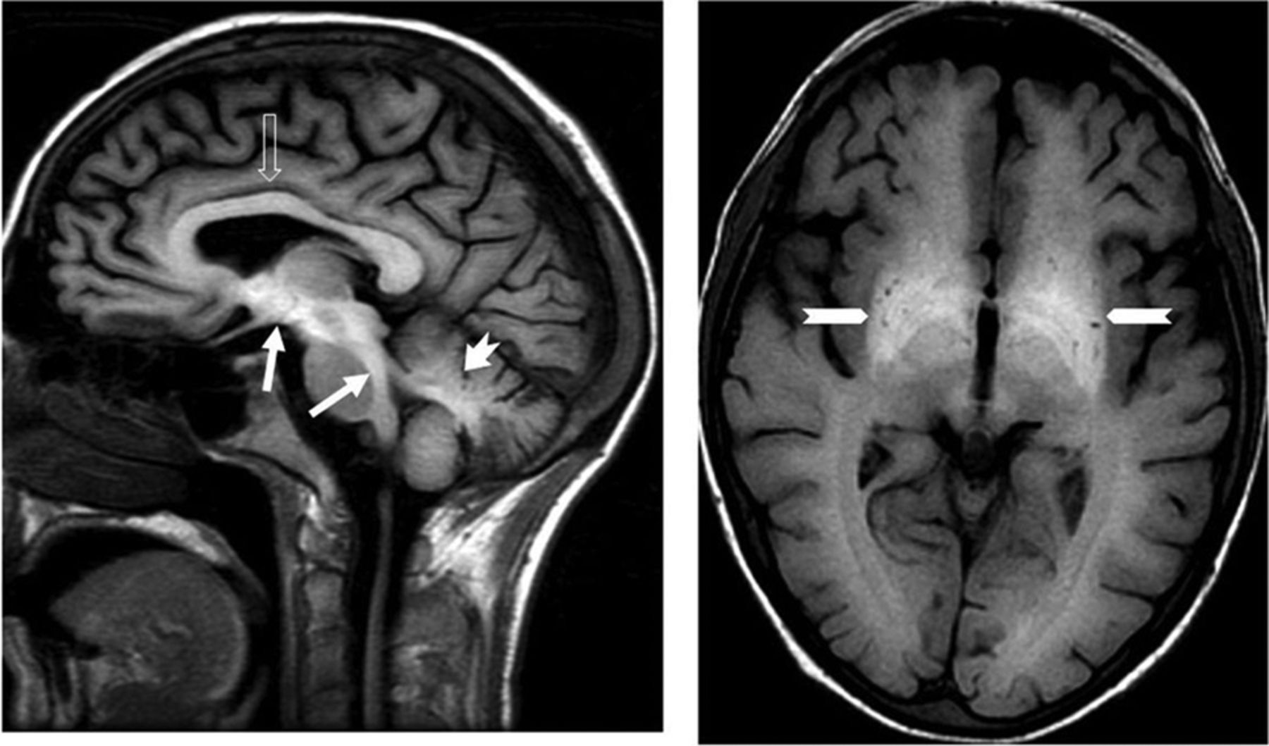
Concerning future brain Mn uptake studies, we find it relevant to address a patient who received MnDPDP in a total dose of 140 µmol/kg during 8 months.3 In this patient with terminal cancer of the colon, MnDPDP (10 µmol/kg) was administered for the protection of normal tissue during 14 cycles of chemotherapy with anticancer agents (oxaliplatin and 5-fluorouracil). The regimen was well-tolerated without the adverse effects of chemotherapy, and there was a notable relief of pain. At end of treatment, mild Parkinson-like symptoms occurred, and brain MR imaging (Fig 1), images not previously disclosed, showed high SI in the basal ganglia (caudate nucleus, globus pallidus, putamen, and thalamus). However, the SI was also high in the corpus callosum, mesencephalon, brain stem, cerebellum, and anterior pituitary gland. In this patient, the first to receive MnDPDP for therapy, an apparent palliation was at the cost of widespread brain deposition of Mn caused by a far-too-high total dose of MnDPDP, advanced liver failure, and, possibly, a BBB weakened by disease or treatment.

Sagittal (T1-weighted-FLAIR) and axial (T1-weighted spin-echo) images show high SI reflecting Mn deposition in the corpus callosum (open arrow), mesencephalon, crus cerebri, dorsal brain stem, medulla oblongata (white arrows), cerebellum (dentate nucleus) (short white arrow with cut), and basal ganglia (globus pallidus and putamen) (long arrows with cut).
Most interesting, SI was maximal in the dentate nucleus and globus pallidus, sites also noted for deposition of gadolinium (Gd) ions released from linear chelates.4 This finding may indicate a mutual, possibly calcium-related, pathway for storage of Mn and Gd adducts in the brain.
Indicates open access to non-subscribers at www.ajnr.org
- © 2020 by American Journal of Neuroradiology


