Advertisement

Main menu

  • Home
  • Content
    • Current Issue
    • Accepted Manuscripts
    • Article Preview
    • Past Issue Archive
    • AJNR Case Collection
    • Case of the Week Archive
    • Classic Case Archive
    • Case of the Month Archive
  • Special Collections
    • Spinal CSF Leak Articles (Jan 2020-June 2024)
    • 2024 AJNR Journal Awards
    • Most Impactful AJNR Articles
  • Multimedia
    • AJNR Podcast
    • AJNR Scantastics
    • Video Articles
  • For Authors
    • Submit a Manuscript
    • Author Policies
    • Fast publishing of Accepted Manuscripts
    • Graphical Abstract Preparation
    • Manuscript Submission Guidelines
    • Imaging Protocol Submission
    • Submit a Case for the Case Collection
  • About Us
    • About AJNR
    • Editorial Board
  • More
    • Become a Reviewer/Academy of Reviewers
    • Subscribers
    • Permissions
    • Alerts
    • Feedback
    • Advertisers
    • ASNR Home
  • Other Publications
    • ajnr

User menu

  • Alerts
  • Log in

Search

  • Advanced search
American Journal of Neuroradiology

American Journal of Neuroradiology

ASHNR American Society of Functional Neuroradiology ASHNR American Society of Pediatric Neuroradiology ASSR
  • Alerts
  • Log in

Advanced Search

  • Home
  • Content
    • Current Issue
    • Accepted Manuscripts
    • Article Preview
    • Past Issue Archive
    • AJNR Case Collection
    • Case of the Week Archive
    • Classic Case Archive
    • Case of the Month Archive
  • Special Collections
    • Spinal CSF Leak Articles (Jan 2020-June 2024)
    • 2024 AJNR Journal Awards
    • Most Impactful AJNR Articles
  • Multimedia
    • AJNR Podcast
    • AJNR Scantastics
    • Video Articles
  • For Authors
    • Submit a Manuscript
    • Author Policies
    • Fast publishing of Accepted Manuscripts
    • Graphical Abstract Preparation
    • Manuscript Submission Guidelines
    • Imaging Protocol Submission
    • Submit a Case for the Case Collection
  • About Us
    • About AJNR
    • Editorial Board
  • More
    • Become a Reviewer/Academy of Reviewers
    • Subscribers
    • Permissions
    • Alerts
    • Feedback
    • Advertisers
    • ASNR Home
  • Follow AJNR on Twitter
  • Visit AJNR on Facebook
  • Follow AJNR on Instagram
  • Join AJNR on LinkedIn
  • RSS Feeds

Welcome to the new AJNR, Updated Hall of Fame, and more. Read the full announcements.


AJNR is seeking candidates for the position of Associate Section Editor, AJNR Case Collection. Read the full announcement.


Research ArticleAdult Brain
Open Access

COVID-19 Neurologic Complication with CNS Vasculitis-Like Pattern

R. Hanafi, P.-A. Roger, B. Perin, G. Kuchcinski, N. Deleval, F. Dallery, D. Michel, L. Hacein-Bey, J.-P. Pruvo, O. Outteryck and J.-M. Constans
American Journal of Neuroradiology August 2020, 41 (8) 1384-1387; DOI: https://doi.org/10.3174/ajnr.A6651
R. Hanafi
aFrom the Department of Neuroradiology (R.H., G.K., J.-P.P., O.O.), University Hospital of Lille, Hôpital Roger Salengro, Lille, France
bDepartments of Interventional Radiology (R.H.), Cardiothoracic, Vascular and Respiratory Intensive Care (P.-A.R.), Neurology and Neurophysiology (B.P.), and Neuroradiology (N.D., F.D., D.M., J.-M.C.), University Hospital of Amiens, Amiens, France
  • Find this author on Google Scholar
  • Find this author on PubMed
  • Search for this author on this site
  • ORCID record for R. Hanafi
P.-A. Roger
bDepartments of Interventional Radiology (R.H.), Cardiothoracic, Vascular and Respiratory Intensive Care (P.-A.R.), Neurology and Neurophysiology (B.P.), and Neuroradiology (N.D., F.D., D.M., J.-M.C.), University Hospital of Amiens, Amiens, France
  • Find this author on Google Scholar
  • Find this author on PubMed
  • Search for this author on this site
  • ORCID record for P.-A. Roger
B. Perin
bDepartments of Interventional Radiology (R.H.), Cardiothoracic, Vascular and Respiratory Intensive Care (P.-A.R.), Neurology and Neurophysiology (B.P.), and Neuroradiology (N.D., F.D., D.M., J.-M.C.), University Hospital of Amiens, Amiens, France
  • Find this author on Google Scholar
  • Find this author on PubMed
  • Search for this author on this site
  • ORCID record for B. Perin
G. Kuchcinski
aFrom the Department of Neuroradiology (R.H., G.K., J.-P.P., O.O.), University Hospital of Lille, Hôpital Roger Salengro, Lille, France
  • Find this author on Google Scholar
  • Find this author on PubMed
  • Search for this author on this site
  • ORCID record for G. Kuchcinski
N. Deleval
bDepartments of Interventional Radiology (R.H.), Cardiothoracic, Vascular and Respiratory Intensive Care (P.-A.R.), Neurology and Neurophysiology (B.P.), and Neuroradiology (N.D., F.D., D.M., J.-M.C.), University Hospital of Amiens, Amiens, France
  • Find this author on Google Scholar
  • Find this author on PubMed
  • Search for this author on this site
  • ORCID record for N. Deleval
F. Dallery
bDepartments of Interventional Radiology (R.H.), Cardiothoracic, Vascular and Respiratory Intensive Care (P.-A.R.), Neurology and Neurophysiology (B.P.), and Neuroradiology (N.D., F.D., D.M., J.-M.C.), University Hospital of Amiens, Amiens, France
  • Find this author on Google Scholar
  • Find this author on PubMed
  • Search for this author on this site
  • ORCID record for F. Dallery
D. Michel
bDepartments of Interventional Radiology (R.H.), Cardiothoracic, Vascular and Respiratory Intensive Care (P.-A.R.), Neurology and Neurophysiology (B.P.), and Neuroradiology (N.D., F.D., D.M., J.-M.C.), University Hospital of Amiens, Amiens, France
  • Find this author on Google Scholar
  • Find this author on PubMed
  • Search for this author on this site
  • ORCID record for D. Michel
L. Hacein-Bey
cDepartments of Neuroradiology and Radiology (L.H.-B.), University of California Davis School of Medicine, Sacramento, California.
  • Find this author on Google Scholar
  • Find this author on PubMed
  • Search for this author on this site
  • ORCID record for L. Hacein-Bey
J.-P. Pruvo
aFrom the Department of Neuroradiology (R.H., G.K., J.-P.P., O.O.), University Hospital of Lille, Hôpital Roger Salengro, Lille, France
  • Find this author on Google Scholar
  • Find this author on PubMed
  • Search for this author on this site
  • ORCID record for J.-P. Pruvo
O. Outteryck
aFrom the Department of Neuroradiology (R.H., G.K., J.-P.P., O.O.), University Hospital of Lille, Hôpital Roger Salengro, Lille, France
  • Find this author on Google Scholar
  • Find this author on PubMed
  • Search for this author on this site
  • ORCID record for O. Outteryck
J.-M. Constans
bDepartments of Interventional Radiology (R.H.), Cardiothoracic, Vascular and Respiratory Intensive Care (P.-A.R.), Neurology and Neurophysiology (B.P.), and Neuroradiology (N.D., F.D., D.M., J.-M.C.), University Hospital of Amiens, Amiens, France
  • Find this author on Google Scholar
  • Find this author on PubMed
  • Search for this author on this site
  • ORCID record for J.-M. Constans

Abstract

SUMMARY: Coronavirus disease 2019 (COVID-19) is a viral infection caused by the Severe Acute Respiratory Syndrome coronavirus 2 (SARS-CoV-2), which spreads rapidly from person to person and manifests in most symptomatic patients as a respiratory illness, similar to prior SARS viruses. Neurologic manifestations of COVID-19 are uncommon; those so far reported include encephalopathy, stroke from large-vessel occlusion, and polyneuropathy. We report a unique neurologic complication of COVID-19 in a patient who had extensive cerebral small-vessel ischemic lesions resembling cerebral vasculitis in a characteristic combined imaging pattern of ischemia, hemorrhage, and punctuate postcontrast enhancement. Also, a characteristic lower extremity skin rash was present in our patient. Our observation lends support to the increasingly suspected mechanism of “endotheliitis” associated with this novel coronavirus.

ABBREVIATIONS:

ACE2
angiotensin converting enzyme 2
COVID-19
coronavirus disease 2019
SARS-CoV-2
Severe Acute Respiratory Syndrome coronavirus 2

Coronavirus disease 2019 (COVID-19) has now spread to the whole world following an outbreak in China in late 2019.1 The virus is so highly contagious that the World Health Organization has declared COVID-19 a pandemic, with close to 3.5 million confirmed cases and 250,000 fatalities worldwide at the time of this report (May 1, 2020).1,2 Similar to other coronaviruses, the Severe Acute Respiratory Syndrome coronavirus 2 (SARS-CoV-2) virus primarily targets the respiratory system. In symptomatic patients, the most common symptoms are fever, fatigue, cough, headache, and shortness of breath. Severely symptomatic patients may present with pneumonia, acute respiratory distress syndrome, acute cardiac dysfunction from myocarditis, and multiorgan failure.3⇓-5 Several mild neurologic manifestations of COVID-19 have now been reported, which include hyposmia or anosmia.6⇓-8 More severe neurologic complications have also been recently reported, which may possibly be associated with COVID-19, including acute hemorrhagic necrotizing encephalopathy,9 acute transverse myelitis and Guillain Barré syndrome,10 and meningitis.11

We report herein a unique neurologic complication of COVID-19 with extensive ischemic lesions resembling CNS vasculitis.

Case Report

A 65-year-old man presented to the emergency department with a 5-day history of fatigue, fever, and cough. Initial blood work showed an inflammatory syndrome with an elevated C-reactive protein (60 mg/L), bicytopenia (normocytic anemia and thrombocytopenia), and lymphopenia. The patient was admitted to the medical ward for further evaluation. Noncontrast chest CT showed ground-glass opacities characteristic of COVID-19 infection. The diagnosis was confirmed by reverse transcription polymerase chain reaction detection of SARS-CoV-2 from a nasopharyngeal swab.

The following day, the patient had a high fever and chills. A lower extremity skin rash was noted. Severe hypoxemia (56 mm Hg) and low oxygen saturation (90.6%) prompted oxygen therapy with a high-concentration mask (15L/min) and antibiotic therapy (spiramycin, amoxicillin, and clavulanic acid). Because oxygen saturation continued to drop to 88%, with worsening of skin rash and tachypnea, the patient was admitted to the pulmonary intensive care unit.

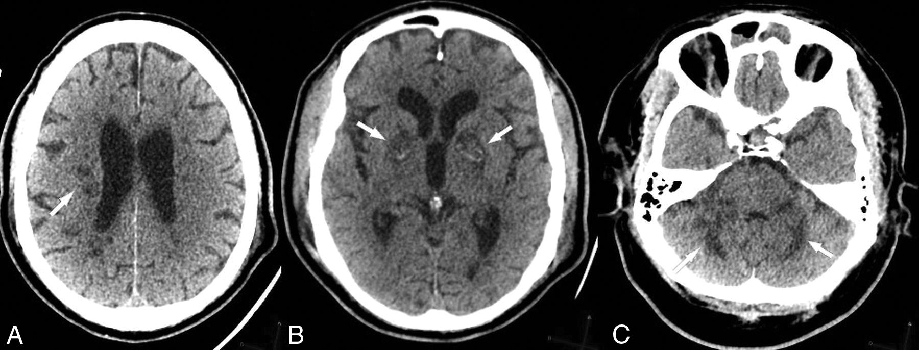
Rapid stabilization of pulmonary function was obtained with oxygen and care in the intensive care unit; however, the patient remained drowsy and unarousable despite discontinuation of sedation. CT and MR imaging of the brain were performed to look for possible neurologic complications such as hypoxic-ischemic injuries or encephalitis. CT showed multiple white matter, basal ganglia, and cerebellar hypodensities and bilateral globus pallidus hyperdensities (Fig 1), suggestive of hemorrhage confirmed on T2*-weighted MR imaging (Fig 2).

Fig 1.
  • Download figure
  • Open in new tab
  • Download powerpoint
Fig 1.

Noncontrast brain CT of presumptive COVID-associated small-vessel injury. Note deep white matter (A), basal ganglia (B), and cerebellar (C) hypodensities (arrows). Bilateral globus pallidus hemorrhage is also present (B, arrows).

Fig 2.
  • Download figure
  • Open in new tab
  • Download powerpoint
Fig 2.

T2*-weighted MR imaging demonstrates bilateral globus pallidus susceptibility, consistent with microhemorrhage (arrows).

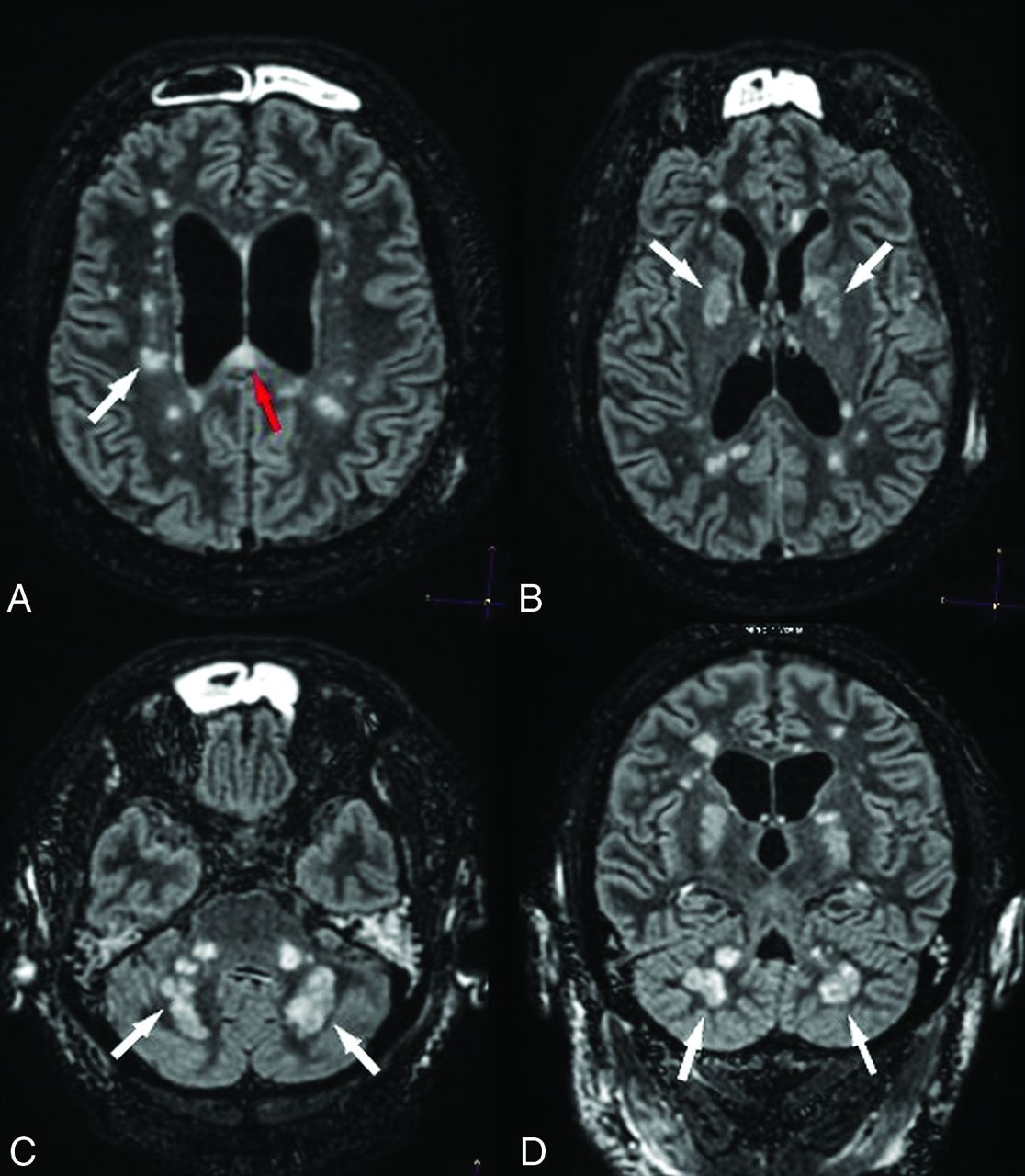
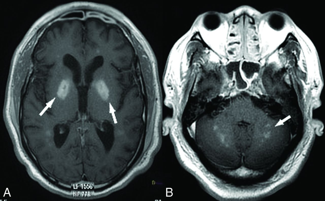
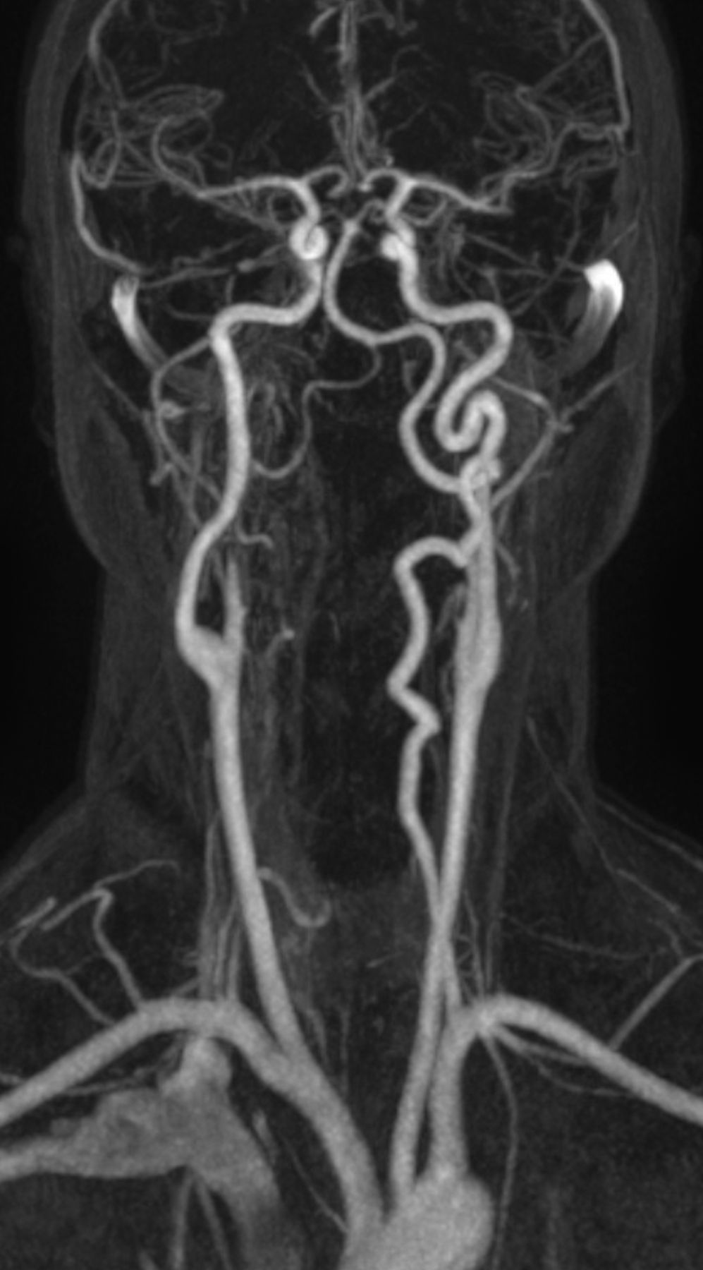
Brain MR imaging showed extensive ischemic lesions with restricted diffusion (Fig 3), involving the centrum semiovale, corpus callosum, basal ganglia, and cerebellum (Fig 4), with patchy/punctuate enhancement (Fig 5). Coronal 3D reconstruction MRA of supra-aortic arteries showed no detectable abnormality of large intra- or extracranial vessels (Fig 6).

Fig 3.
  • Download figure
  • Open in new tab
  • Download powerpoint
Fig 3.

Diffuse deep white matter ischemic injuries. DWI shows numerous focal ischemic lesions within the hemispheric deep white matter (A, white arrow), the corpus callosum (A, red arrow), the basal ganglia, the middle cerebellar peduncles, and the cerebellar hemispheres (B, white arrows).

Fig 4.
  • Download figure
  • Open in new tab
  • Download powerpoint
Fig 4.

Diffuse ischemic lesions on FLAIR imaging. Axial (A–C) and coronal (D) FLAIR images show hyperintense ischemic lesions within the periventricular white matter, basal ganglia, cerebellar peduncles (white arrows), and the corpus callosum (A, red arrow).

Fig 5.
  • Download figure
  • Open in new tab
  • Download powerpoint
Fig 5.

Patchy/punctate postcontrast enhancement pattern with consistent small vessel involvement. Postcontrast T1 MR imaging shows intense patchy enhancement of all lesions, in particular the globus pallidus bilaterally (A, white arrows), with a punctuate pattern in the middle cerebellar peduncles and cerebellar hemispheres (B, arrow).

Fig 6.
  • Download figure
  • Open in new tab
  • Download powerpoint
Fig 6.

Coronal 3D reconstruction MRA of the supra-aortic arteries shows no detectable abnormality of large intra- or extracranial vessels.

DISCUSSION

Neurologic complications are definitely associated with COVID-19, though the mechanism of neurologic injury remains speculative. Encephalopathy, ischemic stroke from large-vessel occlusion,7⇓-9,12 Guillain-Barré syndrome,10 and reversible cranial nerve injuries (Miller Fisher syndrome)13 have all been reported with no evidence thus far that the SARS-CoV-2 can cross the blood-brain barrier.12,13

In our patient, hypoxic-ischemic injury was considered an unlikely mechanism of injury because the patient had remained responsive throughout the period of low oxygen saturation, and only deteriorated several hours after successful stabilization of oxygen saturation; in addition, the patient did not have cardiac arrest. Also, imaging studies showed sparing of the striatum and cortical structures throughout. Similarly, a pro-thrombotic state was not considered a likely causative factor because thorough coagulation panel findings (including the antiphospholipid antigen) were normal.

Neuroimaging findings in our patient suggest the possibility of injury to the small intracranial vasculature in the distribution of distal perforating arteries. The association of ischemic and hemorrhagic lesions to the “patchy/punctate” enhancement pattern is highly suggestive of vasculitis.14 The punctate enhancement pattern has been reported to be characteristic of disease processes that progress from endoluminal (angiocentric lymphoma), vessel wall (vasculitis), or perivascular (progressive multifocal leukoencephalopathy) cellular proliferation.15

SARS-CoV-2 infects the host through its CoV spike glycoprotein, which binds to the angiotensin converting enzyme 2 (ACE2) receptor, which is expressed in the lungs, heart, and kidney (the most frequenty targeted organs in COVID-19), but also endothelial cells.16 Endothelial cell involvement with COVID-19 has recently been demonstrated across vascular beds.17 The expression of the ACE2 receptor in neurons and cerebral endothelial cells indicates a high level of invasiveness for the SARS-CoV-2 in comparison with other coronaviruses (SARS and Middle East Respiratory Syndrome).18 Such an endothelial mechanism of injury (endotheliitis) has been convincingly demonstrated in severe cases of cardiac injury in humans.19 Histologic evidence of COVID-19–induced vasculitis has also been reported in several other organs including the lung, liver, kidney, or skin.20,21 The concomitance of a characteristic skin rash with a “mottled” appearance and such a pattern of CNS lesions in our patient would lend support to a mechanism of virus-related endothelial injury.

CONCLUSIONS

COVID-19–related neurologic complications remain poorly understood. We report a patient whose clinical presentation and neuroimaging findings are supportive of a mechanism of SARS-CoV-2–induced vasculitis/endotheliitis.

Acknowledgments

The authors wish to acknowledge Fanny Ducourneau, radiologic technologist, and Gilles Boulu, radiologist, who kindly agreed to perform the MR imaging study in our patient.

Indicates open access to non-subscribers at www.ajnr.org

References

  1. 1.
    World Health Organization. Coronavirus disease 2019. 2020 April 30. https://www.who.int/emergencies/diseases/novel-coronavirus-2019. Accessed April 30, 2020
  2. 2.
    COVID-19 CORONAVIRUS PANDEMIC. May 20 2020. https://www.worldometers.info/coronavirus. Accessed April 30, 2020
  3. 3.
    2019–20 Coronavirus Pandemic. Public Health Emergency. Coronavirus https://www.who.int/emergencies/diseases/novel-coronavirus-2019. Accessed April 30, 2020
  4. 4.
    US Centers for Disease Control and Prevention. Symptoms of Novel Coronavirus (2019-nCoV). February 10, 2020. https://www.cdc.gov/coronavirus/2019-ncov/symptoms-testing/symptoms.html. Accessed April 30, 2020
  5. 5.
    1. Huang C,
    2. Wang Y,
    3. Li X, et al
    . Clinical features of patients infected with 2019 novel coronavirus in Wuhan, China. Lancet 2020;395:497–506 doi:10.1016/S0140-6736(20)30183-5 pmid:31986264
  6. 6.
    Clinical characteristics of 113 deceased patients with coronavirus disease 2019: retrospective study. BMJ 2020;368:m1295 doi:10.1136/bmj.m1295 pmid:32234718
  7. 7.
    1. Mao L,
    2. Jin H,
    3. Wang M, et al
    . Neurologic manifestations of hospitalized patients with coronavirus disease 2019 in Wuhan, China. JAMA Neurol 2020 April 10. [Epub ahead of print] doi:10.1001/jamaneurol.2020.1127 pmid:32275288
  8. 8.
    1. Avindra N
    . Neurologic complications of coronavirus infections. Neurology 2020;94:809–10 doi:10.1212/WNL.0000000000009455 pmid:32229625
  9. 9.
    1. Poyiadji N,
    2. Shahin G,
    3. Noujaim D, et al
    . COVID-19–associated acute hemorrhagic necrotizing encephalopathy: CT and MRI features. Radiology 2020 Mar 31. [Epub ahead of print] doi:10.1148/radiol.2020201187 pmid:32228363
  10. 10.
    1. Zhao H,
    2. Shen D,
    3. Zhou H, et al
    . Guillain-Barré syndrome associated with SARS-CoV-2 infection: causality or coincidence. Lancet Neurol 2020;19:383–84 doi:10.1016/S1474-4422(20)30109 pmid:32246917
  11. 11.
    1. Moriguchi T,
    2. Harii N,
    3. Goto J, et al
    . A first case of meningitis/encephalitis associated with SARS-coronavirus-2. Int J Infect Dis 2020;94:55–58 doi:10.1016/j.ijid.2020.03.062 pmid:32251791
  12. 12.
    1. Filatov A,
    2. Sharma P,
    3. Hindi F, et al
    . Neurological complications of coronavirus disease (COVID-19): encephalopathy. Cureus 2020;12:e7352 doi:10.7759/cureus.7352 pmid:32328364
  13. 13.
    1. Gutiérrez-Ortiz C,
    2. Méndez A,
    3. Rodrigo-Rey S, et al
    . Miller Fisher syndrome and polyneuritis cranialis in COVID-19. Neurology 2020 Apr 17. [Epub ahead of print] doi:10.1212/WNL.0000000000009619 pmid:32303650
  14. 14.
    1. Gupta RK
    . Small vessel vasculitis-optimism and challenges in imaging diagnosis. Neurol India 2017;65:1219–20 doi:10.4103/0028-3886.217993 pmid:29133683
  15. 15.
    1. Hodel J,
    2. Darchis C,
    3. Outteryck O, et al
    . Punctate pattern: a promising imaging marker for the diagnosis of natalizumab-associated PML. Neurology 2016;86:1516–23 doi:10.1212/WNL.0000000000002586 pmid:27009257
  16. 16.
    1. Monteil V,
    2. Kwon H,
    3. Prado P, et al
    . Inhibition of SARS-CoV-2 infections in engineered human tissues using clinical-grade soluble human ACE2. Cell 2020;181:905–913.e7 doi:10.1016/j.cell.2020.04.004 pmid:32333836
  17. 17.
    1. Varga Z,
    2. Flammer AJ,
    3. Steiger P, et al
    . Endothelial cell infection and endotheliitis in COVID-19. Lancet 2020;395:1417–18 doi:10.1016/S0140-6736(20)30937-5 pmid:32325026
  18. 18.
    1. Natoli S,
    2. Oliveira V,
    3. Calabresi P, et al
    . Does SARS-Cov-2 invade the brain? Translational lessons from animal models. Eur J Neurol 2020 Apr 25. [Epub ahead of print] doi:10.1111/ene.14277 pmid:32333487
  19. 19.
    1. Chen L,
    2. Li X,
    3. Chen M, et al
    . The ACE2 expression in human heart indicates new potential mechanism of heart injury among patients infected with SARS-CoV-2. Cardiovasc Res 2020;116:1097–1100 doi:10.1093/cvr/cvaa078 pmid:32227090
  20. 20.
    1. Xu Z,
    2. Shi L,
    3. Wang Y, et al
    . Pathological findings of COVID-19 associated with acute respiratory distress syndrome. Lancet Respir Med 2020;8:420–22 doi:10.1016/S2213-2600(20)30076-X pmid:2085846
  21. 21.
    1. Yao XH,
    2. Li TY,
    3. He ZC, et al
    . A pathological report of three COVID-19 cases by minimally invasive autopsies [in Chinese]. Zhonghua Bing Li Xue Za Zhi 2020;49:411–17 doi:10.3760/cma.j.cn112151-20200312-00193 pmid:32172546
  • Received April 23, 2020.
  • Accepted after revision May 5, 2020.
  • © 2020 by American Journal of Neuroradiology
Advertisement

Indexed Content

  • Current Issue
  • Accepted Manuscripts
  • Article Preview
  • Past Issues
  • Editorials
  • Editors Choice
  • Fellow Journal Club
  • Letters to the Editor

Cases

  • Case Collection
  • Archive - Case of the Week
  • Archive - Case of the Month
  • Archive - Classic Case

Special Collections

  • Special Collections

Resources

  • News and Updates
  • Turn around Times
  • Submit a Manuscript
  • Author Policies
  • Manuscript Submission Guidelines
  • Evidence-Based Medicine Level Guide
  • Publishing Checklists
  • Graphical Abstract Preparation
  • Imaging Protocol Submission
  • Submit a Case
  • Become a Reviewer/Academy of Reviewers
  • Get Peer Review Credit from Publons

Multimedia

  • AJNR Podcast
  • AJNR SCANtastic
  • Video Articles

About Us

  • About AJNR
  • Editorial Board
  • Not an AJNR Subscriber? Join Now
  • Alerts
  • Feedback
  • Advertise with us
  • Librarian Resources
  • Permissions
  • Terms and Conditions

American Society of Neuroradiology

  • Not an ASNR Member? Join Now

© 2025 by the American Society of Neuroradiology All rights, including for text and data mining, AI training, and similar technologies, are reserved.
Print ISSN: 0195-6108 Online ISSN: 1936-959X

Powered by HighWire