Advertisement

Main menu

  • Home
  • Content
    • Current Issue
    • Accepted Manuscripts
    • Article Preview
    • Past Issue Archive
    • AJNR Case Collection
    • Case of the Week Archive
    • Classic Case Archive
    • Case of the Month Archive
  • Special Collections
    • Spinal CSF Leak Articles (Jan 2020-June 2024)
    • 2024 AJNR Journal Awards
    • Most Impactful AJNR Articles
  • Multimedia
    • AJNR Podcast
    • AJNR Scantastics
    • Video Articles
  • For Authors
    • Submit a Manuscript
    • Author Policies
    • Fast publishing of Accepted Manuscripts
    • Graphical Abstract Preparation
    • Manuscript Submission Guidelines
    • Imaging Protocol Submission
    • Submit a Case for the Case Collection
  • About Us
    • About AJNR
    • Editorial Board
  • More
    • Become a Reviewer/Academy of Reviewers
    • Subscribers
    • Permissions
    • Alerts
    • Feedback
    • Advertisers
    • ASNR Home
  • Other Publications
    • ajnr

User menu

  • Alerts
  • Log in

Search

  • Advanced search
American Journal of Neuroradiology

American Journal of Neuroradiology

ASHNR American Society of Functional Neuroradiology ASHNR American Society of Pediatric Neuroradiology ASSR
  • Alerts
  • Log in

Advanced Search

  • Home
  • Content
    • Current Issue
    • Accepted Manuscripts
    • Article Preview
    • Past Issue Archive
    • AJNR Case Collection
    • Case of the Week Archive
    • Classic Case Archive
    • Case of the Month Archive
  • Special Collections
    • Spinal CSF Leak Articles (Jan 2020-June 2024)
    • 2024 AJNR Journal Awards
    • Most Impactful AJNR Articles
  • Multimedia
    • AJNR Podcast
    • AJNR Scantastics
    • Video Articles
  • For Authors
    • Submit a Manuscript
    • Author Policies
    • Fast publishing of Accepted Manuscripts
    • Graphical Abstract Preparation
    • Manuscript Submission Guidelines
    • Imaging Protocol Submission
    • Submit a Case for the Case Collection
  • About Us
    • About AJNR
    • Editorial Board
  • More
    • Become a Reviewer/Academy of Reviewers
    • Subscribers
    • Permissions
    • Alerts
    • Feedback
    • Advertisers
    • ASNR Home
  • Follow AJNR on Twitter
  • Visit AJNR on Facebook
  • Follow AJNR on Instagram
  • Join AJNR on LinkedIn
  • RSS Feeds

Welcome to the new AJNR, Updated Hall of Fame, and more. Read the full announcements.


AJNR is seeking candidates for the position of Associate Section Editor, AJNR Case Collection. Read the full announcement.


Research ArticleHead and Neck Imaging

Imaging Findings Post-Stereotactic Radiosurgery for Vestibular Schwannoma: A Primer for the Radiologist

Girish Bathla, Parv M. Mehta, John C. Benson, Amit K. Agrwal, Neetu Soni, Michael J. Link, Matthew L. Carlson and John I. Lane
American Journal of Neuroradiology September 2024, 45 (9) 1194-1201; DOI: https://doi.org/10.3174/ajnr.A8175
Girish Bathla
aFrom the Department of Radiology (G.B., P.M.M., J.C.B., J.I.L.), Mayo Clinic, Rochester, Minnesota
  • Find this author on Google Scholar
  • Find this author on PubMed
  • Search for this author on this site
  • ORCID record for Girish Bathla
Parv M. Mehta
aFrom the Department of Radiology (G.B., P.M.M., J.C.B., J.I.L.), Mayo Clinic, Rochester, Minnesota
  • Find this author on Google Scholar
  • Find this author on PubMed
  • Search for this author on this site
  • ORCID record for Parv M. Mehta
John C. Benson
aFrom the Department of Radiology (G.B., P.M.M., J.C.B., J.I.L.), Mayo Clinic, Rochester, Minnesota
  • Find this author on Google Scholar
  • Find this author on PubMed
  • Search for this author on this site
  • ORCID record for John C. Benson
Amit K. Agrwal
bDepartment of Radiology (A.K.A., N.S.), Mayo Clinic, Jacksonville, Florida
  • Find this author on Google Scholar
  • Find this author on PubMed
  • Search for this author on this site
  • ORCID record for Amit K. Agrwal
Neetu Soni
bDepartment of Radiology (A.K.A., N.S.), Mayo Clinic, Jacksonville, Florida
  • Find this author on Google Scholar
  • Find this author on PubMed
  • Search for this author on this site
  • ORCID record for Neetu Soni
Michael J. Link
cDepartment of Neurosurgery (M.J.L.), Mayo Clinic, Rochester, Minnesota
  • Find this author on Google Scholar
  • Find this author on PubMed
  • Search for this author on this site
  • ORCID record for Michael J. Link
Matthew L. Carlson
dDepartment of Otolaryngology (M.L.C.), Mayo Clinic, Rochester, Minnesota.
  • Find this author on Google Scholar
  • Find this author on PubMed
  • Search for this author on this site
  • ORCID record for Matthew L. Carlson
John I. Lane
aFrom the Department of Radiology (G.B., P.M.M., J.C.B., J.I.L.), Mayo Clinic, Rochester, Minnesota
  • Find this author on Google Scholar
  • Find this author on PubMed
  • Search for this author on this site
  • ORCID record for John I. Lane
  • Article
  • Figures & Data
  • Supplemental
  • Info & Metrics
  • Responses
  • References
  • PDF
Loading

Abstract

SUMMARY: Noninvasive tumor control of vestibular schwannomas through stereotactic radiosurgery allows high rates of long-term tumor control and has been used primarily for small- and medium-sized vestibular schwannomas. The posttreatment imaging appearance of the tumor, temporal patterns of growth and treatment response, as well as extratumoral complications can often be both subtle or confusing and should be appropriately recognized. Herein, the authors present an imaging-based review of expected changes as well as associated complications related to radiosurgery for vestibular schwannomas.

ABBREVIATIONS:

FSRT
fractionated stereotactic radiation therapy
ISRS
International Stereotactic Radiosurgery Society
SRS
stereotactic radiosurgery
VS
vestibular schwannoma

Vestibular schwannomas (VSs) are benign, slow-growing tumors that most often arise from the vestibular component of the eighth cranial nerve, with the patient commonly presenting clinically with unilateral hearing loss and tinnitus. Less commonly, disequilibrium, ipsilateral trigeminal hypoesthesia or neuralgia, or hydrocephalus may occur.1,2 Advances and wider availability of MR imaging have allowed an earlier diagnosis of these lesions, at times when patients may have only minimal hearing loss or are asymptomatic.3 This has led to an increasing role of minimally invasive management strategies such as stereotactic radiosurgery (SRS) in VS management.2

The International Stereotactic Radiosurgery Society (ISRS) practice guidelines consider both observation and SRS as reasonable treatment options for newly diagnosed VSs without substantial mass effect.4 On the other hand, microsurgical resection can be used for any-sized tumor and is currently considered the treatment of choice for larger tumors.5 In general, lesions up to 2.5 cm in maximal cerebellopontine angle diameter are considered appropriate for SRS treatment, though more recent studies have also explored SRS for larger lesions with acceptable results.4,6,7 The ISRS practice guidelines recommend a dose of 11–14 Gy to the tumor margin for single-fraction SRS in VSs.4 Fractionated stereotactic radiation therapy (FSRT) may also be used and provides a similar 5-year tumor control rate.2 No randomized trials have evaluated the efficacy of SRS versus FSRT for tumor control in VS. Limited available data, however, do not suggest any definite advantage of one technique over the other.8 Unlike SRS, which involves 1–5 fractions, FSRT delivers the prescribed radiation dose in 25–30 fractions to maximize targeting tumor cells during the radiation-sensitive phase of the cell cycle.8

Acute radiation effects following SRS are not uncommon and have been reported in up to 22%–24% of patients. These generally include vertigo, hemifacial spasm, gait disturbance, and exacerbation of pre-existing hearing loss and may appear in the first few months.9,10 SRS, however, can also lead to myriad imaging appearances in VS, which may be more apparent over the ensuing months to years. Herein, we present an imaging-based review of both the common (and often expected) as well as atypical appearances in VS post-SRS. An increased awareness of these findings may help avoid imaging pitfalls as well as recognize complications at an earlier stage.

Pseudoprogression

VS treated with SRS may show transient tumor enlargement in the first 3 years after SRS, often referred to as pseudoprogression (Fig 1).5 In such cases, the tumor will develop early swelling or growth but then stabilize or shrink. In contrast, true progression (ie, radiation failure) implies sustained tumor growth over serial MR imaging studies. The incidence of pseudoprogression is variable, ranging between 5% and 74% in different studies.11⇓-13 This wide variability is due to considerable heterogeneity in imaging techniques, measurement methodology (2D versus 3D), and the criteria used to define pseudoprogression, which is variably considered as a 10%–20% increase in the volume of VS over baseline, with some studies even considering any increase in VS volume over baseline as pseudoprogression.13 Regardless, findings are often accompanied by variable loss of central enhancement on MR imaging. Even though several studies have not shown any correlation between pseudoprogression and post-SRS clinical deterioration, a few others have noted a higher incidence of cranial nerve impairment, an increased risk of hydrocephalus requiring shunt placement, and loss of serviceable hearing.13⇓-15

FIG 1.
  • Download figure
  • Open in new tab
  • Download powerpoint
FIG 1.

Pseudoprogression in a VS treated with SRS. The tumor volume on the planning scan (A) was 3.9 mL, which increased to 4.4 mL 6 months posttreatment. A follow-up study at 4 years (C) showed tumor regression with a volume of 0.6 mL.

During pseudoprogression, the increase in tumor volume can be quite impressive, with a reported median of 20%–88% over baseline and up to 800% in some cases.13,16 The underlying risk factors for pseudoprogression remain unclear, though 1 study noted higher probability with solid VSs (OR = 2.79; P = .017).12

Pseudoprogression generally peaks around 6 months post-SRS and resolves between 12 and 18 months. However, these timelines are approximate at best, and up to 17% of patients may show late pseudoprogression peaking around 3–4 years, while some may show delayed resolution during the next 2–3 years.11,13 Given the wide variabilities in timelines for pseudoprogression and overlap with treatment failure, there are no well-defined timelines for adjudicating treatment failure. In general, pseudoprogression should be considered when tumor volumes increase in the first 3 years post-SRS.4 A recent systematic review of 300 patients who underwent microsurgical decompression post-SRS also noted that the mean time to surgery post-SRS was 39.4 months, with the overwhelming indication (92%) being tumor growth post-SRS.17

Loss of central lesion enhancement often follows a similar timeline but likely has a different pathogenesis from pseudoprogression (Fig 2). Some authors believe it reflects an early effect of radiation and is not necessarily indicative of long-term tumor control. The reported prevalence varies between 45% and 93% in the literature.11,13 In authors’ personal experience, the loss of central enhancement can vary considerably in individual cases, reaching up to 50%–60% of the tumor volume.

FIG 2.
  • Download figure
  • Open in new tab
  • Download powerpoint
FIG 2.

Loss of central enhancement. Postcontrast images obtained pre- (A) and 6 months post-SRS (B) show near-complete loss of central enhancement in the right VS.

Treatment Response and Tumor Growth

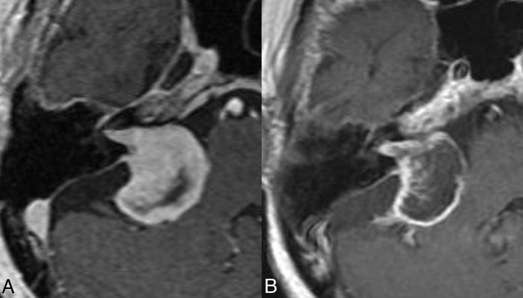
SRS can achieve long-term tumor control in >90% of cases with a relatively low risk of neurologic deterioration or facial nerve palsy.6,8,18,19 Tumor control is broadly considered as lesion regression or stability, obviating need for additional intervention (Figs 3 and 4).2,19 Kawashima et al,18 in their cohort, noted tumor regression in 62%, stable tumor in 31%, and enlargement in 7% of patients post-SRS. Treatment failures may be more common with larger or fast-growing tumors at baseline (Fig 5).7,20

FIG 3.
  • Download figure
  • Open in new tab
  • Download powerpoint
FIG 3.

Favorable treatment response post-SRS. Axial postcontrast images obtained pre-SRS (A) and at 8 years post-SRS (B) show considerable lesion regression, consistent with a response.

FIG 4.
  • Download figure
  • Open in new tab
  • Download powerpoint
FIG 4.

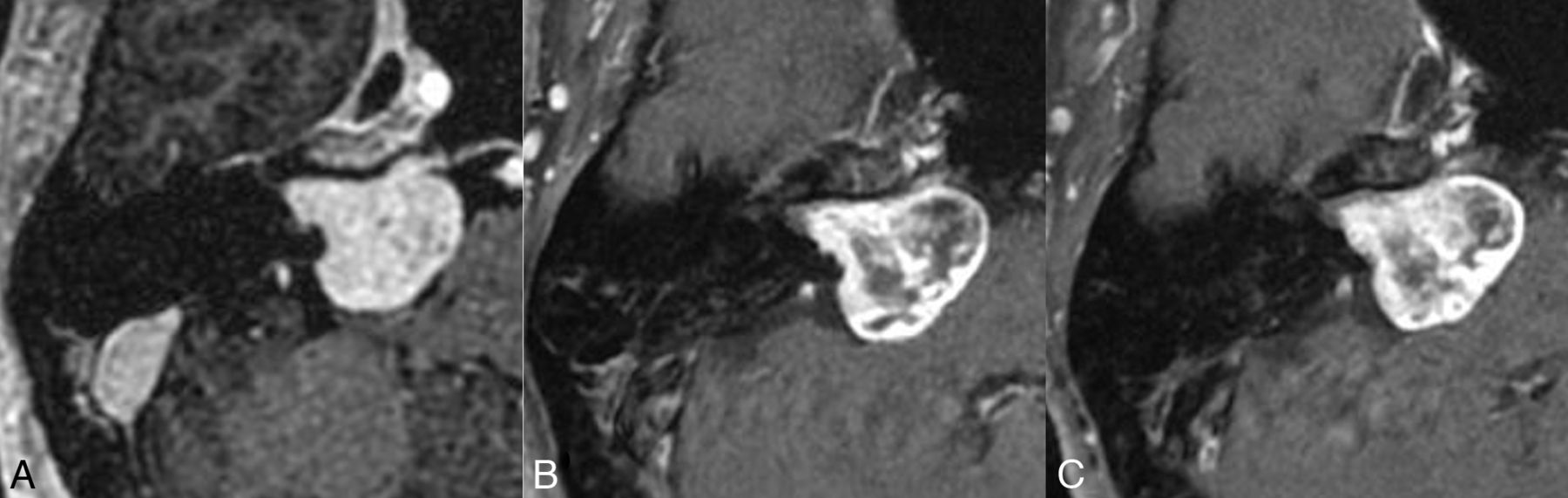
Tumor control post-SRS. Axial postcontrast images obtained pre-SRS (A) and at 1 (B) and 3 years (C) post-SRS show stable tumor size, despite considerable loss of central enhancement in the post-SRS period.

FIG 5.
  • Download figure
  • Open in new tab
  • Download powerpoint
FIG 5.

Treatment failure post-SRS. Axial postcontrast images obtained pre-SRS (A), and at 1 (B), and 2 (C) years posttherapy show a progressive increase in tumor volume from 1.9 mL at baseline to 3.2 mL at 2 years, accompanied by worsening disequilibrium clinically.

As noted earlier, tumors may also occasionally show a delayed pseudoprogression, and tumor enlargement within the first 3-4 years is generally not used as a sole criterion for salvage therapy.16 Because differentiation between tumor regrowth and delayed pseudoprogression may be challenging on conventional MR imaging, some authors recommend a documented increase in tumor size for 3 consecutive annual scans before adjudicating treatment failure, except in cases with new symptoms or larger tumors.11 Table 1 outlines the various forms of treatment outcomes that may be seen in VS post-SRS.

View this table:
  • View inline
  • View popup
Table 1:

Treatment outcomes in VS post-SRS

Surrounding Parenchymal Changes

Development of peritumoral edema postradiosurgery is well-recognized with meningiomas and has been reported in 28%–50% of cases.21 Similar changes may also be observed along the pons and cerebellar peduncles in patients with VSs treated with SRS and have been reported in about 24% of patients, developing at a median of 6 months postradiosurgery (range, 4–24 months).9 These may be associated with contrast enhancement and generally resolve on follow-up imaging (Fig 6). The exact etiology is unclear but is presumably related to radiation effects, given the temporal association with SRS. Hayhurst et al9 noted that the development of edema was significantly associated with nonauditory complications such as hydrocephalus (6%), ataxia (12.5%), trigeminal (21%) or facial nerve dysfunction (3.75%) (P = .001, OR = 7.27; range, 2.33–22.66). Overall, there is scarce literature on these findings, which may not always be clinically symptomatic but can, nevertheless, be mistaken for inflammation and/or tumor extension into the inner ear structures.

FIG 6.
  • Download figure
  • Open in new tab
  • Download powerpoint
FIG 6.

Parenchymal edema and enhancement post-SRS. Axial FLAIR (A) and postcontrast (B) images show a left-sided vestibular schwannoma abutting the left brachium pontis without edema. Post-SRS, axial FLAIR (C), and postcontrast T1WI (D) reveal parenchymal edema (C) and enhancement (arrow, D).

Intracochlear hemorrhage secondary to SRS has been previously reported and is rare, generally presenting with acute onset hearing loss.22 Additionally, an increased dose to the cochlea may be associated with post-SRS hearing loss, and keeping the cochlear radiation dose below 4 Gy when possible is generally recommended.23

Development of Cysts Post-SRS

Intra- or peritumoral cysts may occur de novo, or enlarge post-SRS, with the incidence of delayed cyst formation reported at about 2.3% (Online Supplemental Data).24⇓-26 The thin-walled peritumoral cysts may histopathologically demonstrate arachnoid cells without any tumor cells.26 Enlarging or symptomatic cysts (secondary to mass effect) may require surgical decompression or may spontaneously regress with time.27

Contrast Leakage

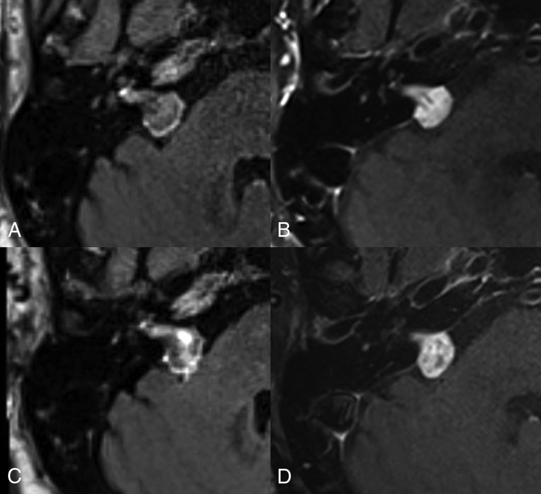
Peripheral contrast leakage, manifesting as a peritumoral halo may be seen in about 90% of patients with treatment-naïve VS. In such cases however, the contrast is only seen along the periphery of the VS.28 In our experience, early or delayed intratumoral contrast leakage in VSs treated with SRS may also be seen in a large percentage of cases (Fig 7). The exact incidence or clinical relevance of the latter remains unclear, but the phenomenon is likely secondary to slow diffusion of the contrast into the necrotic/extracellular spaces. Similar findings have also been described in metastatic brain lesions post-SRS as well as in cardiac imaging.29 On imaging, this may manifest as increased signal within the tumor core on post-contrast FLAIR images as well as increased extracellular, extravascular gadolinium in the internal auditory canal and vicinity of tumor. Whether this intra- or peri-tumoral contrast leakage correlates with elevated CSF protein post-SRS remains unclear.

FIG 7.
  • Download figure
  • Open in new tab
  • Download powerpoint
FIG 7.

Contrast leakage post-SRS. Axial postcontrast FLAIR (A and C) and T1WI (B and D) obtained pre- (A and B), and 1 year post-SRS (C and D). On the pre-SRS images, there is a thin peritumoral halo on the FLAIR imaging (A) without any contrast leakage centrally. Post-SRS FLAIR (C) shows contrast leakage within the VS more centrally.

Hydrocephalus

Even though hydrocephalus may be seen with untreated VSs in about 3.7%–18% of cases, it may additionally develop post-SRS in 2%–3% of patients (Fig 8).30⇓-32 The precise causal relationship remains controversial, and potential mechanisms include obstruction of the fourth ventricle, protein shedding from the tumor leading to plugging of arachnoid granulations, as well as alterations in CSF flow dynamics in the basal cisterns.30,32,33 Patients may develop communicating hydrocephalus 4–18 months post-SRS and often show elevated CSF protein levels and may have normal opening pressure.30,33 Risk factors include larger tumor size and female sex.34

FIG 8.
  • Download figure
  • Open in new tab
  • Download powerpoint
FIG 8.

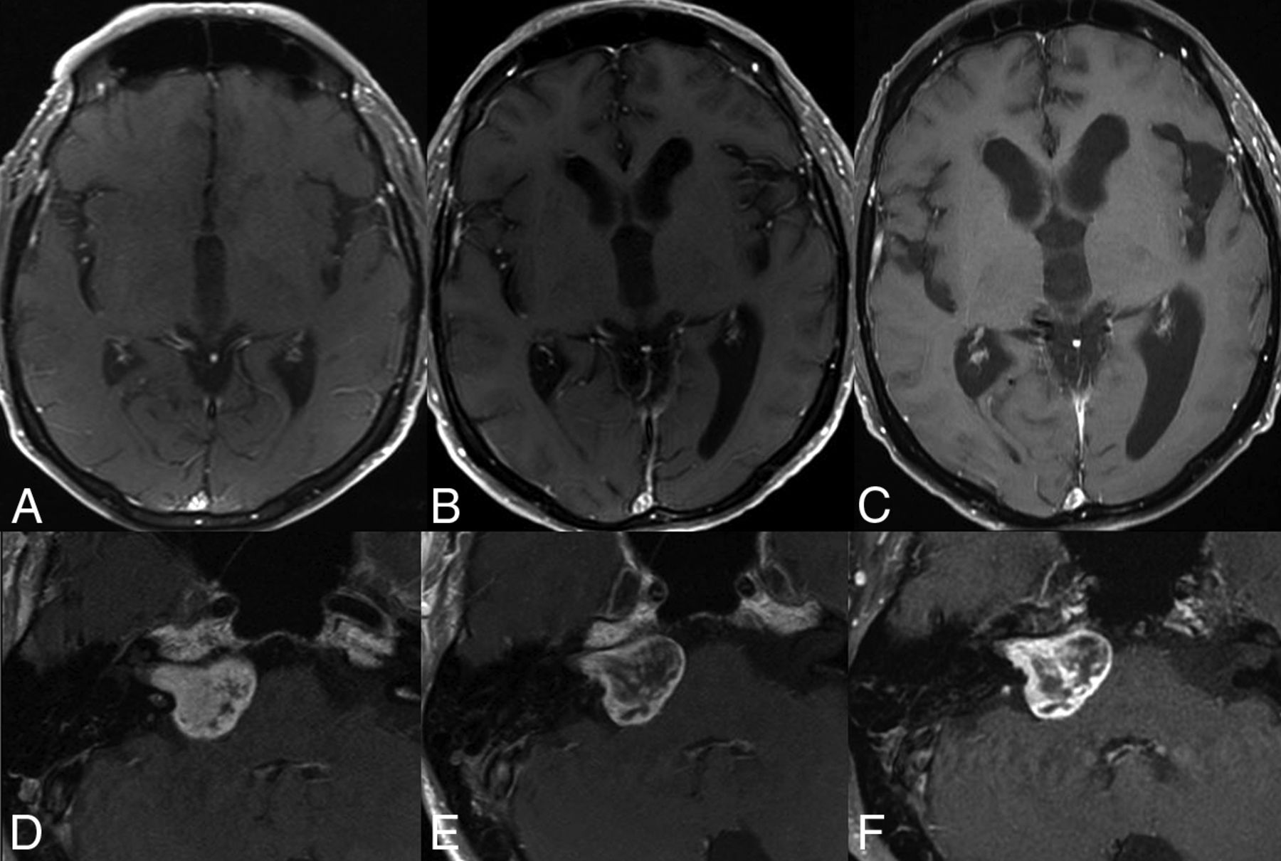
Hydrocephalus post-SRS. Axial postcontrast images at the level of third ventricle (A–C) and the VS (D–F) obtained at baseline (A and D), at 1 (B and E) and 2 years (C and F) post-SRS show progressive enlargement in ventricular dimensions and disproportionate enlargement of left Sylvian fissure. The underlying VS (D–F) remained stable in size and showed loss of central enhancement. The patient was diagnosed with normal pressure hydrocephalus and underwent ventricular shunting.

Patients may also present with signs of elevated intracranial pressure or gait disturbances and urinary incontinence as seen with normal pressure hydrocephalus.35 Surgical CSF diversion may be required in most patients to alleviate symptoms.31 Table 2 summarizes the various reported post-SRS complications in patients with VS.

View this table:
  • View inline
  • View popup
Table 2:

Post-SRS complications in patients with VS

Tumors Post-SRS

The risk of a secondary malignant CNS tumor developing post-SRS is considered low, with the overall risk estimated at approximately 0.04% at 15 years.36 However, these can, nevertheless, still occur and should be carefully considered, especially in young patients and those with underlying tumor-predisposition syndromes such as neurofibromatosis.37

Tumors reported post-SRS include malignant gliomas (including astrocytoma, glioblastoma, and ependymomas), sarcomas, meningiomas, as well as dedifferentiation of primary VS into malignant nerve sheath tumor. In the context of VS, malignant nerve sheath tumor and glioblastoma appear to be overall more common (Figs 9 and 10).36,37

FIG 9.
  • Download figure
  • Open in new tab
  • Download powerpoint
FIG 9.

Glioblastoma post-SRS. Axial postcontrast, pretreatment images reveal a left VS (A). The post-SRS left temporal lobe at 2 years (B) is without any lesions. The patient subsequently presented with seizures 3 years post-SRS with a new left temporal intra-axial mass on imaging (C), which was subsequently resected and diagnosed as a glioblastoma.

FIG 10.
  • Download figure
  • Open in new tab
  • Download powerpoint
FIG 10.

Malignant transformation of a VS post-SRS. Pre-SRS (A) contrast-enhanced image shows a left-sided VS. Post-SRS image after 5 years (B) shows a mild overall increase in tumor size. However, the tumor showed increased growth at 7 years post-SRS (C). A subtotal resection was performed (D), and pathology revealed a malignant peripheral nerve sheath tumor. Follow-up imaging after 4 months (E) shows considerable recurrent tumor burden with involvement of adjacent structures.

The mean latency period for stereotactic radiosurgery–induced neoplasms is about 7.9 years and is generally shorter for malignant neoplasms (7.1 years) compared with benign secondary neoplasms (14.25 years). Secondary neoplasms may occur either inside or outside the original lesion, as well as in regions receiving high- or low-dose radiation. The size of the original tumor often tends to be >2 cm.36,37

CONCLUSIONS

Post-SRS VSs can present with a variable appearances, besides the rare occasional complications such as secondary tumor and hydrocephalus. Familiarity with the various imaging findings may be helpful to avoid incorrect interpretation and recognize early complications.

Footnotes

  • Disclosure forms provided by the authors are available with the full text and PDF of this article at www.ajnr.org.

References

  1. 1.↵
    1. Bathla G,
    2. Case BM,
    3. Berbaum K, et al
    . Vestibular schwannomas: do linear and volumetric parameters on MRI correlate with hearing loss? Otol Neurotol 2016;37:1168–73 doi:10.1097/MAO.0000000000001150 pmid:27466888
    CrossRefPubMed
  2. 2.↵
    1. Buss EJ,
    2. Wang TJ,
    3. Sisti MB
    . Stereotactic radiosurgery for management of vestibular schwannoma: a short review. Neurosurg Rev 2021;44:901–04 doi:10.1007/s10143-020-01279-2 pmid:32170501
    CrossRefPubMed
  3. 3.↵
    1. Marinelli JP,
    2. Lohse CM,
    3. Carlson ML
    . Incidence of vestibular schwannoma over the past half-century: a population-based study of Olmsted County, Minnesota. Otolaryngol Head Neck Surg 2018;159:717–23 doi:10.1177/0194599818770629 pmid:29712512
    CrossRefPubMed
  4. 4.↵
    1. Tsao MN,
    2. Sahgal A,
    3. Xu W, et al
    . Stereotactic radiosurgery for vestibular schwannoma: International Stereotactic Radiosurgery Society (ISRS) Practice Guideline. J Radiosurg SBRT 2017;5:5–24 pmid:29296459
    PubMed
  5. 5.↵
    1. Carlson ML,
    2. Link MJ,
    3. Driscoll CL, et al
    . Working toward consensus on sporadic vestibular schwannoma care: a modified Delphi Study. Otol Neurotol 2020;41:e1360–71 doi:10.1097/MAO.0000000000002917 pmid:33492814
    CrossRefPubMed
  6. 6.↵
    1. Tosi U,
    2. Lavieri MET,
    3. An A, et al
    . Outcomes of stereotactic radiosurgery for large vestibular schwannomas: a systematic review and meta-analysis. Neurooncol Pract 2021;8:405–16 doi:10.1093/nop/npab011 pmid:34277019
    CrossRefPubMed
  7. 7.↵
    1. Yeole U,
    2. Prabhuraj AR,
    3. Arivazhagan A, et al
    . Gamma knife radiosurgery for large vestibular schwannoma more than 10 cm3: a single-center Indian study. J Neurol Surg B Skull Base 2022;83:e343–52 doi:10.1055/s-0041-1729977 pmid:35832954
    CrossRefPubMed
  8. 8.↵
    1. Persson O,
    2. Bartek J Jr.,
    3. Shalom NB, et al
    . Stereotactic radiosurgery vs. fractionated radiotherapy for tumor control in vestibular schwannoma patients: a systematic review. Acta Neurochir (Wien) 2017;159:1013–21 doi:10.1007/s00701-017-3164-6 pmid:28409393
    CrossRefPubMed
  9. 9.↵
    1. Hayhurst C,
    2. Monsalves E,
    3. Bernstein M, et al
    . Predicting nonauditory adverse radiation effects following radiosurgery for vestibular schwannoma: a volume and dosimetric analysis. Int J Radiat Oncol Biol Phys 2012;82:2041–46 doi:10.1016/j.ijrobp.2011.02.017 pmid:21531086
    CrossRefPubMed
  10. 10.↵
    1. Tuleasca C,
    2. George M,
    3. Faouzi M, et al
    . Acute clinical adverse radiation effects after gamma knife surgery for vestibular schwannomas. J Neurosurg 2016;125:73–82 doi:10.3171/2016.7.GKS161496 pmid:27903185
    CrossRefPubMed
  11. 11.↵
    1. Fouard O,
    2. Daisne JF,
    3. Wanet M, et al
    . Long-term volumetric analysis of vestibular schwannomas following stereotactic radiotherapy: practical implications for follow-up. Clin Transl Radiat Oncol 2022;33:1–6 doi:10.1016/j.ctro.2021.12.003 pmid:34977365
    CrossRefPubMed
  12. 12.↵
    1. Kim JH,
    2. Jung HH,
    3. Chang JH, et al
    . Predictive factors of unfavorable events after gamma knife radiosurgery for vestibular schwannoma. World Neurosurg 2017;107:175–84 doi:10.1016/j.wneu.2017.07.139 pmid:28826715
    CrossRefPubMed
  13. 13.↵
    1. Rueß D,
    2. Schütz B,
    3. Celik E, et al
    . Pseudoprogression of vestibular schwannoma after stereotactic radiosurgery with cyberknife: proposal for new response criteria. Cancers (Basel) 2023;15:1496 doi:10.3390/cancers15051496 pmid:36900290
    CrossRefPubMed
  14. 14.↵
    1. Aoyama H,
    2. Onodera S,
    3. Takeichi N, et al
    . Symptomatic outcomes in relation to tumor expansion after fractionated stereotactic radiation therapy for vestibular schwannomas: single-institutional long-term experience. Int J Radiat Oncol Biol Phys 2013;85:329–34 doi:10.1016/j.ijrobp.2012.05.003 pmid:22704983
    CrossRefPubMed
  15. 15.↵
    1. Nagano O,
    2. Higuchi Y,
    3. Serizawa T, et al
    . Transient expansion of vestibular schwannoma following stereotactic radiosurgery. J Neurosurg 2008;109:811–16 doi:10.3171/JNS/2008/109/11/0811 pmid:18976069
    CrossRefPubMed
  16. 16.↵
    1. Breshears JD,
    2. Chang J,
    3. Molinaro AM, et al
    . Temporal dynamics of pseudoprogression after gamma knife radiosurgery for vestibular schwannomas: a retrospective volumetric study. Neurosurgery 2019;84:1:23–31 doi:10.1093/neuros/nyy019 pmid:29518221
    CrossRefPubMed
  17. 17.↵
    1. Whitmeyer M,
    2. Brahimaj BC,
    3. Beer-Furlan A, et al
    . Resection of vestibular schwannomas after stereotactic radiosurgery: a systematic review. J Neurosurg 2020;135:881–89 doi:10.3171/2020.7.JNS2044 pmid:34331121
    CrossRefPubMed
  18. 18.↵
    1. Kawashima M,
    2. Hasegawa H,
    3. Shin M, et al
    . Outcomes of stereotactic radiosurgery in young adults with vestibular schwannomas. J Neurooncol 2021;154:93–100 doi:10.1007/s11060-021-03803-w pmid:34241770
    CrossRefPubMed
  19. 19.↵
    1. Ogino A,
    2. Lunsford LD,
    3. Long H, et al
    . Stereotactic radiosurgery as the first-line treatment for intracanalicular vestibular schwannomas. J Neurosurg 2021;135:1051–57 doi:10.3171/2020.9.JNS202818 pmid:34600434
    CrossRefPubMed
  20. 20.↵
    1. Langenhuizen P,
    2. Zinger S,
    3. Hanssens PE, et al
    . Influence of pretreatment growth rate on gamma knife treatment response for vestibular schwannoma: a volumetric analysis. J Neurosurg 2018 Nov 1:1–8. [Epub ahead of print]
  21. 21.↵
    1. Milano MT,
    2. Grimm J,
    3. Niemierko A, et al
    . Radiation-induced edema after single-fraction or multifraction stereotactic radiosurgery for meningioma: a critical review. Int J Radiat Oncol Biol Phys 2018;110:68–86 doi:10.1016/j.ijrobp.2018.03.026 pmid:29726362
    CrossRefPubMed
  22. 22.↵
    1. Franco-Vidal V,
    2. Songu M,
    3. Blanchet H, et al
    . Intracochlear hemorrhage after gamma knife radiosurgery. Otol Neurotol 2007;28:240–44 doi:10.1097/01.mao.0000244360.57379.d4 pmid:17159493
    CrossRefPubMed
  23. 23.↵
    1. Massager N,
    2. Nissim O,
    3. Delbrouck C, et al
    . Irradiation of cochlear structures during vestibular schwannoma radiosurgery and associated hearing outcome. J Neurosurg 2007;107:733–39 doi:10.3171/JNS-07/10/0733 pmid:17937216
    CrossRefPubMed
  24. 24.↵
    1. Hasegawa T,
    2. Kida Y,
    3. Kato T, et al
    . Long-term safety and efficacy of stereotactic radiosurgery for vestibular schwannomas: evaluation of 440 patients more than 10 years after treatment with gamma knife surgery. J Neurosurg 2013;118:557–65 doi:10.3171/2012.10.JNS12523 pmid:23140152
    CrossRefPubMed
  25. 25.↵
    1. Kwon Y,
    2. Khang SK,
    3. Kim CJ, et al
    . Radiologic and histopathologic changes after gamma knife radiosurgery for acoustic schwannoma. Stereotact Funct Neurosurg 1999;72(Suppl 1):2–10 doi:10.1159/000056433 pmid:10681685
    CrossRefPubMed
  26. 26.↵
    1. Murakami K,
    2. Jokura H,
    3. Kawagishi J, et al
    . Development of intratumoral cyst or extratumoral arachnoid cyst in intracranial schwannomas following gamma knife radiosurgery. Acta Neurochir (Wien) 2011;153:1201–09 doi:10.1007/s00701-011-0972-y pmid:21369949
    CrossRefPubMed
  27. 27.↵
    1. Lee TK,
    2. Kim YJ,
    3. Jung TY, et al
    . From the perspective of pseudo-progression rather than treatment failure, how long should we wait before considering treatment failure if large cystic enlargement occurs after gamma knife radiosurgery for vestibular schwannoma? Insight into pseudoprogression based on two case reports. Acta Neurochir (Wien) 2023;165:2105–09 doi:10.1007/s00701-023-05684-6 pmid:37421424
    CrossRefPubMed
  28. 28.↵
    1. Benson JC,
    2. Carlson ML,
    3. Lane JI
    . Peritumoral signal on postcontrast FLAIR images: description and proposed biomechanism in vestibular schwannomas. AJNR Am J Neuroradiol 2023;44:1171–75 doi:10.3174/ajnr.A7979 pmid:37652582
    Abstract/FREE Full Text
  29. 29.↵
    1. Wagner S,
    2. Gufler H,
    3. Eichner G, et al
    . Characterisation of lesions after stereotactic radiosurgery for brain metastases: impact of delayed contrast magnetic resonance imaging. Clin Oncol (R Coll Radiol) 2017;29:143–50 doi:10.1016/j.clon.2016.09.021 pmid:27777145
    CrossRefPubMed
  30. 30.↵
    1. Cauley KA,
    2. Ratkovits B,
    3. Braff SP, et al
    . Communicating hydrocephalus after gamma knife radiosurgery for vestibular schwannoma: an MR imaging study. AJNR Am J Neuroradiol 2009;30:992–94 doi:10.3174/ajnr.A1379 pmid:19039040
    Abstract/FREE Full Text
  31. 31.↵
    1. De Sanctis P,
    2. Green S,
    3. Germano I
    . Communicating hydrocephalus after radiosurgery for vestibular schwannomas: does technique matter? A systematic review and meta-analysis. J Neurooncol 2019;145:365–73 doi:10.1007/s11060-019-03305-w pmid:31621039
    CrossRefPubMed
  32. 32.↵
    1. Nakahara M,
    2. Imahori T,
    3. Sasayama T, et al
    . Refractory communicating hydrocephalus after radiation for small vestibular schwannoma with asymptomatic ventriculomegaly: a case report. Radiol Case Rep 2020;15:1023–28 doi:10.1016/j.radcr.2020.04.063 pmid:32435322
    CrossRefPubMed
  33. 33.↵
    1. Shimizu Y,
    2. Miyamori T,
    3. Yamano J
    . Hydrocephalus after gamma knife radiosurgery for schwannoma. Asian J Neurosurg 2019;14:487–90 doi:10.4103/ajns.AJNS_278_18 pmid:31143267
    CrossRefPubMed
  34. 34.↵
    1. Lee SH,
    2. Seol HJ,
    3. Kong DS, et al
    . Risk factors and tumor response associated with hydrocephalus after gamma knife radiosurgery for vestibular schwannoma. Acta Neurochir (Wien) 2012;154:1679–84 doi:10.1007/s00701-012-1350-0 pmid:22535199
    CrossRefPubMed
  35. 35.↵
    1. Park Y,
    2. Yoo BG,
    3. Jeong S, et al
    . Normal pressure hydrocephalus after gamma knife radiosurgery in a patient with vestibular schwannoma. J Neurocrit Care 2020;13:127–31 doi:10.18700/jnc.200014
    CrossRef
  36. 36.↵
    1. Patel TR,
    2. Chiang VL
    . Secondary neoplasms after stereotactic radiosurgery. World Neurosurg 2014;81:594–99 doi:10.1016/j.wneu.2013.10.043 pmid:24148883
    CrossRefPubMed
  37. 37.↵
    1. Balasubramaniam A,
    2. Shannon P,
    3. Hodaie M, et al
    . Glioblastoma multiforme after stereotactic radiotherapy for acoustic neuroma: case report and review of the literature. Neuro Oncol 2007;9:447–53 doi:10.1215/15228517-2007-027 pmid:17704364
    CrossRefPubMed
  • Received November 28, 2023.
  • Accepted after revision January 3, 2024.
  • © 2024 by American Journal of Neuroradiology
Back to top
Advertisement
Print
Download PDF
Email Article

Thank you for your interest in spreading the word on American Journal of Neuroradiology.

NOTE: We only request your email address so that the person you are recommending the page to knows that you wanted them to see it, and that it is not junk mail. We do not capture any email address.

Enter multiple addresses on separate lines or separate them with commas.
Imaging Findings Post-Stereotactic Radiosurgery for Vestibular Schwannoma: A Primer for the Radiologist
(Your Name) has sent you a message from American Journal of Neuroradiology
(Your Name) thought you would like to see the American Journal of Neuroradiology web site.
CAPTCHA
This question is for testing whether or not you are a human visitor and to prevent automated spam submissions.
Cite this article
Girish Bathla, Parv M. Mehta, John C. Benson, Amit K. Agrwal, Neetu Soni, Michael J. Link, Matthew L. Carlson, John I. Lane
Imaging Findings Post-Stereotactic Radiosurgery for Vestibular Schwannoma: A Primer for the Radiologist
American Journal of Neuroradiology Sep 2024, 45 (9) 1194-1201; DOI: 10.3174/ajnr.A8175

Citation Manager Formats

  • BibTeX
  • Bookends
  • EasyBib
  • EndNote (tagged)
  • EndNote 8 (xml)
  • Medlars
  • Mendeley
  • Papers
  • RefWorks Tagged
  • Ref Manager
  • RIS
  • Zotero
0 Responses
Respond to this article
Share
Bookmark this article
Post-SRS Imaging for Vestibular Schwannoma
Girish Bathla, Parv M. Mehta, John C. Benson, Amit K. Agrwal, Neetu Soni, Michael J. Link, Matthew L. Carlson, John I. Lane
American Journal of Neuroradiology Sep 2024, 45 (9) 1194-1201; DOI: 10.3174/ajnr.A8175
del.icio.us logo Twitter logo Facebook logo Mendeley logo
  • Tweet Widget
  • Facebook Like
  • Google Plus One
Purchase

Jump to section

  • Article
    • Abstract
    • ABBREVIATIONS:
    • Surrounding Parenchymal Changes
    • CONCLUSIONS
    • Footnotes
    • References
  • Figures & Data
  • Supplemental
  • Info & Metrics
  • Responses
  • References
  • PDF

Related Articles

  • PubMed
  • Google Scholar

Cited By...

  • No citing articles found.
  • Crossref
  • Google Scholar

This article has not yet been cited by articles in journals that are participating in Crossref Cited-by Linking.

More in this TOC Section

  • Vestibular Schwannoma: PostGad Signal & Hearing Loss
  • CT facial recess and hard cochlear implantation
  • Imaging Hyrtl Fissure: Congenital CSF Otorrhea
Show more Head and Neck Imaging

Similar Articles

Advertisement

Indexed Content

  • Current Issue
  • Accepted Manuscripts
  • Article Preview
  • Past Issues
  • Editorials
  • Editors Choice
  • Fellow Journal Club
  • Letters to the Editor

Cases

  • Case Collection
  • Archive - Case of the Week
  • Archive - Case of the Month
  • Archive - Classic Case

Special Collections

  • Special Collections

Resources

  • News and Updates
  • Turn around Times
  • Submit a Manuscript
  • Author Policies
  • Manuscript Submission Guidelines
  • Evidence-Based Medicine Level Guide
  • Publishing Checklists
  • Graphical Abstract Preparation
  • Imaging Protocol Submission
  • Submit a Case
  • Become a Reviewer/Academy of Reviewers
  • Get Peer Review Credit from Publons

Multimedia

  • AJNR Podcast
  • AJNR SCANtastic
  • Video Articles

About Us

  • About AJNR
  • Editorial Board
  • Not an AJNR Subscriber? Join Now
  • Alerts
  • Feedback
  • Advertise with us
  • Librarian Resources
  • Permissions
  • Terms and Conditions

American Society of Neuroradiology

  • Not an ASNR Member? Join Now

© 2025 by the American Society of Neuroradiology All rights, including for text and data mining, AI training, and similar technologies, are reserved.
Print ISSN: 0195-6108 Online ISSN: 1936-959X

Powered by HighWire