
Abstract
Summary: Eighteen patients with vertebral lesions located in the thoracic or lumbar spine underwent percutaneous biopsy performed via a transpedicular approach under fluoroscopic guidance. This technique led to an accurate diagnosis in 16 cases (89%). No complications were encountered. For percutaneous lumbar and thoracic vertebral biopsy, the transpedicular approach is a safe and accurate alternative to the posterolateral approach.
The most common approach to biopsy of the thoracic or lumbar spine is usually posterolateral, under fluoroscopic or CT guidance (1–6). The transpedicular approach has seldom been used for these biopsies (3, 7). We retrospectively studied 18 patients with thoracic or lumbar vertebral disease in whom a biopsy was performed via the transpedicular pathway under fluoroscopic guidance in a setting in which rapid CT scanning was not available.
Technique
Between April 1996 and September 1997, 18 patients with vertebral lesions (10 women and eight men aged 20–87 years; average age, 57 years) underwent percutaneous biopsy via the transpedicular approach.
Biopsies were indicated after a review of all available diagnostic images, including plain film radiographs, CT scans, bone scintigrams, and MR images. All patients were examined by MR imaging.
Indications for the biopsies in this series were to confirm or rule out the presence of metastatic lesions in patients with known primary tumor (five cases) (these patients had vertebral lesions that were not typical of metastasis on MR images); to determine the presence of metastatic lesions in patients without any known primary tumor (10 cases); and to diagnose spondylitis (three cases). Biopsy was contraindicated in patients with uncorrected bleeding diathesis.
Biopsy sites were in the thoracic spine in 11 cases and in the lumbar spine in seven. In 16 patients, biopsies were performed under local anesthesia and intravenous sedation. In the remaining two, severe back pain required general anesthesia. Biopsies were performed with the patient in the prone position, using the trephine biopsy kits for thoracic or lumbar biopsy described by Laredo and Bard (4) (Fig 1). Preoperative CT scans or MR images determined the side of the approach. All biopsies were performed with fluoroscopic guidance, using a biplane C-arm digitalized radiographic system. The puncture point was determined under fluoroscopic control. After local anesthesia with 1% lidocaine, the thin needle of the trephine set was introduced through a small skin incision and advanced under biplane fluoroscopic guidance until it contacted the periosteum (Fig 2). This thin needle was used to anesthetize the deep soft-tissue planes and the periosteum. The fine needle was then replaced by the guidewire. Over the guidewire, the graduated external sheath mounted on the handle was positioned in the middle portion of the pedicle. The handle and guide were removed, and biopsies were performed with the cutting cannula. For cytologic evaluation, biopsy specimens were prepared on glass slides and fixed; for histologic evaluation, core biopsy samples were fixed in formalin solution; and for microbiological examination, biopsy material was collected in sterile dry tubes. The procedure lasted 30 to 60 minutes.

Trephine set for biopsy of the thoracic spine. 1 = fine needle (external diameter, 1.5 mm); 2 = rigid guidewire (length, 32 cm); 3 = graduated external sheath (external diameter, 2.8 mm); 4 = hollow intermediate piece with handle; 5 = serrated cannula (internal diameter, 1.6 mm; external diameter, 2.2 mm); 6 = obturator. In the set for biopsy of the lumbar spine, the rigid guidewire is 40 cm long. The graduated external sheath has an external diameter of 4 mm, and the serrated cannula, an internal diameter of 2.2 mm

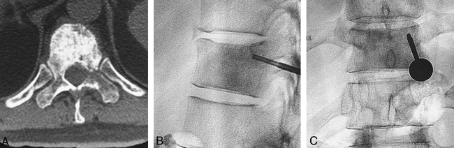
Percutaneous transpedicular biopsy at the thoracic (T11) level.
A, CT scan shows a heterogeneous sclerotic and lytic lesion at T11.
B and C, Posteroanterior (B) and lateral (C) views show the transpedicular pathway of the trephine needle.
Biopsy material was obtained in all cases. No biopsy finding was false negative. Percutaneous transpedicular biopsy led to an accurate diagnosis in 16 cases (89%). In the remaining two cases, conclusive results were not obtained. In one of these, the biopsy material permitted a histologic evaluation that led to a satisfactory diagnosis of subacute chronic inflammatory or infectious spondylitis, but the bacteriologic studies were negative. In this case, positive cultures for tuberculosis were obtained by open biopsy. In the remaining case, the material obtained by percutaneous biopsy led to a diagnosis of lymphoma; however, it was not sufficient to establish its type precisely, and open biopsy was therefore needed, which led to an accurate diagnosis and appropriate treatment.
The final diagnoses were osteoporotic collapse (eight cases, 44%); metastasis (five cases, 28%); infectious spondylitis (four cases, 22%); and lymphoma (one case, 6%). Of the eight patients who had osteoporotic collapse, four had a known primary neoplasm. The other four had no such neoplasm, but the lesions were considered suspicious on MR images. In these eight cases, clinical follow-up confirmed that the lesions were benign.
No complications were encountered in this small series. Neurologic examinations were unchanged after the biopsy procedure, which was well tolerated by all patients. No back or radicular pain was observed, either immediately after biopsy or during the days that followed, and no pulmonary complications were observed. The postoperative hospital stay lasted at least 24 hours.
Discussion
The efficacy of percutaneous biopsy in the management of spinal lesions has been evaluated extensively (1–7). The posterolateral approach is widely used for the thoracic and lumbar spine (1–6). At the thoracic level, this approach can lead to pulmonary complications, especially pneumothorax or, occasionally, pneumonia. To avoid such complications, we decided to perform biopsies via the transpedicular approach under fluoroscopic guidance. Renfrew et al (7) reported six cases of percutaneous transpedicular biopsy for thoracic or lumbar spine lesions under CT guidance. In 1994, Langer-Cherbit et al (3), who reported a large series of 76 percutaneous vertebral biopsies, used this approach for eight thoracic or lumbar biopsies but did not evaluate its results precisely.
The risk involved in percutaneous vertebral biopsy has been variously estimated at 0% (4), 2.2% (5), 7.6% (1), and 26% (2). The most frequently reported complications were pulmonary, neurologic, and infective disorders. In our series, the transpedicular approach avoided pulmonary complications without increasing the rate of neurologic complications. During the follow-up of these 18 patients, no clinical data or radiologic examinations, when available, indicated the presence of pedicular fractures. We attribute this to the fact that the diameter of the external sheath we used was much smaller than the diameter of the pedicle. Furthermore, the use of a coaxial system enabled us to do two or three biopsies with only one transpedicular tract. This approach is currently used for vertebroplasty, with no reported subsequent complications (8). It is therefore safe, provided the technique is performed with rigorous exactitude.
The accuracy of vertebral biopsy using the posterolateral approach has been estimated at 50% to 91% (1–6). With our technique, biopsies were accurate in 16 (89%) of 18 cases. The accuracy of percutaneous biopsies using the transpedicular approach therefore compares favorably with that of biopsies obtained via the posterolateral approach. Biopsies were performed under fluoroscopic control because we do not presently have access to rapid CT scanning. With the development of MR imaging, vertebral lesions are being identified earlier and in larger numbers. The specificity of MR images is often, however, insufficient to define an adequate strategy of treatment. Therefore, vertebral biopsies are increasingly needed, and this technique should be available in centers with limited access to rapid CT scanning. In addition, fluoroscopy allows real-time control of the position of the needles in both the anteroposterior and cephalocaudal directions. With the transpedicular approach it is important to be sure that the sheath and cannulas are in a central position in the pedicle, and not near the cortical bone. Biplane fluoroscopy is useful for the transpedicular approach, because it permits permanent and simultaneous control of the position of the needle, as seen in both the frontal and lateral views.
Indications for using either the posterolateral or transpedicular approach depend on the location of the lesion. If the lesion is located predominantly in the disk space, as in cases of infectious disease, the posterolateral approach should be used. This approach is also mandatory when a lesion is located in the lower part of the vertebral body; however, if the lesion is located in the posterior half of the vertebral body or if the pedicle is involved, the transpedicular approach is an effective method of biopsy. In case of lesions of the entire vertebral body, we usually prefer the transpedicular approach.
Footnotes
↵1 Address reprint requests to Laurent Pierot, MD.
References
- Received January 14, 1998.
- Copyright © American Society of Neuroradiology


