
Article Figures & Data
Figures
fig.
1. CNSV was diagnosed on the basis of perfusion imaging findings in a patient with acute and profound neurologic symptoms related to the left hemisphere. The clinical impression was acute stroke caused by occlusion of the left internal carotid and cerebral arteries. The angiogram (not shown) was negative for CNSV. The patient made a complete recovery a few days after initiation of steroid therapy. A, FLAIR image shows nonspecific white matter disease of both hemispheres. B, Perfusion image shows extensive abnormality involving the left hemisphere, including decreased cerebral blood flow (hyperintensity on rMTT map). C, Perfusion image shows decreased cerebral blood volume (hypointensity on the rCBV map).
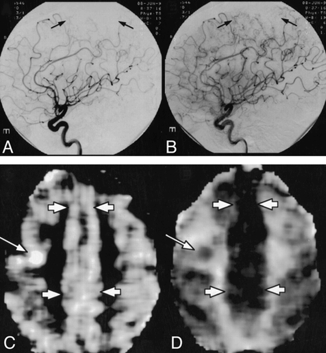
fig. 2. CNSV was excluded on the basis of perfusion imaging findings in a patient with remote and recent brain infarctions and a clinical suspicion of CNSV. Brain biopsy along the left anterior cerebral artery (ACA) distribution was performed immediately after the negative perfusion findings that depicted normal vasculature were obtained.
A and B, Early (A) and late (B) arterial phase of the left internal carotid artery angiogram shows a paucity of arterial staining in the parietal lobe along the ACA distribution (arrows). Similar findings were also noted on the angiogram of the right internal carotid artery (not shown). C and D, Perfusion MR image was obtained concurrent with the CBV map (C) and the MTT map (D) and showed normal perfusion of brain parenchyma along the ACA distribution (short arrows). An incidental finding of a recent infarction (long arrows) in the middle cerebral artery distribution is also noted.
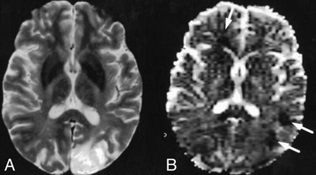
fig. 3.
CNSV was diagnosed by diffusion imaging. Findings were consistent with multiple, small-vessel ischemic disease in that they did not depict vascular distribution, a characteristic finding in CNSV. A, T2-weighted MR image shows an old left occipital infarction that is not specific for the diagnosis of CNSV. B, The corresponding ADC map shows three additional acute ischemic lesions (arrows).


In this issue


Related Articles
Cited By...
- No citing articles found.