Advertisement

Main menu

  • Home
  • Content
    • Current Issue
    • Accepted Manuscripts
    • Article Preview
    • Past Issue Archive
    • Video Articles
    • AJNR Case Collection
    • Case of the Week Archive
    • Case of the Month Archive
    • Classic Case Archive
  • Special Collections
    • AJNR Awards
    • Low-Field MRI
    • Alzheimer Disease
    • ASNR Foundation Special Collection
    • Photon-Counting CT
    • View All
  • Multimedia
    • AJNR Podcasts
    • AJNR SCANtastic
    • Trainee Corner
    • MRI Safety Corner
    • Imaging Protocols
  • For Authors
    • Submit a Manuscript
    • Submit a Video Article
    • Submit an eLetter to the Editor/Response
    • Manuscript Submission Guidelines
    • Statistical Tips
    • Fast Publishing of Accepted Manuscripts
    • Graphical Abstract Preparation
    • Imaging Protocol Submission
    • Author Policies
  • About Us
    • About AJNR
    • Editorial Board
    • Editorial Board Alumni
  • More
    • Become a Reviewer/Academy of Reviewers
    • Subscribers
    • Permissions
    • Alerts
    • Feedback
    • Advertisers
    • ASNR Home

User menu

  • Alerts
  • Log in

Search

  • Advanced search
American Journal of Neuroradiology

American Journal of Neuroradiology

ASHNR American Society of Functional Neuroradiology ASHNR American Society of Pediatric Neuroradiology ASSR
  • Alerts
  • Log in

Advanced Search

  • Home
  • Content
    • Current Issue
    • Accepted Manuscripts
    • Article Preview
    • Past Issue Archive
    • Video Articles
    • AJNR Case Collection
    • Case of the Week Archive
    • Case of the Month Archive
    • Classic Case Archive
  • Special Collections
    • AJNR Awards
    • Low-Field MRI
    • Alzheimer Disease
    • ASNR Foundation Special Collection
    • Photon-Counting CT
    • View All
  • Multimedia
    • AJNR Podcasts
    • AJNR SCANtastic
    • Trainee Corner
    • MRI Safety Corner
    • Imaging Protocols
  • For Authors
    • Submit a Manuscript
    • Submit a Video Article
    • Submit an eLetter to the Editor/Response
    • Manuscript Submission Guidelines
    • Statistical Tips
    • Fast Publishing of Accepted Manuscripts
    • Graphical Abstract Preparation
    • Imaging Protocol Submission
    • Author Policies
  • About Us
    • About AJNR
    • Editorial Board
    • Editorial Board Alumni
  • More
    • Become a Reviewer/Academy of Reviewers
    • Subscribers
    • Permissions
    • Alerts
    • Feedback
    • Advertisers
    • ASNR Home
  • Follow AJNR on Twitter
  • Visit AJNR on Facebook
  • Follow AJNR on Instagram
  • Join AJNR on LinkedIn
  • RSS Feeds

AJNR Awards, New Junior Editors, and more. Read the latest AJNR updates

Research ArticlePediatrics

Syndromes of Bilateral Symmetrical Polymicrogyria

A. James Barkovich, Robert Hevner and Renzo Guerrini
American Journal of Neuroradiology November 1999, 20 (10) 1814-1821;
A. James Barkovich
  • Find this author on Google Scholar
  • Find this author on PubMed
  • Search for this author on this site
Robert Hevner
  • Find this author on Google Scholar
  • Find this author on PubMed
  • Search for this author on this site
Renzo Guerrini
  • Find this author on Google Scholar
  • Find this author on PubMed
  • Search for this author on this site
  • Article
  • Figures & Data
  • Info & Metrics
  • Responses
  • References
  • PDF
Loading

Article Figures & Data

Figures

  • Tables
  • fig 1.
    • Download figure
    • Open in new tab
    • Download powerpoint
    fig 1.

    Case 1: 3-year-old girl with bifrontal polymicrogyria. Axial spin-echo (SE) (600/20) MR image shows shallow sulci with irregularity or the cortical–white matter junction, consistent with polymicrogyria, involving the entire frontal cortex posteriorly to the central sulcus.

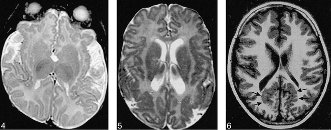
    fig 2. Case 15: 10-month-old girl with bilateral holosylvian polymicrogyria.

    A, Axial reformation from 3D Fourier transformation (3DFT) gradient-echo (GRE) (35/7) image shows thickened, irregular cortex involving the entirety of the cortex surrounding the sylvian fissures and widening of the fissures.

    B, Sagittal reformation from 3DFT GRE (35/7) image shows that the entire perisylvian cortex (arrows) is abnormal.

  • fig 3.
    • Download figure
    • Open in new tab
    • Download powerpoint
    fig 3.

    Case 12: 31-year-old man with bilateral posterior sylvian polymicrogyria.

    A, Axial SE (2500/80) image shows normal anterior perisylvian cortex, with thickening of the cortex (arrows) posteriorly.

    B, Sagittal SE (600/20) image shows that the abnormal posterior perisylvian cortex extends superiorly (arrows) to the parietal convexity.

  • fig 4.
    • Download figure
    • Open in new tab
    • Download powerpoint
    fig 4.

    Case 19: 14-month-old boy with bilateral frontal and sylvian polymicrogyria. Axial SE (3000/120) image, obtained at age 3 months, shows polymicrogyria involving the orbital and medial surfaces of the frontal lobes and along the insular cortex. The opercula are too wide.

    fig 5. Case 16: 10-month-old girl with bilateral lateral parietal polymicrogyria. Axial SE (3000/120) image shows polymicrogyria over the parietal convexities (arrows) bilaterally.

    fig 6. Case 17: 6-year-old boy with bilateral parasagittal parieto-occipital polymicrogyria. Axial inversion-recovery (1600/16, IR = 400) image shows the irregular cortex (arrows) in a parasagittal location involving the parietal and occipital lobes.

  • fig 7.
    • Download figure
    • Open in new tab
    • Download powerpoint
    fig 7.

    Case 20: 6-year-old boy with bilateral perisylvian and parasagittal parieto-occipital polymicrogyria.

    A, Axial SE (2800/80) image shows polymicrogyria (arrows) involving the posterior perisylvian cortex and extending posteriorly and medially into the parietal parasagittal region.

    B, Sagittal SE (550/11) image shows polymicrogyria continuing from the posterior sylvian area (white arrows) into the parieto-occipital area (black arrows).

  • fig 8.
    • Download figure
    • Open in new tab
    • Download powerpoint
    fig 8.

    Case 21: 7-year-old girl with bilateral perisylvian, lateral parietal, and parieto-occipital polymicrogyria.

    A, Axial SE (2000/40) image shows polymicrogyria (arrows) involving the parasagittal parieto-occipital cortex and the lateral parietal cortex.

    B, Sagittal SE (600/20) image shows the polymicrogyria involving the entire perisylvian cortex (solid white arrows) and extending posteriorly into the parietal (open black arrows) and occipital (open white arrows) lobes.

Tables

  • Figures
  • Table1

    Findings in 21 patients with polymicrogyria

PreviousNext
Back to top

In this issue

American Journal of Neuroradiology
Vol. 20, Issue 10
1 Nov 1999
  • Table of Contents
  • Index by author
Advertisement
Print
Download PDF
Email Article

Thank you for your interest in spreading the word on American Journal of Neuroradiology.

NOTE: We only request your email address so that the person you are recommending the page to knows that you wanted them to see it, and that it is not junk mail. We do not capture any email address.

Enter multiple addresses on separate lines or separate them with commas.
Syndromes of Bilateral Symmetrical Polymicrogyria
(Your Name) has sent you a message from American Journal of Neuroradiology
(Your Name) thought you would like to see the American Journal of Neuroradiology web site.
CAPTCHA
This question is for testing whether or not you are a human visitor and to prevent automated spam submissions.
Cite this article
A. James Barkovich, Robert Hevner, Renzo Guerrini
Syndromes of Bilateral Symmetrical Polymicrogyria
American Journal of Neuroradiology Nov 1999, 20 (10) 1814-1821;

Citation Manager Formats

  • BibTeX
  • Bookends
  • EasyBib
  • EndNote (tagged)
  • EndNote 8 (xml)
  • Medlars
  • Mendeley
  • Papers
  • RefWorks Tagged
  • Ref Manager
  • RIS
  • Zotero
0 Responses
Respond to this article
Share
Bookmark this article
Syndromes of Bilateral Symmetrical Polymicrogyria
A. James Barkovich, Robert Hevner, Renzo Guerrini
American Journal of Neuroradiology Nov 1999, 20 (10) 1814-1821;
del.icio.us logo Twitter logo Facebook logo Mendeley logo
  • Tweet Widget
  • Facebook Like
  • Google Plus One

Jump to section

  • Article
    • Abstract
    • Methods
    • Results
    • Discussion
    • Conclusion
    • Footnotes
    • References
  • Figures & Data
  • Info & Metrics
  • Responses
  • References
  • PDF

Related Articles

  • No related articles found.
  • PubMed
  • Google Scholar

Cited By...

  • Teaching NeuroImage: Drug Refractory Epilepsy With Developmental Dysarthria Due to Bilateral Perisylvian Polymicrogyria
  • Subcortical heterotopic gray matter brain malformations: Classification study of 107 individuals
  • Cerebral cortex expansion and folding: what have we learned?
  • Malformations of Cortical Development and Epilepsy
  • Syndrome of Megalencephaly, Polydactyly, and Polymicrogyria Lacking Frank Hydrocephalus, with Associated MR Imaging Findings
  • Bilateral mesial temporal polymicrogyria: a case report
  • Disorders of Cortical Formation: MR Imaging Features
  • Bilateral mesial temporal polymicrogyria: a case report
  • A familial syndrome of unilateral polymicrogyria affecting the right hemisphere
  • A developmental and genetic classification for malformations of cortical development
  • Genetics of the polymicrogyria syndromes
  • Bilateral generalized polymicrogyria (BGP): A distinct syndrome of cortical malformation
  • Classification system for malformations of cortical development: Update 2001
  • Crossref
  • Google Scholar

This article has not yet been cited by articles in journals that are participating in Crossref Cited-by Linking.

More in this TOC Section

  • Early Ultrasonic Monitoring of Brain Growth and Later Neurodevelopmental Outcome in Very Preterm Infants
  • Diagnostic Value of Sylvian Fissure Hyperechogenicity in Fetal SAH
  • Feasibility and Added Value of Fetal DTI Tractography in the Evaluation of an Isolated Short Corpus Callosum: Preliminary Results
Show more Pediatrics

Similar Articles

Advertisement

Indexed Content

  • Current Issue
  • Accepted Manuscripts
  • Article Preview
  • Past Issues
  • Editorials
  • Editor's Choice
  • Fellows' Journal Club
  • Letters to the Editor
  • Video Articles

Cases

  • Case Collection
  • Archive - Case of the Week
  • Archive - Case of the Month
  • Archive - Classic Case

More from AJNR

  • Trainee Corner
  • Imaging Protocols
  • MRI Safety Corner
  • Book Reviews

Multimedia

  • AJNR Podcasts
  • AJNR Scantastics

Resources

  • Turnaround Time
  • Submit a Manuscript
  • Submit a Video Article
  • Submit an eLetter to the Editor/Response
  • Manuscript Submission Guidelines
  • Statistical Tips
  • Fast Publishing of Accepted Manuscripts
  • Graphical Abstract Preparation
  • Imaging Protocol Submission
  • Evidence-Based Medicine Level Guide
  • Publishing Checklists
  • Author Policies
  • Become a Reviewer/Academy of Reviewers
  • News and Updates

About Us

  • About AJNR
  • Editorial Board
  • Editorial Board Alumni
  • Alerts
  • Permissions
  • Not an AJNR Subscriber? Join Now
  • Advertise with Us
  • Librarian Resources
  • Feedback
  • Terms and Conditions
  • AJNR Editorial Board Alumni

American Society of Neuroradiology

  • Not an ASNR Member? Join Now

© 2025 by the American Society of Neuroradiology All rights, including for text and data mining, AI training, and similar technologies, are reserved.
Print ISSN: 0195-6108 Online ISSN: 1936-959X

Powered by HighWire