Advertisement

Main menu

  • Home
  • Content
    • Current Issue
    • Accepted Manuscripts
    • Article Preview
    • Past Issue Archive
    • Video Articles
    • AJNR Case Collection
    • Case of the Week Archive
    • Case of the Month Archive
    • Classic Case Archive
  • Special Collections
    • AJNR Awards
    • Low-Field MRI
    • Alzheimer Disease
    • ASNR Foundation Special Collection
    • Photon-Counting CT
    • View All
  • Multimedia
    • AJNR Podcasts
    • AJNR SCANtastic
    • Trainee Corner
    • MRI Safety Corner
    • Imaging Protocols
  • For Authors
    • Submit a Manuscript
    • Submit a Video Article
    • Submit an eLetter to the Editor/Response
    • Manuscript Submission Guidelines
    • Statistical Tips
    • Fast Publishing of Accepted Manuscripts
    • Graphical Abstract Preparation
    • Imaging Protocol Submission
    • Author Policies
  • About Us
    • About AJNR
    • Editorial Board
    • Editorial Board Alumni
  • More
    • Become a Reviewer/Academy of Reviewers
    • Subscribers
    • Permissions
    • Alerts
    • Feedback
    • Advertisers
    • ASNR Home

User menu

  • Alerts
  • Log in

Search

  • Advanced search
American Journal of Neuroradiology

American Journal of Neuroradiology

ASHNR American Society of Functional Neuroradiology ASHNR American Society of Pediatric Neuroradiology ASSR
  • Alerts
  • Log in

Advanced Search

  • Home
  • Content
    • Current Issue
    • Accepted Manuscripts
    • Article Preview
    • Past Issue Archive
    • Video Articles
    • AJNR Case Collection
    • Case of the Week Archive
    • Case of the Month Archive
    • Classic Case Archive
  • Special Collections
    • AJNR Awards
    • Low-Field MRI
    • Alzheimer Disease
    • ASNR Foundation Special Collection
    • Photon-Counting CT
    • View All
  • Multimedia
    • AJNR Podcasts
    • AJNR SCANtastic
    • Trainee Corner
    • MRI Safety Corner
    • Imaging Protocols
  • For Authors
    • Submit a Manuscript
    • Submit a Video Article
    • Submit an eLetter to the Editor/Response
    • Manuscript Submission Guidelines
    • Statistical Tips
    • Fast Publishing of Accepted Manuscripts
    • Graphical Abstract Preparation
    • Imaging Protocol Submission
    • Author Policies
  • About Us
    • About AJNR
    • Editorial Board
    • Editorial Board Alumni
  • More
    • Become a Reviewer/Academy of Reviewers
    • Subscribers
    • Permissions
    • Alerts
    • Feedback
    • Advertisers
    • ASNR Home
  • Follow AJNR on Twitter
  • Visit AJNR on Facebook
  • Follow AJNR on Instagram
  • Join AJNR on LinkedIn
  • RSS Feeds

AJNR Awards, New Junior Editors, and more. Read the latest AJNR updates

Research ArticleBrain

Evaluation of Cerebral Aneurysms with High-Resolution MR Angiography Using a Section-Interpolation Technique: Correlation with Digital Subtraction Angiography

Tae-Sub Chung, Jin-Yang Joo, Seung-Koo Lee, Daisy Chien and Gerhard Laub
American Journal of Neuroradiology February 1999, 20 (2) 229-235;
Tae-Sub Chung
  • Find this author on Google Scholar
  • Find this author on PubMed
  • Search for this author on this site
Jin-Yang Joo
  • Find this author on Google Scholar
  • Find this author on PubMed
  • Search for this author on this site
Seung-Koo Lee
  • Find this author on Google Scholar
  • Find this author on PubMed
  • Search for this author on this site
Daisy Chien
  • Find this author on Google Scholar
  • Find this author on PubMed
  • Search for this author on this site
Gerhard Laub
  • Find this author on Google Scholar
  • Find this author on PubMed
  • Search for this author on this site
  • Article
  • Figures & Data
  • Info & Metrics
  • Responses
  • References
  • PDF
Loading

Article Figures & Data

Figures

  • Tables
  • fig 1.
    • Download figure
    • Open in new tab
    • Download powerpoint
    fig 1.

    Case 25: 28-year-old man with SAH.

    A and B, DSA (A) and MR angiogram (B) immediately after SAH reveal a 1-mm aneurysm (arrow) at the paraclinoid portion of the internal cerebral artery, which was missed at initial examination and was detected on review at a follow-up examination 2 weeks later.

    C and D, Two-week follow-up examination with DSA (C) and MR angiography (D) reveals a 3-mm aneurysm (arrow) in the same area. The size of the aneurysm grew rapidly during those 2 weeks. The aneurysm was confirmed and clipped during surgery.

  • fig 2.
    • Download figure
    • Open in new tab
    • Download powerpoint
    fig 2.

    Case 9: 53-year-old man with an unruptured aneurysm on a screening test.

    A, DSA image reveals a small aneurysm less than 2 mm (arrow) at the superior border of the AcomA.

    B, MIP MR angiogram shows a small aneurysmal sac (arrow) at the AcomA.

    C, Magnified MPR image shows a small outpouching of vessel lumen (arrow) at the same site of the AcomA that was confirmed during surgery to be an unruptured aneurysm.

  • fig 3.
    • Download figure
    • Open in new tab
    • Download powerpoint
    fig 3.

    Case 21: 39-year-old woman with SAH.

    A, DSA image reveals a 1-mm aneurysmal sac (arrow) at the superior border of the right side bifurcation area of the MCA.

    B, Magnified MIP MR angiogram shows superior direction of the small aneurysmal sac (arrow) at the same site.

    C, Magnified MPR image shows a small projection of vessel lumen (arrow) at the same site of the right bifurcation of the MCA.

  • fig 4.
    • Download figure
    • Open in new tab
    • Download powerpoint
    fig 4.

    Case 12: 69-year-old woman with SAH.

    A, DSA image reveals a 7-mm aneurysm with daughter sac (arrow) at the left AcomA.

    B, MIP MR angiogram shows a multilobulated aneurysmal sac (arrow) at the left AcomA.

    C, 3-mm-thick MPR image shows a multilobulated aneurysmal sac with aneurysmal neck and parent artery more clearly than the DSA image.

  • fig 5.
    • Download figure
    • Open in new tab
    • Download powerpoint
    fig 5.

    Case 10: 58-year-old woman with SAH.

    A, DSA image reveals an 8-mm aneurysmal sac (arrow) at the left AcomA. The neck of the aneurysm and the relationship to the parent vessel were not clearly identified with DSA.

    B, Aneurysmal sac is not clearly seen on routine MR angiogram of both ICAs and ACAs.

    C, Magnified MIP MR angiogram shows a good relationship among the aneurysmal sac, the A2 portion of the right ACA (small arrows), the A2 portion of the left ACA (arrowheads), and the A1 portion of the left ACA (large arrow).

    D, Magnified MPR image shows the broad neck of an aneurysm at the AcomA, and good visualization of the right (arrow) and left (arrowheads) A2 portions of the ACAs. Decreased signal intensity in the aneurysmal sac because of turbulent flow was noted on the MPR image.

Tables

  • Figures
  • Table1

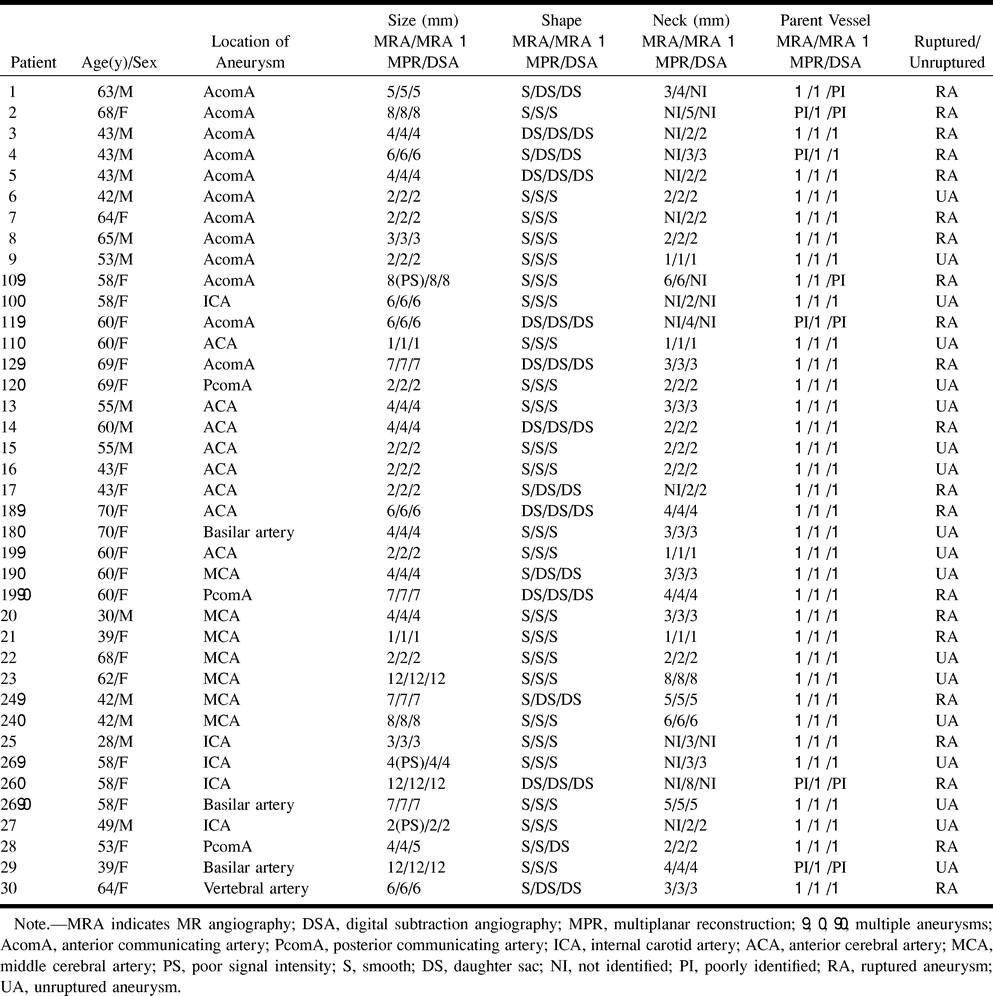
    Findings of MR angiography and DSA in patients with aneurysms

PreviousNext
Back to top

In this issue

American Journal of Neuroradiology
Vol. 20, Issue 2
1 Feb 1999
  • Table of Contents
  • Index by author
Advertisement
Print
Download PDF
Email Article

Thank you for your interest in spreading the word on American Journal of Neuroradiology.

NOTE: We only request your email address so that the person you are recommending the page to knows that you wanted them to see it, and that it is not junk mail. We do not capture any email address.

Enter multiple addresses on separate lines or separate them with commas.
Evaluation of Cerebral Aneurysms with High-Resolution MR Angiography Using a Section-Interpolation Technique: Correlation with Digital Subtraction Angiography
(Your Name) has sent you a message from American Journal of Neuroradiology
(Your Name) thought you would like to see the American Journal of Neuroradiology web site.
CAPTCHA
This question is for testing whether or not you are a human visitor and to prevent automated spam submissions.
Cite this article
Tae-Sub Chung, Jin-Yang Joo, Seung-Koo Lee, Daisy Chien, Gerhard Laub
Evaluation of Cerebral Aneurysms with High-Resolution MR Angiography Using a Section-Interpolation Technique: Correlation with Digital Subtraction Angiography
American Journal of Neuroradiology Feb 1999, 20 (2) 229-235;

Citation Manager Formats

  • BibTeX
  • Bookends
  • EasyBib
  • EndNote (tagged)
  • EndNote 8 (xml)
  • Medlars
  • Mendeley
  • Papers
  • RefWorks Tagged
  • Ref Manager
  • RIS
  • Zotero
0 Responses
Respond to this article
Share
Bookmark this article
Evaluation of Cerebral Aneurysms with High-Resolution MR Angiography Using a Section-Interpolation Technique: Correlation with Digital Subtraction Angiography
Tae-Sub Chung, Jin-Yang Joo, Seung-Koo Lee, Daisy Chien, Gerhard Laub
American Journal of Neuroradiology Feb 1999, 20 (2) 229-235;
del.icio.us logo Twitter logo Facebook logo Mendeley logo
  • Tweet Widget
  • Facebook Like
  • Google Plus One

Jump to section

  • Article
    • Abstract
    • Methods
    • Results
    • Discussion
    • Conclusion
    • Footnotes
    • References
  • Figures & Data
  • Info & Metrics
  • Responses
  • References
  • PDF

Related Articles

  • No related articles found.
  • PubMed
  • Google Scholar

Cited By...

  • Comparison of High-Resolution MR Imaging and Digital Subtraction Angiography for the Characterization and Diagnosis of Intracranial Artery Disease
  • Risk Factors Associated With the Presence of Unruptured Intracranial Aneurysms
  • 3D Computerized Occlusion Rating of Embolized Experimental Aneurysms Using Noninvasive 1.5T MR Imaging
  • Complex Bilobular, Bisaccular, and Broad-Neck Microsurgical Aneurysm Formation in the Rabbit Bifurcation Model for the Study of Upcoming Endovascular Techniques
  • Intracranial Aneurysm Enlargement on Serial Magnetic Resonance Angiography: Frequency and Risk Factors
  • Improved Image Quality of Intracranial Aneurysms: 3.0-T versus 1.5-T Time-of-Flight MR Angiography
  • Current theory in imaging of intracranial vascular disease
  • Crossref
  • Google Scholar

This article has not yet been cited by articles in journals that are participating in Crossref Cited-by Linking.

More in this TOC Section

  • Usefulness of Quantitative Susceptibility Mapping for the Diagnosis of Parkinson Disease
  • White Matter Alterations in the Brains of Patients with Active, Remitted, and Cured Cushing Syndrome: A DTI Study
  • Qualitative and Quantitative Analysis of MR Imaging Findings in Patients with Middle Cerebral Artery Stroke Implanted with Mesenchymal Stem Cells
Show more Brain

Similar Articles

Advertisement

Indexed Content

  • Current Issue
  • Accepted Manuscripts
  • Article Preview
  • Past Issues
  • Editorials
  • Editor's Choice
  • Fellows' Journal Club
  • Letters to the Editor
  • Video Articles

Cases

  • Case Collection
  • Archive - Case of the Week
  • Archive - Case of the Month
  • Archive - Classic Case

More from AJNR

  • Trainee Corner
  • Imaging Protocols
  • MRI Safety Corner
  • Book Reviews

Multimedia

  • AJNR Podcasts
  • AJNR Scantastics

Resources

  • Turnaround Time
  • Submit a Manuscript
  • Submit a Video Article
  • Submit an eLetter to the Editor/Response
  • Manuscript Submission Guidelines
  • Statistical Tips
  • Fast Publishing of Accepted Manuscripts
  • Graphical Abstract Preparation
  • Imaging Protocol Submission
  • Evidence-Based Medicine Level Guide
  • Publishing Checklists
  • Author Policies
  • Become a Reviewer/Academy of Reviewers
  • News and Updates

About Us

  • About AJNR
  • Editorial Board
  • Editorial Board Alumni
  • Alerts
  • Permissions
  • Not an AJNR Subscriber? Join Now
  • Advertise with Us
  • Librarian Resources
  • Feedback
  • Terms and Conditions
  • AJNR Editorial Board Alumni

American Society of Neuroradiology

  • Not an ASNR Member? Join Now

© 2025 by the American Society of Neuroradiology All rights, including for text and data mining, AI training, and similar technologies, are reserved.
Print ISSN: 0195-6108 Online ISSN: 1936-959X

Powered by HighWire