
Article Figures & Data
Figures
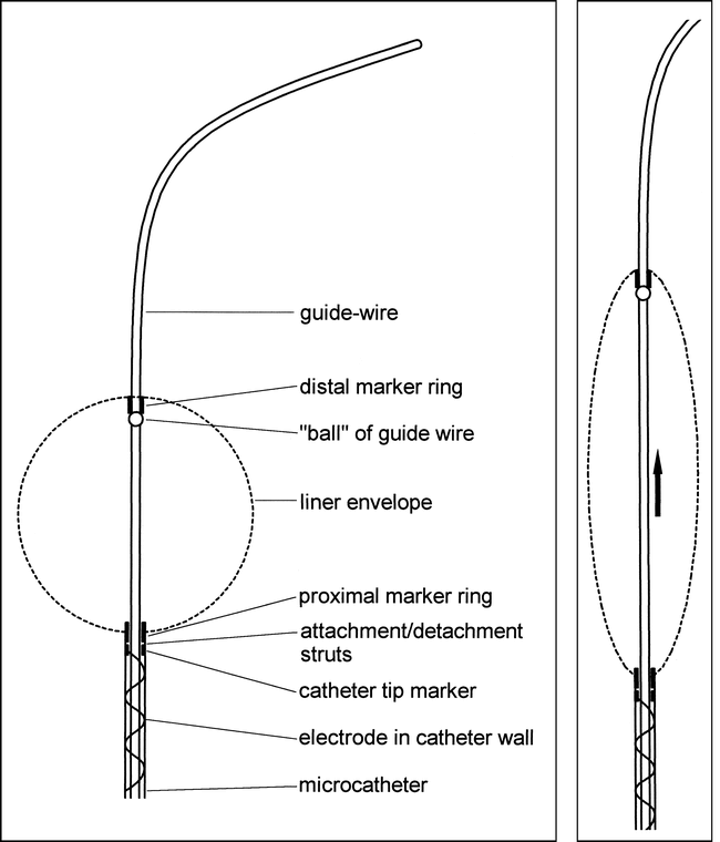
- fig 1.
Line diagrams of the liner, catheter, and guidewire combination.
A, The liner is a bag of highly porous, strong, stretchy fabric detachably mounted on the tip of a microcatheter. It has proximal and distal marker rings, allowing the passage of a guidewire. The catheter has an electrical contact close to its hub and a conducting wire incorporated into its braiding. The proximal marker ring is attached to the catheter tip marker ring by two thin stainless steel struts. It is detached by electrolysis of these struts.
B, The “ball” of the guidewire is too large to pass through the distal marker ring. When the guidewire is advanced in the catheter, the ball engages with the distal ring and stretches the liner into a long, thin configuration to facilitate endovascular navigation.
- fig 2.
Prototype liner, catheter, guidewire combination with electrolytically detachable catheter tip. The detachment zone is shown (small arrow). A conducting wire is incorporated into the catheter braiding. A stainless-steel contact is shown just distal to its hub (large arrow)
- fig 3.
In vitro experiment. The liner has been deployed in a model wide-necked side-wall aneurysm and filled with three GDCs. The liner has retained the coils in the neck. It remains attached to its delivery catheter.
fig 4. The liner, containing three coils, was retrieved from the in vitro aneurysm model. The first few millimeters of the first coil have penetrated the liner but the remaining coils are contained. The distal marker ring of the liner is visible as a dark dot in the upper third of the specimen.
- fig 5.
Angiogram shows the appearance of the side-wall venous-pouch aneurysm in a pig 21 days after embolization with a liner and one GDC. The appearances are consistent with complete obliteration of the aneurysm with healing of the neck. The distal marker ring of the liner can be seen as a dot to the left of the coil mass.
fig 6. Gross specimen of the venous-pouch side-wall aneurysm. (The specimens are supported, for photography, by collars of modeling clay.)
A, The carotid artery has been opened to show the healed neck of the aneurysm represented by a small dimple (arrow).
B, The aneurysm has been sectioned longitudinally to show peripheral granulation tissue (arrow) and central unresolved blood clot within the interstices of the GDC. The liner material cannot be seen in the gross specimen.
- fig 7.
Wide-necked dog carotid bifurcation aneurysm, measuring 24 mm in length and 11 mm in width, occluded with one aneurysm liner filled with two GDCs. The coils are well contained within the neck. The prototype liner detached from the catheter prematurely, precluding tighter coil packing
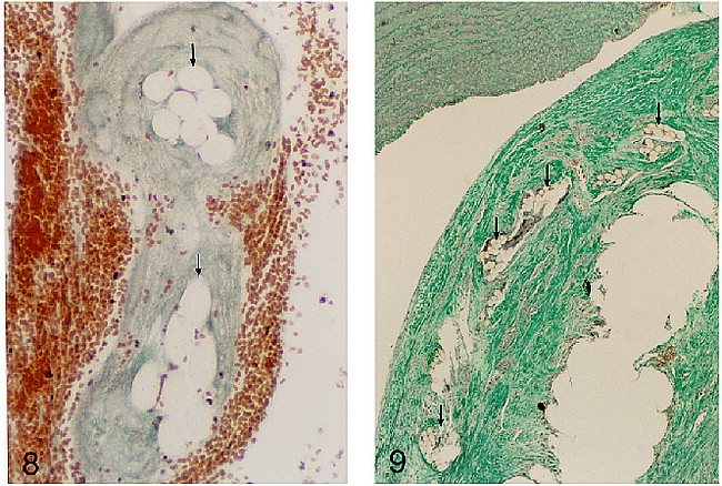
- fig 8.
Microscopic appearance of the threads of an aneurysm liner, inserted into a pig aneurysm and harvested within 3 hours of insertion. The threads (arrows) are surrounded by fibrin, which bridges the spaces between threads and probably forms a substrate for subsequent fibrosis (Masson trichrome, original magnification approximately ×400).
fig 9. Microscopic appearance of the neck region of an aneurysm in a dog 30 days after insertion of an aneurysm liner and GDCs. The specimen was sectioned after removal of the GDCs and the resulting vacuole is in the right lower corner. Fibrous tissue surrounds the site of the coil. Sectioned threads of the liner material (arrows) are shown, overlain by a thin continuous layer of fibrous tissue, separating the liner from the luminal surface. There is a continuous layer of flattened cells lining the neck of the aneurysm. The parent arterial wall is seen at the top of this image (Masson trichrome, original magnification approximately ×90).


