Advertisement

Main menu

  • Home
  • Content
    • Current Issue
    • Accepted Manuscripts
    • Article Preview
    • Past Issue Archive
    • AJNR Case Collection
    • Case of the Week Archive
    • Classic Case Archive
    • Case of the Month Archive
  • Special Collections
    • Spinal CSF Leak Articles (Jan 2020-June 2024)
    • 2024 AJNR Journal Awards
    • Most Impactful AJNR Articles
  • Multimedia
    • AJNR Podcast
    • AJNR Scantastics
    • Video Articles
  • For Authors
    • Submit a Manuscript
    • Author Policies
    • Fast publishing of Accepted Manuscripts
    • Graphical Abstract Preparation
    • Manuscript Submission Guidelines
    • Imaging Protocol Submission
    • Submit a Case for the Case Collection
  • About Us
    • About AJNR
    • Editorial Board
  • More
    • Become a Reviewer/Academy of Reviewers
    • Subscribers
    • Permissions
    • Alerts
    • Feedback
    • Advertisers
    • ASNR Home
  • Other Publications
    • ajnr

User menu

  • Alerts
  • Log in

Search

  • Advanced search
American Journal of Neuroradiology

American Journal of Neuroradiology

ASHNR American Society of Functional Neuroradiology ASHNR American Society of Pediatric Neuroradiology ASSR
  • Alerts
  • Log in

Advanced Search

  • Home
  • Content
    • Current Issue
    • Accepted Manuscripts
    • Article Preview
    • Past Issue Archive
    • AJNR Case Collection
    • Case of the Week Archive
    • Classic Case Archive
    • Case of the Month Archive
  • Special Collections
    • Spinal CSF Leak Articles (Jan 2020-June 2024)
    • 2024 AJNR Journal Awards
    • Most Impactful AJNR Articles
  • Multimedia
    • AJNR Podcast
    • AJNR Scantastics
    • Video Articles
  • For Authors
    • Submit a Manuscript
    • Author Policies
    • Fast publishing of Accepted Manuscripts
    • Graphical Abstract Preparation
    • Manuscript Submission Guidelines
    • Imaging Protocol Submission
    • Submit a Case for the Case Collection
  • About Us
    • About AJNR
    • Editorial Board
  • More
    • Become a Reviewer/Academy of Reviewers
    • Subscribers
    • Permissions
    • Alerts
    • Feedback
    • Advertisers
    • ASNR Home
  • Follow AJNR on Twitter
  • Visit AJNR on Facebook
  • Follow AJNR on Instagram
  • Join AJNR on LinkedIn
  • RSS Feeds

Welcome to the new AJNR, Updated Hall of Fame, and more. Read the full announcements.


AJNR is seeking candidates for the position of Associate Section Editor, AJNR Case Collection. Read the full announcement.


Research ArticleBRAIN

Tagged MR Imaging of Intracranial Aneurysm Models

Haruo Isoda, Yasutomi Kinosada, Satoshi Isogai, Yasuo Takehara and Tatsuhiko Ito
American Journal of Neuroradiology May 1999, 20 (5) 807-811;
Haruo Isoda
  • Find this author on Google Scholar
  • Find this author on PubMed
  • Search for this author on this site
Yasutomi Kinosada
  • Find this author on Google Scholar
  • Find this author on PubMed
  • Search for this author on this site
Satoshi Isogai
  • Find this author on Google Scholar
  • Find this author on PubMed
  • Search for this author on this site
Yasuo Takehara
  • Find this author on Google Scholar
  • Find this author on PubMed
  • Search for this author on this site
Tatsuhiko Ito
  • Find this author on Google Scholar
  • Find this author on PubMed
  • Search for this author on this site
  • Article
  • Figures & Data
  • Info & Metrics
  • Responses
  • References
  • PDF
Loading

Abstract

Summary: We used tagged MR imaging to investigate the flow in two lateral and two terminal saccular intracranial aneurysm models of different neck sizes. Imaging was performed with a 1.5-T superconducting MR system using 2D fast spatial modulation of magnetization (SPAMM) sequences with an intersegmental delay of 25 milliseconds. The flow in the saccular aneurysm models varied with the shape and size of the neck: flow was faster in wider-necked aneurysms than in those with narrower necks.

MR angiography is widely used in the evaluation of intracranial abnormalities, including intracranial aneurysms (1, 2). However, MR angiography sometimes fails to depict intracranial aneurysms. The main reason for decreased signal in aneurysms is thought to be slow or disturbed flow (3, 4). Previous work by Isoda et al (4) has documented the signal intensity in models of lateral and terminal saccular aneurysms with different neck sizes using 3D time-of-flight (TOF) MR angiography with various imaging parameters. In that study, noncontrast 3D TOF MR angiography showed terminal saccular aneurysms, or aneurysms with wider necks, or both better than it did lateral saccular aneurysms, aneurysms with narrower necks or both. Flow dynamics have also been presumed to have a significant effect on the signal intensity of aneurysms.

The purpose of this study was to use tagged MR imaging to depict the flow in lateral and terminal saccular intracranial aneurysm models of different neck sizes.

Methods

Imaging was performed using a 1.5-T superconducting MR system with a commercially available head coil. Teflon tubes with inside diameters of 10 mm were used as models for two lateral and two terminal saccular aneurysms with pulsatile flow. Their height and fundus diameters were 10 mm and their neck diameters were 2.5 mm and 10 mm, respectively (Fig 1). Pulsatile flow was created with a bellows pump and a constant flow pump with a pulsation period of 1500 milliseconds. Tap water was used as fluid in a closed-circuit system. After adjusting the flow volume of each pump, we measured the flow velocity and waveform with 2D phase mapping MR imaging defined by the following parameters: 40/7.9/2 (TR/TE/excitations); flip angle, 20°; field of view, 20 cm; section thickness, 5 mm; matrix, 256 × 128; number of phases, 16; imaging time, 6 minutes 26 seconds. The sequence was gated from the phantom trigger. Peak systolic velocity was 120 cm/s, and end-diastolic velocity was 40 cm/s (Fig 2).

fig 1.
  • Download figure
  • Open in new tab
  • Download powerpoint
fig 1.

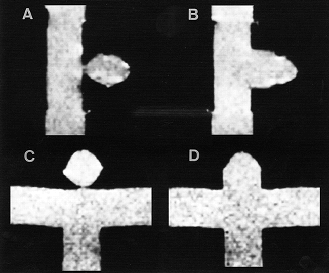
A–D, Spin-echo T1-weighted images (300/15/1) of lateral and terminal saccular aneurysm models with fluid but without active flow. The height and fundus diameter of the models were 10 mm; the neck diameters were 2.5 mm (A, C) and 10.0 mm (B, D).

fig 2.
  • Download figure
  • Open in new tab
  • Download powerpoint
fig 2.

Flow velocity during cardiac cycle.

Tagged MR imaging was performed using 2D fast spatial modulation of magnetization (SPAMM) sequences (5) with the following parameters: 25/5/1; flip angle, 20 °; bandwidth, 16 kHz; field of view, 20 to 26 cm; section thickness, 7 mm; matrix, 256 × 160 to 192; imaging time, 4 minutes 6 seconds to 4 minutes 54 seconds; tag space, 5 mm; intersegmental delay, 25 milliseconds; number of phases, 53. The sequence was gated from the phantom trigger. Tag stripes were placed in a transaxial direction to the parent vessel models for each aneurysm model.

Results

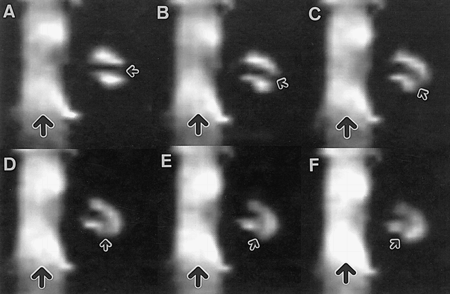
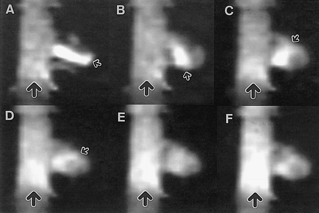
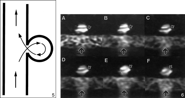
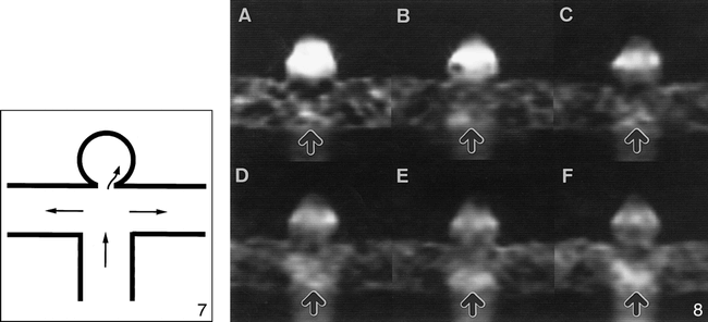
Tag stripes or a high-signal band between tags in the lateral saccular aneurysm models rotated like a waterwheel rotates in a stream (Figs 3–5⇓⇓). Tag stripes in the lateral saccular aneurysm model with a neck size of 10 mm rotated faster than those in the aneurysm model with a neck size of 2.5 mm (Figs 3 and 4). Although a tag stripe in the terminal saccular aneurysm model with a neck size of 2.5 mm rotated slowly (Figs 6 and 7), no distinct tag stripe in the model with a neck size of 10 mm was recognized (Fig 8), probably because of the very fast flow. Tag stripes in the terminal saccular aneurysm models with a neck size of 10 mm rotated faster than those in the lateral saccular aneurysm models with a neck size of 10 mm (Figs 4 and 8).

fig 3.
  • Download figure
  • Open in new tab
  • Download powerpoint
fig 3.

Tagged MR images of the lateral saccular aneurysm model with a neck size of 2.5 mm.

A–F, The time difference between images is 25 milliseconds (large arrows indicate the flow direction of the parent vessel; small arrows, the tag stripe).

fig 4.
  • Download figure
  • Open in new tab
  • Download powerpoint
fig 4.

Tagged MR images of the lateral saccular aneurysm model with a neck size of 10 mm.

A–F, The time difference between images is 25 milliseconds (large arrows indicate the flow direction of the parent vessel; small arrows, the high signal band between tag stripes).

fig 5.
  • Download figure
  • Open in new tab
  • Download powerpoint
fig 5.

Diagram of circulation in lateral saccular aneurysm models.fig 6. Tagged MR images of the terminal saccular aneurysm model with a neck size of 2.5 mm.

A–F, The time difference between images is 25 milliseconds (large arrows indicate the flow direction of the parent vessel; small arrows, the tag stripe).

fig 7.
  • Download figure
  • Open in new tab
  • Download powerpoint
fig 7.

Diagram of circulation in a terminal saccular aneurysm model with a neck size of 2.5 mm.fig 8. Tagged MR images of the terminal saccular aneurysm model with a neck size of 10 mm.

A–F, The time difference between images is 25 milliseconds (arrows indicate the flow direction of the parent vessel model).

Discussion

Previously reported methods for visualizing aneurysmal flow include conventional angiography (6), sonography (6), dye injection (7, 8), double refraction (8), laser-induced fluorescence (9), and computer simulation (10–12). As far as we know, tagged MR imaging has not been used in the evaluation of intracranial aneurysm models. We found this method useful in evaluating both flow rate and flow direction in the aneurysm model.

Intraaneurysmal flow dynamics are thought to play an important role in developing intracranial aneurysms and in their rupture mechanisms (7). We expect that the technique of tagged MR imaging will be applied to humans for the evaluation of intracranial aneurysms, leading to further information about the mechanism of aneurysmal development and rupture.

In this study, tag lines moved more rapidly in the terminal saccular aneurysm models or models with wider necks than in the lateral saccular aneurysm models or models with narrower necks. These findings are thought to reflect the fact that the flow in the terminal saccular aneurysm models or models with wider necks was faster than the flow in the lateral saccular aneurysm models or models with narrower necks. On the basis of the findings in this and a previous study by Isoda et al (4) using 3D TOF MR angiography, this angiographic technique better delineates the aneurysm models with a faster flow than those with slower flow.

Limitations of the study were that our intracranial models were hard, and that the section direction of tagged MR images was perpendicular to the main parent vessels. Real aneurysms are elastic, and aneurysmal walls are expected to move according to pulsation during the cardiac cycle. Therefore, there may be some differences in flow between our hard-walled intracranial aneurysm models and real intracranial aneurysms, especially in the case of terminal saccular aneurysms. As the section direction of the tagged MR images used in this study was unidirectional, the flow rate and direction were only partly revealed. However, from our data we were able to successfully estimate flow characteristics in the various aneurysm models.

Conclusion

Tagged MR imaging was found to be useful in evaluating the flow in models of intracranial aneurysms. Flow in the aneurysm models varied with the shape and size of the neck. The flow in the saccular aneurysm models with wider necks was faster than that in those with narrower necks.

Footnotes

  • ↵1 Presented at the annual meeting of the American Society of Neuroradiology, Philadelphia, May 1998.

  • ↵2 Address reprint requests to Haruo Isoda, MD, Department of Radiology, Hamamatsu University School of Medicine, 3600 Handa-cho, Hamamatsu, 431–3192 Japan.

References

  1. ↵
    Korogi Y, Takahashi M, Mabuchi N, et al. Intracranial aneurysms: diagnostic accuracy of three-dimensional, Fourier transform, time-of-flight MR angiography. Radiology 1994;193:181-186
    PubMed
  2. Huston J III, Nichols DA, Luetmer PH, et al. Blinded prospective evaluation of sensitivity of MR angiography to known intracranial aneurysms: importance of aneurysm size. AJNR Am J Neuroradiol 1994;15:1607-1614
    Abstract/FREE Full Text
  3. ↵
    Bosmans H, Wilms G, Marchal G, Demaerel P, Baert AL. Characterization of intracranial aneurysms with MR angiography. Neuroradiology 1995;37:262-266
    PubMed
  4. ↵
    Isoda H, Ramsey RG, Takehara Y, Takahashi M, Kaneko M. MR angiography of aneurysm models of various shapes and neck sizes. AJNR Am J Neuroradiol 1997;18:1463-1472
    Abstract
  5. ↵
    Wayte SC, Redpath TW. Cine magnetic resonance imaging of pulsatile cerebrospinal fluid flow using CSPAMM. Br J Radiol 1994;67:1088-1095
    Abstract/FREE Full Text
  6. ↵
    Strother CM, Graves VB, Rappe A. Aneurysm hemodynamics: an experimental study. AJNR Am J Neuroradiol 1992;13:1089-1095
    Abstract/FREE Full Text
  7. ↵
    Kerber CW, Hecht ST, Knox K, Buxton RB, Meltzer HS. Flow dynamics in a fatal aneurysm of the basilar artery. AJNR Am J Neuroradiol 1996;17:1417-1421
    Abstract
  8. ↵
    Steiger HJ, Poll A, Liepsch D, Reulen H-J. Basic flow structure in saccular aneurysms: a flow visualization study. Heart Vessels 1987;3:55-65
    CrossRefPubMed
  9. ↵
    Lieber BB, Stancampiano AP, Wakhloo AK. Alteration of hemodynamics in aneurysm models by stenting: influence of stent porosity. Ann Biomed Eng 1997;25:460-469
    CrossRefPubMed
  10. ↵
    Perktold K, Gruber K, Kenner T, Florian H. Calculation of pulsatile flow and particle paths in an aneurysm-model. Basic Res Cardiol 1984;79:253-261
    CrossRefPubMed
  11. Perktold K, Kenner T, Hilbert D, Spork B, Florian H. Numerical blood flow analysis: arterial bifurcation with a saccular aneurysm. Basic Res Cardiol 1988;83:24-31
    CrossRefPubMed
  12. Gonzalez CF, Cho YI, Ortega HV, Moret J. Intracranial aneurysms: flow analysis of their origin and progression. AJNR Am J Neuroradiol 1992;13:181-188
    Abstract/FREE Full Text
  • Received August 7, 1998.
  • Accepted after revision January 7, 1999.
  • Copyright © American Society of Neuroradiology
PreviousNext
Back to top

In this issue

American Journal of Neuroradiology
Vol. 20, Issue 5
1 May 1999
  • Table of Contents
  • Index by author
Advertisement
Print
Download PDF
Email Article

Thank you for your interest in spreading the word on American Journal of Neuroradiology.

NOTE: We only request your email address so that the person you are recommending the page to knows that you wanted them to see it, and that it is not junk mail. We do not capture any email address.

Enter multiple addresses on separate lines or separate them with commas.
Tagged MR Imaging of Intracranial Aneurysm Models
(Your Name) has sent you a message from American Journal of Neuroradiology
(Your Name) thought you would like to see the American Journal of Neuroradiology web site.
CAPTCHA
This question is for testing whether or not you are a human visitor and to prevent automated spam submissions.
Cite this article
Haruo Isoda, Yasutomi Kinosada, Satoshi Isogai, Yasuo Takehara, Tatsuhiko Ito
Tagged MR Imaging of Intracranial Aneurysm Models
American Journal of Neuroradiology May 1999, 20 (5) 807-811;

Citation Manager Formats

  • BibTeX
  • Bookends
  • EasyBib
  • EndNote (tagged)
  • EndNote 8 (xml)
  • Medlars
  • Mendeley
  • Papers
  • RefWorks Tagged
  • Ref Manager
  • RIS
  • Zotero
0 Responses
Respond to this article
Share
Bookmark this article
Tagged MR Imaging of Intracranial Aneurysm Models
Haruo Isoda, Yasutomi Kinosada, Satoshi Isogai, Yasuo Takehara, Tatsuhiko Ito
American Journal of Neuroradiology May 1999, 20 (5) 807-811;
del.icio.us logo Twitter logo Facebook logo Mendeley logo
  • Tweet Widget
  • Facebook Like
  • Google Plus One

Jump to section

  • Article
    • Abstract
    • Methods
    • Results
    • Discussion
    • Conclusion
    • Footnotes
    • References
  • Figures & Data
  • Info & Metrics
  • Responses
  • References
  • PDF

Related Articles

  • No related articles found.
  • PubMed
  • Google Scholar

Cited By...

  • Preliminary Study of Tagged MR Image Velocimetry in a Replica of an Intracranial Aneurysm
  • Crossref
  • Google Scholar

This article has not yet been cited by articles in journals that are participating in Crossref Cited-by Linking.

More in this TOC Section

  • Evaluating the Effects of White Matter Multiple Sclerosis Lesions on the Volume Estimation of 6 Brain Tissue Segmentation Methods
  • White Matter Alterations in the Brains of Patients with Active, Remitted, and Cured Cushing Syndrome: A DTI Study
  • Enhanced Axonal Metabolism during Early Natalizumab Treatment in Relapsing-Remitting Multiple Sclerosis
Show more Brain

Similar Articles

Advertisement

Indexed Content

  • Current Issue
  • Accepted Manuscripts
  • Article Preview
  • Past Issues
  • Editorials
  • Editors Choice
  • Fellow Journal Club
  • Letters to the Editor

Cases

  • Case Collection
  • Archive - Case of the Week
  • Archive - Case of the Month
  • Archive - Classic Case

Special Collections

  • Special Collections

Resources

  • News and Updates
  • Turn around Times
  • Submit a Manuscript
  • Author Policies
  • Manuscript Submission Guidelines
  • Evidence-Based Medicine Level Guide
  • Publishing Checklists
  • Graphical Abstract Preparation
  • Imaging Protocol Submission
  • Submit a Case
  • Become a Reviewer/Academy of Reviewers
  • Get Peer Review Credit from Publons

Multimedia

  • AJNR Podcast
  • AJNR SCANtastic
  • Video Articles

About Us

  • About AJNR
  • Editorial Board
  • Not an AJNR Subscriber? Join Now
  • Alerts
  • Feedback
  • Advertise with us
  • Librarian Resources
  • Permissions
  • Terms and Conditions

American Society of Neuroradiology

  • Not an ASNR Member? Join Now

© 2025 by the American Society of Neuroradiology All rights, including for text and data mining, AI training, and similar technologies, are reserved.
Print ISSN: 0195-6108 Online ISSN: 1936-959X

Powered by HighWire