
Article Figures & Data
Figures
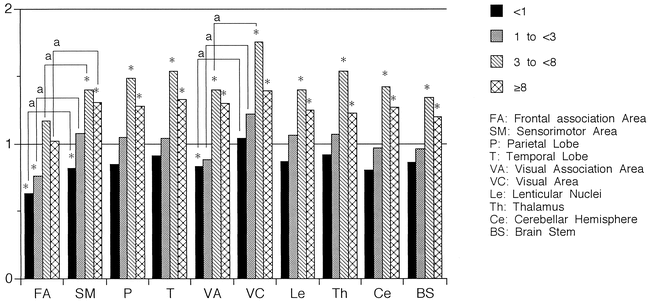
- fig 1.
Ratio of rCBF values to those in adults distributed by age. rCBF values increased with development in all areas. The last increase was observed in the frontal association area. Compared with rCBF values in adults, low values were observed in the frontal and visual association areas and the sensorimotor area in the <1-year-old group, and in the frontal association area in the 1- to <3-year-old group, whereas rCBF values were significantly increased in all areas in the 3- to <8-year-old group, except for the frontal association area. rCBF values were always lower in the association areas than in the primary areas, except for those in the visual association area and the visual area in the ≥8-year-old group. Asterisk indicates Mann-Whitney U-test, P < .05; a, paired t-test, P < .05
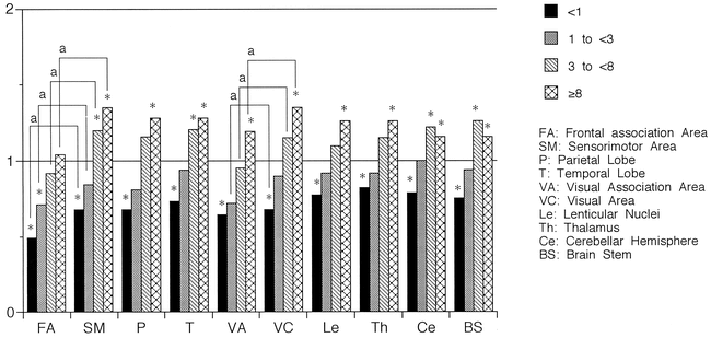
- fig 2.
The ratio of rCMRO2 values to those in adults distributed by age. rCMRO2 values increased with development in all areas but the ratio of increase was smaller than that of rCBF. The last increase was observed in the frontal association area. The ratio increased to more than one at a later age compared with that of rCBF. Compared with rCMRO2 values in adults, low values were observed in all areas in the <1-year-old age group and in the frontal association area in the 1- to <3-year-old group, whereas rCMRO2 values were significantly increased in the sensorimotor area, visual area, temporal lobe, cerebellar cortex, and brain stem in the 3- to <8-year-old group and in all areas in the ≥8-year-old group, except for the frontal association area. rCMRO2 values were always lower in the association areas than in the primary areas, except for those in the visual association area and the visual area in the <1-year-old group. Asterisk indicates Mann-Whitney U-test, P < .05; a, paired t-test, P < .05
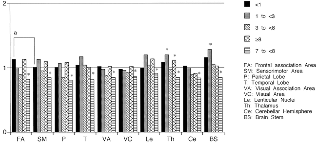
- fig 3.
The ratio of rOEF values to those in adults distributed by age. Regardless of age, rOEF values were within or nearly in the range of adult values. Compared with rOEF values in adults, statistically significant differences were observed only in the thalamus and brain stem in the 1- to <3-year-old age group, in the parietal lobe in the 3- to <8-year-old group, and in the thalamus in the ≥8-year-old group. rOEF values of the primary cerebral areas did not differ from those of the association areas in any age group, except in the frontal association and sensorimotor areas in the <1-year-old group. Asterisk indicates Mann-Whitney U-test, P < .05; a, paired t-test, P < .05


Tables

Summary of 30 PET studies in children 10 days to 16 years old

In this issue


Jump to section
Related Articles
Cited By...
- Macrovascular blood flow and microvascular cerebrovascular reactivity are regionally coupled in adolescence
- Regional patterns of human cortex development correlate with underlying neurobiology
- Resting-State Functional MRI and PET Imaging as Noninvasive Tools to Study (Ab)Normal Neurodevelopment in Humans and Rodents
- Cerebral Blood Flow Increases Across Early Childhood
- Impact of puberty on the evolution of cerebral perfusion during adolescence
- Gene expression-based modeling of human cortical synaptic density
- Resolving the transition from negative to positive blood oxygen level-dependent responses in the developing brain
- Posterior Circulation and High Prevalence of Ischemic Stroke among Young Pediatric Patients with Moyamoya Disease: Evidence of Angiography-Based Differences by Age at Diagnosis
- Cerebral Blood Flow Measurement in Children With Sickle Cell Disease Using Continuous Arterial Spin Labeling at 3.0-Tesla MRI
- Developmental Changes in Cerebral and Visceral Blood Flow Velocity in Healthy Neonates and Infants