
Article Figures & Data
Figures
- fig 1.
Schematic diagram of aneurysm measurements.
A, The dimensions measured on the angiographic tracings are the neck width (N), the dome diameter (D), the dome height (H), and the dome semi-axis height (S).
B, For BB aneurysms, the angle, β, at which the dome tilts with respect to the parent vessel is measured.
- fig 2.
The effect of D/H and H/S ratios on general shape.
A, For a D/H ratio value less than 1, the observed shape is an ellipse, with the major axis in the vertical direction; for a value of 1, a circle; and for values greater than 1, an ellipse, with the major axis in the horizontal direction.
B, For an H/S value less than 2, the aneurysm has a pear shape; for a value of 2, a circle; and for a value greater than 2, a beehive shape.
- fig 3.
Sample angiogram with markers. For estimating true in vivo size, the marker diameters (black arrows) were measured to determine the magnification factor. The diameter of the internal carotid was then measured (white arrow) and divided by the magnification factor in order to obtain the absolute in vivo size
- fig 4.
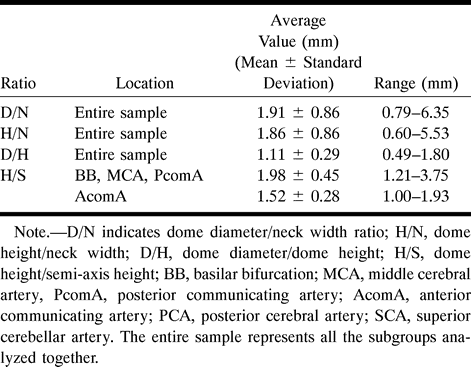
Distribution of ratios.
A, Distribution of dome diameter/dome height (D/H) for all locations considered.
B, Distribution of dome height/semi-axis height (H/S) for aneurysms located on the BB, MCA, and PcomA.
C, Distribution of dome height/semi-axis height (H/S) for aneurysms located on the AcomA.
- fig 5.
Distribution of the angle at the BB, β (mean value, 1.6, SD, 23)
- fig 6.
Correlations of absolute in vivo aneurysm dimensions.
A, Dome diameter vs neck width (D vs N); r = .82, D = 1.4 N + 1.7, P < .0001.
B, Dome height vs neck width (H vs N); r = .79, H = 1.3 N + 1.9, P < .0001.
C, Dome height vs dome diameter (H vs D), r = .88, H = 0.8 D + 1.3, P < .0001.
- fig 7.
3D representations of the typical simple-lobed aneurysm.
A, Two views of the aneurysm arising at the BB, MCA, and PcomA.
B, Two views of the aneurysm arising from the AcomA.


Tables
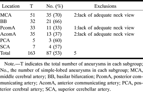
 Table 1:
Summary of the simple-lobed aneurysms available for study, divided into subgroups according to location, and the exclusions made in order to arrive at a sample size of 82
- Table 2:
Summary of the literature values used in calculating the average internal carotid artery diameter
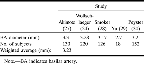
- Table 3:
Summary of the literature values used in calculating the average basilar artery diameter
- Table 5:
Mean values and ranges of absolute sizes of N, D, and H for aneurysms of the MCA, AcomA, PcomA, and BB (mean ± SD)
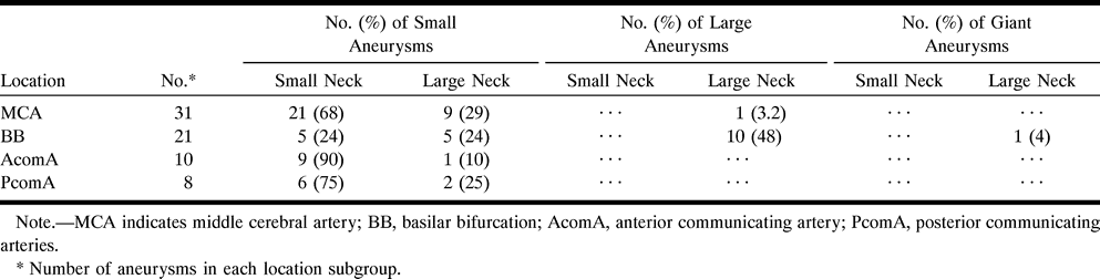
- Table 6:
Summary of aneurysm size classification based on neck width and dome height
- Table 7:
Summary of results for intact and ruptured simple-lobed aneurysms
- Table 8:
Distribution of saccular aneurysms at the MCA, BB, AcomA, PcomA, PCA, and SCA as observed previously and in the present study


In this issue


Jump to section
Related Articles
Cited By...
- Efficacy of Skull Plain Films in Follow-up Evaluation of Cerebral Aneurysms Treated with Detachable Coils: Quantitative Assessment of Coil Mass
- Hemodynamics and Anatomy of Elastase-Induced Rabbit Aneurysm Models: Similarity to Human Cerebral Aneurysms?
- Difficult Aneurysms for Endovascular Treatment: Overwide or Undertall?
- Comparison of 2D Digital Subtraction Angiography and 3D Rotational Angiography in the Evaluation of Dome-to-Neck Ratio
- Elastase-Induced Saccular Aneurysms in Rabbits: Comparison of Geometric Features with Those of Human Aneurysms