
Article Figures & Data
Figures
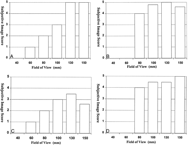
- fig 1.
Histogram of the subjective primary image quality scores (left axis) at varying fields of view. The line plot represented on the right axis shows the mean value for calculated image contrast from six normal data sets
- fig 2.
Samples of primary images from a healthy volunteer obtained using FOVs of 60 mm (left) and 130 mm (right). Note the improvement in S/N ratio with the larger FOV and the separate demonstration of the scala vestibuli (long arrow) and scala tympani (short arrow) in the basal turn of the cochlea
- fig 3.
Histograms show the effect of changing FOV size on the subjective image quality scores for each visualization technique: method 1, MIP (A); method 2, ray casting with transparent voxels (B); method 3, ray casting with opaque voxels (C); and method 4, isosurface rendering (D). Scores are the average of six image sets, each scored by two observers
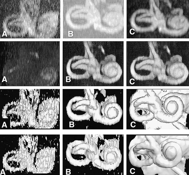
- fig 4.
The effect of increasing S/N ratio on each of the four visualization techniques. Images labeled A (left column) are renderings from studies obtained with an FOV of 60 mm, images labeled B (middle column) are from studies obtained with an FOV of 100 mm, and images labeled C (right column) are from studies obtained with an FOV of 130 mm. Row 1 were rendered using method 1 (MIP), row 2 using method 2 (ray casting with transparent voxels), row 3 using method 3 (ray casting with opaque voxels), and row 4 with method 4 (isosurface rendering). The images show rapid degradation of visualization obtained with methods 1 and 4, as FOV and S/N ratio are reduced. Method 2 continues to produce excellent renderings at an FOV of 100 mm, with no apparent degradation as compared with images acquired at 130 mm
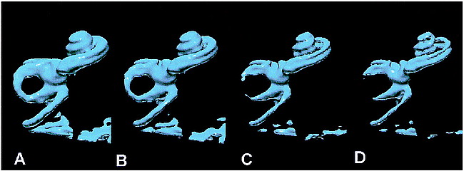
- fig 5.
The results of the optimal combination of imaging and visualization technique in a single healthy subject (5000/250; FOV, 130 mm2; matrix, 2562). All renderings were derived from the same image data set.
A, Method 1: MIP.
B, Method 2: ray casting with transparent voxels.
C, Method 3: ray casting with opaque voxels.
D, Method 4: isosurface rendering (white arrow indicates the posterior semicircular canal; solid black arrow, the lateral semicircular canal; open arrow, the basal turn of the cochlea).
- fig 6.
The effect of decreasing voxel opacity (right to left) using ray tracing. The images are volume renderings of the same normal data set using method 2 (ray casting with transparent voxels). The opacity values of the voxels have been progressively decreased from left to right in order to demonstrate more internal details of the cochlea. The scala vestibuli and scala tympani are clearly seen in the middle and right-hand images, although their margins are rather poorly defined
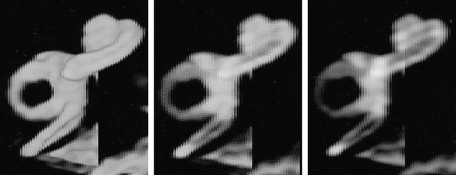
- fig 7.
The effect of increasing the isosurface threshold with isosurface rendering (left to right). The images are isosurface renderings of the same normal data set. As the images pass from right to left, progressively higher threshold values have been applied to extract the isosurface. The isosurface in B corresponds to 65% of the peak signal intensity in the vestibule and is the value used for standardizations of isosurface renderings in the current study. Other renderings were obtained at 55% (A), 75% (B), and 85% (C). The scala vestibuli and scale tympani are clearly and separately depicted throughout their length in C and D.
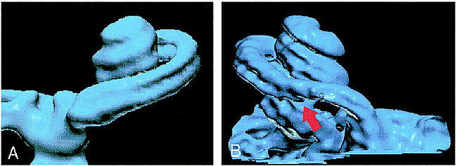
- fig 8.
A and B, Normal (A) and abnormal (B) cochlea are shown for comparison. The scala tympani (arrow, B) is occluded as a sequela of meningitis
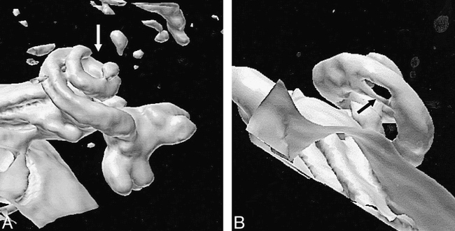
- fig 9.
A and B, Two patients with abnormalities of the cochlea, caused by meningitis. Neither of the abnormalities was considered a contraindication to implantation, which was successfully conducted in both cases.
A, Patient with an occlusion of the terminal portion of the cochlea (arrow).
B, Patient with an abnormality just distal to the basal turn (arrow). Since CT findings were normal, this is assumed to represent a fibrotic narrowing of the cochlear fluid channels.


Tables
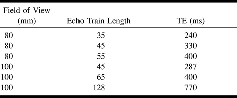
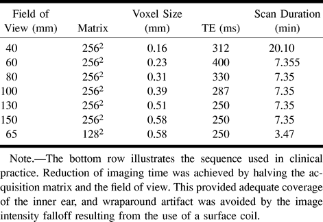
 Table 1:
Imaging parameters used in all high-resolution T2-weighted images


