
Article Figures & Data
Figures
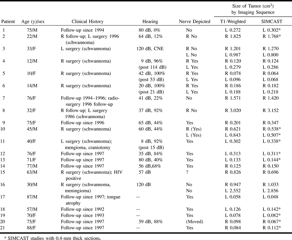
- fig 1.
Case 19: Slightly oblique axial reformatted images of a small mass.
A, SIMCAST image acquired in 5:05 minutes with parameters of 12.3/2.3, 40° flip angle, 22 × 16-cm FOV, 512 × 256 matrix, 32-kHz receiver bandwidth, four RF phase stepping loops (14), and 32 0.8-mm-thick sections interpolated to 0.4 mm.
B, Contrast-enhanced study acquired in 4:07 minutes with parameters of 40/4.2/1, 30° flip angle, identical FOV and matrix size, 15.6-kHz receiver bandwidth, and 32 1.6-mm-thick sections interpolated to 0.8 mm.
Both images (A and B) depict the superior portion of the IAC. On the SIMCAST image (A), a continuous dark line extending from the brain stem to the fundus of the IAC is seen in the anterior portion of the IAC, presumably the facial nerve (solid arrows). Only a portion of the facial nerve is seen on the T1-weighted image (B). With SIMCAST, the tumor mass (open arrow) is clearly seen in the posterior portion of the IAC, displacing the facial nerve anteriorly and leading to a tortuous course around the tumor. Tumor volumes of 0.082 cm3 and 0.078 cm3 were measured on the SIMCAST and T1-weighted images, respectively. The tumor size is 6 mm along the IAC with both sequences, but may appear slightly wider across the IAC on the T1-weighted image than on the SIMCAST image, possibly due to partial volume effects or to tumor involvement of the nerve, leading to signal enhancement. Note that small vessels (dashed arrows) have low signal on the SIMCAST image and high signal on the T1-weighted images, due to the presence of contrast agent in the vessels. With the SIMCAST sequence, using zero-order motion-compensated gradients over TR, no signal loss is observed with slow motion, such as is present in CSF or in the IAC (v < 0.6 cm/s; phantom study by Schmalbrock P, presented at the International Society for Magnetic Resonance in Medicine, 1998). Higher-order motion components are present for faster vascular flow and result in dephasing and signal decrease, accounting for the low vascular signal.
- fig 2.
Case 11.
A and B, Reformatted axial (A) and coronal (B) SIMCAST images acquired in 5:05 minutes with parameters of 12.3/2.3, 40° flip angle, 22 × 16-cm FOV, 512 × 256 matrix, 32-kHz receiver bandwidth, four RF phase stepping loops (14), and 32 0.8-mm-thick axial sections interpolated to 0.4 mm. A was magnified by a factor of three from the original data and reformatted along an axially oriented curved surface from the brain stem to the superior portion of the IAC containing the facial nerve (fn). Only a portion of the superior vestibular nerve (svn) is seen in this view. B was magnified sixfold and reformatted along a coronally oriented curved surface following the facial nerve in the anterior portion of the IAC. Other labeled structures include the pons (pons), the pontomedullary junction (pmj), and the superior aspect of the porus acousticus (spa). The curved views permit continuous depiction of the facial nerve from the brain stem to the fundus of the IAC. On the curved axial image (A) the facial nerve is pushed anteriorly by the tumor (open arrow). On the curved coronal image (B), the nerve courses superiorly to the tumor. There is remodeling of the IAC. In this patient, surgery preserved hearing.
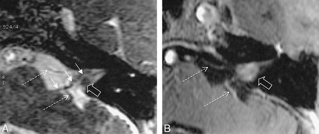
- fig 3.
Case 10: IAC mass (right side) with a tumor volume of 0.6 cm3.
A–D, On SIMCAST images (A and C), acquired in 5:05 minutes with parameters of 12.3/2.3, 40° flip angle, 22 × 16-cm FOV, 512 × 256 matrix, 32-kHz receiver bandwidth, four RF phase stepping loops (14), and 32 0.8-mm-thick sections interpolated to 0.4 mm, the tumor is isointense with brain tissue; nevertheless its boundary may be estimated by using vascular structures (arrows) as reference points. On contrast-enhanced images (B and D), acquired in 5:20 minutes with parameters of 30/4.2/1, 30° flip angle, 20 × 20-cm FOV, 512 × 288 matrix, 15.6-kHz receiver bandwidth, and 60 1.5-mm-thick sections, the hyperintense tumor is easily distinguishable from the adjacent flocculus (fl) and cerebral peduncle.


