
Article Figures & Data
Figures
- fig 1.
Coronal neurosonogram of a newborn term baby with duodenal atresia. The echogenic surface of a frontal gyrus (arrow) and the width of a sulcus (arrowhead) are measured
- fig 2.
Coronal neurosonogram of a healthy 4-week-old baby shows a frontal sulcus of 2 mm on the left (arrows); however, on the right, a faulty measurement of 5.6 mm (asterisk) would have been obtained if the measurement criteria described in the text were not respected
- fig 3.
A and B, Histograms of observed distribution of the readings of single measurements (surface, A) and double measurements (sulci, B) with the expected normal distribution computed with the SD around the mean. The normal, symmetrical, expected distribution does not satisfactorily fit the observed data of the single and double measurements. Kolmogorov-Smirnov test: D = .15094 for single measurements, and D = .15830 for double measurements; P < .05 for both measurements. Lilliefors test: P < .01 for both measurements. All tests were significant, demonstrating an insufficient goodness-of-fit of the expected normal distribution with the observed frequencies
- fig 4.
Scatterplot of single and double measurements, with third-degree polynomial regression and its 95% confidence limits and an ellipse of the 95% confidence limits around the common mean of both measurements. A number of readings are outside the 95% confidence limits. The scales of the measurements differ for graphic reasons. The correlation coefficient is statistically significant (r = .435, P < .001). Most outliers are located at the higher end of the measurements
- fig 5.
Histogram of the cumulated frequencies of the surface and sulci measurements on the same scale. Because the number of subjects is 100, the cumulated frequencies are equivalent to percentile distribution. For illustrative purposes, the 5th and 95th percentiles are delineated. The median is equal to the 50th percentile. Smoothing of the graph can be done by hand by joining the center of each consecutive column of cumulated frequency
- fig 6.
Coronal sonogram of a 3½-week-old infant with pneumococcal meningitis. The surface meninges are of normal thickness (0.8 mm). The subarachnoid space, the interhemispheric fissure, and the sulci are filled with echogenic material (pus and proteins by lumbar puncture). The frontal sulcus is enlarged (2.4 mm) and its undersurface is poorly defined


Tables

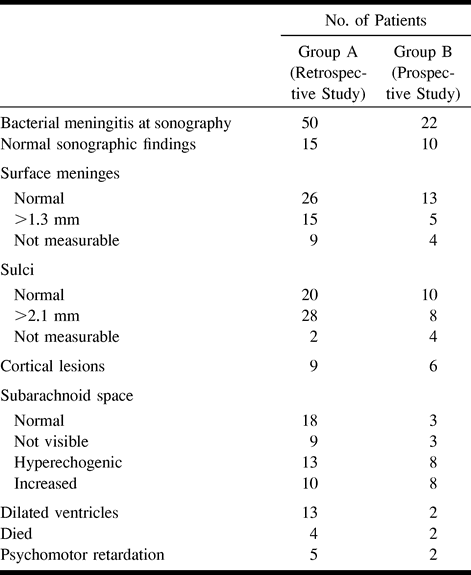
- TABLE 2:
Sonographic findings (and clinical outcome) in infants with bacterial meningitis


