
Article Figures & Data
Figures
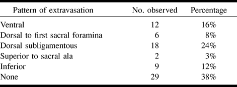
- fig 1.
“Normal” right SIJ arthrograms of a 32-year-old woman with right-sided lower back and hip pain after a motor vehicle accident.
A, Anteroposterior view. Note the characteristic coin-shaped inferior recess (arrowhead) and the bead of contrast within the joint margin (closed arrow). Symbols: S, sacrum; IL, ilium; 3, S3 dorsal sacral foramina.
B, Right anterior oblique projection delineates the full extent of the contrast medium within the joint space (wavy arrow). The closed arrow is directed to the needle tip.
C, Left anterior oblique view. This “en face” projection reveals the auricular configuration of the SIJ surface (dark arrows). Symbols: 4, pedicle of L4; 5, pedicle of L5; 1, pedicle of S1.
D, The ventral capsule of the joint is well demarcated (arrows) in this lateral, plain-film arthrogram.
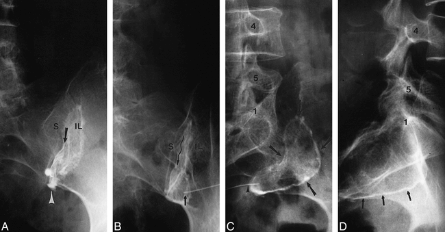
- fig 2.
Images of a 36-year-old man with left-sided lower back, posterior hip, and thigh pain, who sustained an offshore work-related lifting injury.
A, Anteroposterior arthrogram of the inferior aspect of the left SIJ shows a normal inferior recess (r), bead of contrast within the joints margins (arrow), and collection of contrast medium escaping through a ventral tear (VT).
B, Lateral view confirms that the collection of contrast medium escaping through a ventral tear (arrow) is remote to the needle tip, which is in the inferior aspect of the capsule.Symbols: 1, body of S1; 2, body of S2; 3, body of S3.
C, Same projection as that shown in B except with a wider field of view. Arrow indicates ventral tear.
D, Offset opposite lateral arthrogram discloses an intact ventral capsule (arrowheads) on the contralateral side compared with a disrupted capsule with a ventral tear (arrows). White arrow points to the needle in inferior aspect of right SIJ.
E, Post-arthrography axial CT scan obtained at the S2 level (bone window/level settings), with contrast medium in both SIJs. Presacral collection of contrast medium is evidence of a left ventral capsular tear (open arrow). Contrast medium contracts the lumbosacral plexus elements (arrowheads).
F, Line drawing of the ventral hemipelvis, which allows contrast medium to escape and contact the neural elements of the sacral plexus (interrupted lines). The inset shows the torn fibers of the ventral capsule allowing contrast medium to leak slowly.
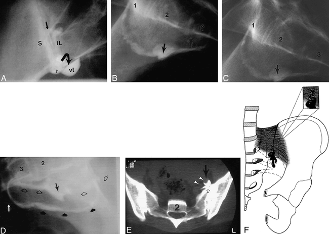
- fig 3.
Capsular attention (ie, focal capsular bulge) and schism in a 28-year-old man with an insidious onset of lower back pain.
A, Concentric area of ventral capsular attenuation is displayed at soft-tissue settings (arrowheads) of this postarthrography axial CT scan obtained at the S2 level.
B, Markedly attenuated area of the ventral capsule and sacroiliac ligament allows seepage of contrast medium in a feathery, wispy dispersal pattern (ie, schism with indistinct margins) into the presacral region (arrow).
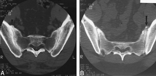
- fig 4.
A 42-year-old laborer with back pain and occasional posterior right lower extremity pain to the calf. A potential pathway between the SIJ and anterior ramus of S1 is present.
A, Arthrogram of the anteroposterior view reveals contrast medium extending (dotted line) into the S1 dorsal foramina (1). Symbols: 2, S2 dorsal foramina; 5, pedicle of L5.
B and C, Axial and direct coronal postarthrography CT scan through the S1 foramina (soft-tissue window/level settings). Contrast is observed in the right S1 dorsal foramina on both scans (arrowheads).
D, Compare this axial CT scan at the S1 level in another patient after bilateral SIJ arthrography to the above case. The arrowheads point to the S1 anterior rami bilaterally. Notice on the right how contrast medium encircles the S1 segmental root.
E, Line drawing of the dorsal view of the pelvis and L5−S1 motion segment. Small arrowheads on the right indicate where the ilium has been “resected” to reveal the SIJ; the dorsal ligaments have been stripped away. The fine wavy arrow indicates contrast medium tracking subligamentously from the SIJ to the S1 dorsal-sacral foramina. The small curved arrow (at the top of the right SIJ) indicates contrast medium extravasating from the superior aspect of the right SIJ into the right L5 root canal. The discontinuous lines indicate the L5 segmental nerve roots.
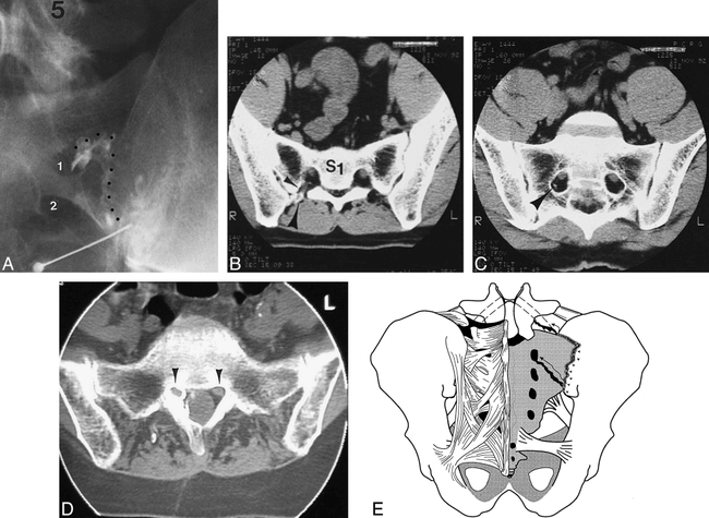
- fig 5.
Another pathway between the SIJ and neural structures was found in this 33-year-old patient with lower back pain and left lower extremity paresthesias.
A, Arthrogram of the anteroposterior view allowed visualization of contrast medium extravasating from the superior recess (wavy line) of the SIJ toward the L5 root canal. Symbols: 5, L5 vertebral body; S, sacral ala; IL, ilium.
B, Postarthrography axial CT scan obtained at the L5−S1 level reveals contrast medium extending to the L5 epiradicular sheath (arrows). The arrowhead points to the contralateral L5 anterior ramus. Symbols: S, sacral ala; IL, ilium. Refer to figure 4E for a comparative line drawing.


Tables
 TABLE 1:
Observed contrast extravasation patterns from 76 sacroiliac joints
In this issue


Jump to section
Related Articles
Cited By...
- Effect of Sacropelvic Hardware on Axis and Center of Rotation of the Sacroiliac Joint: A Finite Element Study
- Does the presence of cranial contrast spread during a sacroiliac joint injection predict short-term outcome?
- International Society for the Advancement of Spine Surgery Policy 2020 Update--Minimally Invasive Surgical Sacroiliac Joint Fusion (for Chronic Sacroiliac Joint Pain): Coverage Indications, Limitations, and Medical Necessity
- Sacroiliac pain in a dialysis patient
- Ilial Anterior Rotation Hypermobility in a Female Collegiate Tennis Player