
Article Figures & Data
Figures
- fig 1.
Ruptured aneurysm originating from anterior communicating artery.
A, DSA image, oblique projection, right internal carotid injection, shows the aneurysm (arrowhead) before embolization.
B, DSA image, semiaxial projection, right internal carotid injection 3 months after treatment with electrolytically detachable coils, shows remnant cavity (arrows) in the neck area.
C, MR angiogram, targeted coronal MIP reconstruction, shows faint signal intensity between the proximal anterior cerebral arteries (large arrow), which was not interpreted as an aneurysmal remnant. Note the local encroachment of the anterior cerebral arteries (small arrows).
- fig 2.
Unruptured aneurysm of the right carotid termination.
A, DSA image, anterior projection, right internal carotid injection, shows the aneurysm (arrowhead) before embolization.
B, Four months after treatment, the aneurysm is subtotally packed with electrolytically detachable coils. The remnant cavity (arrow) is faintly seen through the subtracted coil mass.
C, MR angiogram, targeted parasagittal MIP reconstruction, clearly shows the remnant cavity (arrow).
- fig 3.
Giant unruptured aneurysm originating from the bifurcation of the right ophthalmic artery.
A, DSA image, oblique projection, right internal carotid injection 4 months after treatment, shows the aneurysm is subtotally packed with electrolytically detachable coils. A remnant cavity (arrowheads) is seen in the neck area.
B, MR angiogram, targeted coronal MIP reconstruction, shows scattered high signal intensity in the upper part of the aneurysm (small arrows) in addition to the remnant cavity flow (large arrow) in the neck area.
C, T1-weighted MR image, sagittal view, shows high signal intensity in the upper part of the coiled aneurysm (arrows), which does not represent flow but T1 shortening caused by thrombosis between the coil material.


Tables
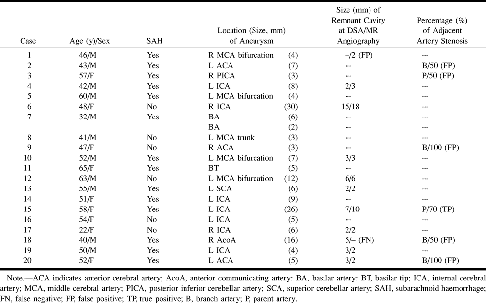
 TABLE 1:
Patient data and aneurysmal characteristics for 20 patients (21 aneurysms) followed up with DSA and MR angiography
In this issue


Jump to section
Related Articles
Cited By...
- MRA versus DSA for the follow-up imaging of intracranial aneurysms treated using endovascular techniques: a meta-analysis
- MRA Versus DSA for Follow-Up of Coiled Intracranial Aneurysms: A Meta-Analysis
- Outcomes of Endovascular Treatments of Aneurysms: Observer Variability and Implications for Interpreting Case Series and Planning Randomized Trials
- MR Angiographic Follow-Up of Intracranial Aneurysms Treated with Detachable Coils: Evaluation of a Blood-Pool Contrast Medium
- Intracranial Aneurysms Treated With Guglielmi Detachable Coils: Imaging Follow-Up With Contrast-Enhanced MR Angiography
- Matrix Detachable Coils for the Endovascular Treatment of Intracranial Aneurysms: Analysis of Early Angiographic and Clinical Outcomes
- CT and MR Imaging Findings and Their Implications in the Follow-up of Patients with Intracranial Aneurysms Treated with Endosaccular Occlusion with Onyx
- Utility of CT Angiography and MR Angiography for the Follow-up of Experimental Aneurysms Treated with Stents or Guglielmi Detachable Coils