
Abstract
BACKGROUND AND PURPOSE: Thiopentone reduces CBF and metabolic rate. Still, it is widely used for sedation during MR spectroscopy. We investigated whether barbiturate anesthesia and preanesthetic fasting have an effect on metabolic ratios in proton MR spectroscopy of the brain.
METHODS: Eight healthy, consenting, male volunteers were studied twice in a random, crossover fashion. The study sessions were conducted during fasting (F) and nonfasting (nonF), with glucose infusion mimicking the fed state. During both sessions, two sets of spectroscopic data were collected, one during the awake state (F or nonF) and one under barbiturate anesthesia (F+B or nonF+B), using TEs of 135 and 270. Spectral areas of N-acetylaspartate (NAA), choline-containing compounds (Cho), and creatine plus phosphocreatine (Cr) were calculated, and the presence of lactate or lipid was noted. Venous blood samples for glucose, β-hydroxybutyrate, lactate, and electrolytes were collected.
RESULTS: Barbiturate anesthesia caused a 42% reduction in blood lactate levels during fasting, but not during glucose infusion. There were no differences in NAA/Cho, NAA/Cr, or in Cho/Cr between the groups F, nonF, F+B, or nonF+B. No lactate or lipid resonances were detected.
CONCLUSION: Barbiturate anesthesia with preanesthetic fasting can be used for proton spectroscopy at TEs of 135 or 270 without interference from NAA/Cho, NAA/Cr, or Cho/Cr or from the appearance of lactate or lipid.
MR spectroscopy enables noninvasive in vivo measurement of brain biochemistry (1–4). Children and other uncooperative patients often need sedation or general anesthesia during the data collection. The metabolic state of the brain is affected by anesthetic agents (5) and possibly also by preanesthetic fasting (6). We wanted to determine whether the fasting preceding anesthesia, or barbiturate anesthesia itself, had any effect on the major spectral metabolites N-acetylaspartate (NAA), choline (Cho), creatine plus phosphocreatine (Cr), lactate, or lipids.
Methods
Subjects and Study Protocol
Eight healthy male volunteers (20 to 32 years old) were studied. None of them had a history of alcohol and/or drug abuse and none were currently using any medication. The study was approved by the Institutional Ethics Committee of Turku University Hospital and written informed consent was obtained prior to participation in the study. Each subject was examined twice in a random crossover fashion. The study sessions during fasting and nonfasting were 1 week apart. The person analyzing the spectra was kept unaware of the treatment.
At every session, baseline MR spectroscopy with the subject awake was performed first, after which anesthesia was induced with 5 to 8 mg/kg thiopentone IV and MR spectroscopy was repeated. The subjects were kept asleep by consecutive 50-mg doses of thiopentone. They were breathing spontaneously, and the adequacy of ventilation was monitored with pulse oximetry (SpO2).
In the nonfasting session, food was allowed until 6 hours before the examination and an infusion of 10% glucose solution 100 mL/h was started 2 hours before the spectroscopy. In the fasting session, volunteers fasted for 18 to 20 hours before the examination and slow isotonic saline infusion was started for maintaining the IV line. Another IV cannula was inserted into the contralateral arm for blood sampling. Venous blood samples for analysis of glucose, β-hydroxybutyrate, lactate, and electrolytes were collected before the start of the glucose infusion, at the end of the spectroscopic examination performed with the subjects awake (F or nonF), and at the end of spectroscopic data collection with the subjects under anesthesia (F+B or nonF+B).
MR Spectroscopic Protocol
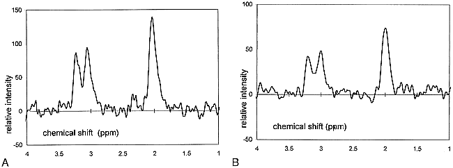
Imaging and spectroscopy were performed with a 1.5-T MR imager (Siemens Magnetom 63 SP 4000 and NUMARIS version A2.7, Erlangen, Germany) with a standard circular head coil and spectroscopic package. Transverse axial T2-weighted spin-echo images 2700/22,90/1 (TR/TE/excitations) with 5-mm section thickness, 192 × 256 matrix size, and 250-mm field of view were collected for localization of the 8-cm3 spectroscopic volume in the left basal ganglia, avoiding the CSF spaces. A double spin-echo sequence with chemical-shift selective water suppression was performed with parameters of 1500/135,270 (TR/TE), 256 acquisitions, and 1024 data points to acquire the spectra from a cubic 8-mL volume. Four spectra were acquired during each session with the two TEs and with the volunteer awake and under barbiturate anesthesia (Fig 1).

A and B, Sample spectra acquired with TE = 135 after fasting (A) and TE = 270 under barbiturate anesthesia (B)
Data Postprocessing
Data postprocessing was performed with the NUMARIS A2.7 spectroscopic software package, and the integrated areas of each metabolite spectral line, the CH3 group of NAA at 2.0 ppm, the N-CH3 group of Cr at 3.0 ppm, and the N-CH3 group of Cho at 3.2 ppm, were determined manually. The baseline of the spectrum was corrected with a second-order polynomial spline function. Metabolic ratios were calculated from the integrated peak areas of spectra recorded with TEs of 135 and 270.
Statistical Analysis
A paired t-test was used to compare the differences in blood glucose, β-hydroxybutyrate, and lactate between the F and nonF, and F+B and nonF+B sessions, respectively. A single-factor ANOVA was used to compare the differences in the other blood parameters and in spectroscopic metabolite ratios between the four treatment groups: fasting and awake (F), fasting and under anesthesia (F+B), glucose infusion (mimicking nonfasting) and awake (nonF), and glucose infusion and under anesthesia (nonF+B).
Results
There were no differences between the total doses of thiopentone administered during the two sessions (F+B vs nonF+B, 738 mg vs 778 mg). No significant decrease in SpO2 occurred during any of the study sessions.
Physiological Data (Table 1)
Physiological data
The 10% glucose infusion resulted in a slightly elevated blood glucose at the end of the infusion (nonF and nonF+B). Prolonged fasting induced significant elevation of blood β-hydroxybutyrate (F and F+B). The induction of barbiturate anesthesia caused a 42% decrease in blood lactate in the F group, whereas in the nonF group blood lactate remained unchanged during anesthesia. Plasma sodium, plasma kalium, and serum calcium remained unchanged during the experiment.
Spectroscopic Data (Table 2)
Spectroscopic data
There were no differences in the mean NAA/Cho, NAA/Cr, or Cho/Cr (P > .33) between the four treatment groups. No lactate or lipid resonances were detected.
Discussion
Selection of anesthetic agents for sedation during MR spectroscopy has been based mainly on anesthetic considerations (7, 8). To what degree these agents may affect the results has not been ascertained. We studied thiopentone because it is widely used for sedation during MR imaging, especially in children, who form the largest patient group for metabolic MR spectroscopic studies. For ethical reasons we were restricted to young adults as study subjects. Because of the obvious risks, we could not feed the subjects before the nonfasting sessions. Instead, we decided to use a continuous glucose infusion to mimic the postprandial state; glucose was infused at a rate corresponding to the average metabolic rate (9, 10). This glucose infusion rate resulted in normal plasma β-hydroxybutyrate levels and mildly elevated blood glucose levels typical for the postprandial state.
A dose-dependent reduction in CBF and cerebral metabolic rate (CMR), approximately paralleling CNS depression, occurs with barbiturates. With the onset of anesthesia, CBF and CMR of oxygen utilization (CMRO2) are reduced by about 30% (11). The impact of barbiturates upon brain carbohydrate chemistry has been studied extensively (12). Pentobarbital transiently reduces the rate of glucose incorporation into amino acids. Barbiturates may also transiently reduce the CMR for glucose more than CMRO2 is reduced. At clinical doses, barbiturates alter the carbohydrate metabolism and the bioenergetic profile of the brain so that concentrations of cellular glucose, glycogen, and energy storage compounds (ATP, ADP, phosphocreatine) are increased and lactate concentrations are reduced. Very high doses of thiopentone have been shown to lead to a decrease in brain phosphocreatine in 31P NMR spectroscopy in sheep, indicating a decreased metabolic rate (13). This finding suggests that thiopentone might affect the Cr resonance detected in proton spectroscopy. However, our results did not show any systematic effect from barbiturate anesthesia on the metabolic ratios of NAA, Cho, and Cr, or on the appearance of lactate or lipid.
Detection of the lactate signal in proton MR spectroscopy is important in estimating the metabolic state of the brain in various local and diffuse disorders (14–16). Blood and tissue lactate concentrations vary under physiological conditions, being lowest after fasting (17). The normal concentration of lactate in the brain is 0.4 to 1.0 mmol/L, and intracellular concentrations of lactate in the brain are thought to be twice as high as those in the venous blood (17). Direct lactate signal from circulation is considered to be attenuated in MR spectroscopy owing to flow velocity effects and thought not to contribute to the spectra. Patients who are scheduled for MR imaging under anesthesia fast for 4 to 6 hours prior to the examination. Fasting is known to alter blood levels of several substrates and thus to potentially affect the metabolic state of the brain (6). The 18-hour fast used in this study resulted in elevated plasma β-hydroxybutyrate levels, indicating fat oxidation typical for prolonged fasting (Table 1). Reduced brain lactate levels during barbiturate anesthesia have been linked to inhibition of phosphofructokinase activity (12). Our finding of the 42% reduction in blood lactate during barbiturate infusion is in line with this; however, we were unable to discover why blood lactate reduction did not occur in the nonfasting group. This difference in blood lactate between the awake state and barbiturate anesthesia is not reflected in brain tissue lactate, at least with the present sensitivity of the method.
Conclusion
Our study shows that if fasting is needed in a spectroscopic study for anesthetic reasons, the results will be comparable to those measured during the fed state. Also, barbiturate anesthesia does not interfere with the main NMR-visible brain metabolites measured during long-TE proton spectroscopy. These results may be important in spectroscopic studies of patients with mitochondrial disease, in which detection of brain lactate is crucial.
Footnotes
↵1 Address reprint requests to Nina Lundbom, MD, PhD, Attending Radiologist, Department of Radiology/Meilahti Clinics, University Hospital of Helsinki, Haartmanink. 4, 00290 Helsinki, Finland.
References
- Received October 14, 1998.
- Accepted after revision April 9, 1999.
- Copyright © American Society of Neuroradiology


