
Article Figures & Data
Figures
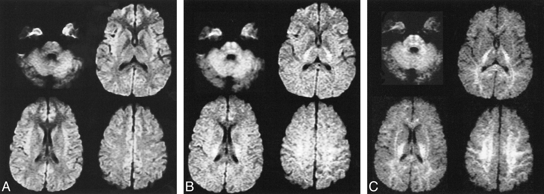
- fig 1.
Isotropic diffusion-weighted images with varying b values.
A, Image obtained with a b value of 1000 and 72/1 (TE/excitations). There is hyperintensity of cortical gray matter, particularly in the insular and medial frontal regions.
B, Image obtained with a b value of 2000 and 84/2. Near isointensity of gray and white matter is seen with slight hyperintensity of posterior capsular white matter and pontine regions.
C, Image obtained with a b value of 3500 and 96/4. Progressive hyperintensity of white matter is present with diminished gray matter signal intensity.
- fig 2.
Mean ADC values with SDs for ROI in six participants. Echo time was fixed at 96 ms for all acquisitions. The numbers of excitation were varied to improve signal-to-noise ratios at higher b values: numbers of excitation were one, two, and four for b values of 1000, 2000, and 3000, respectively. ADC values diminish with increasing b values. ADC SDs decrease with multiple numbers of excitaiton
- fig 3.
ADC maps at varying b values. Improved gray-white differentiation is noted with increased b value. A, b = 1000; B, b = 2000; C, b = 2500; and D, b = 3500
- fig 4.
Clinical case of multiple sclerosis.
A, Fast spin-echo axial T2-weighted image (4000/102/2 [TR/TE/excitations]) shows a dominant subcortical right cingulate gyrus lesion with marked hyperintensity.
B, Fast-FLAIR image (10000/145/2200/1 [TR/TE/excitations]) highlights additional subcortical and periventricular plaques, including a large lesion in the right side of the splenium.
C, Diffusion-weighted image (10000/72/1) obtained with b = 1000 s/mm2 reveals slight hyperintensity in the right cingulate lesion, similar to the contralateral mesial frontal region, and hyperintensity in the right splenium lesion.
D, Diffusion-weighted image (10000/96/1) obtained with b = 3500 s/mm2 reveals marked hypointensity in the right cingulate lesion, likely reflecting reduced T2 weighting and less T2 shine-through. Persistent hyperintensity in the right splenium lesion is likely due to the normal anisotropy of the splenium and inconspicuity of hypointense lesions adjacent to the CSF spaces. This emphasizes the complementary relationship of the diffusion-weighted acquisitions obtained at standard and high b values.
- fig 5.
Reversal of gray-white matter contrast with increasing b values. Relative signal intensity values obtained from equation 2 in the text


Tables
 TABLE 1:
Gray-white matter DWI signal intensity ratios and average values for 6 subjects: fixed TE (96 ms) and variable number of excitations; putamen and centrum semiovale regions of interest were chosen as described in the text
- TABLE 2:
Mean ADC and SD for six subjects; fixed TE (96 ms) and variable number of excitations
- TABLE 3:
Mean ADC and SD for single subject: fixed number of excitations (2) and TE (96 ms)
- TABLE 4:
Mean ADC and SD for single subject with fixed number of excitations (2) and variable TE (72 and 96 ms) at b = 1000 s/mm2


In this issue


Jump to section
Related Articles
Cited By...
- Enhanced Structural Brain Connectivity Analyses Using High Diffusion-weighting Strengths
- Quantification of alterations in diffusion measures of white matter integrity associated with healthy aging
- Diffusion Tensor Model links to Neurite Orientation Dispersion and Density Imaging at high b-value in Cerebral Cortical Gray Matter
- Differentiation of Recurrent Tumor and Posttreatment Changes in Head and Neck Squamous Cell Carcinoma: Application of High b-Value Diffusion-Weighted Imaging
- Correlation of MRI-Derived Apparent Diffusion Coefficients in Newly Diagnosed Gliomas with [18F]-Fluoro-L-Dopa PET: What Are We Really Measuring with Minimum ADC?
- Distinguishing between Germinomas and Pineal Cell Tumors on MR Imaging
- Apparent Diffusion Coefficient with Higher b-Value Correlates Better with Viable Cell Count Quantified from the Cavity of Brain Abscess
- Correlation of 18F-FDG Uptake with Apparent Diffusion Coefficient Ratio Measured on Standard and High b Value Diffusion MRI in Head and Neck Cancer
- High-b-Value Diffusion MR Imaging and Basal Nuclei Apparent Diffusion Coefficient Measurements in Variant and Sporadic Creutzfeldt-Jakob Disease
- Enhanced Detection of Diffusion Reductions in Creutzfeldt-Jakob Disease at a Higher B Factor