Advertisement

Main menu

  • Home
  • Content
    • Current Issue
    • Accepted Manuscripts
    • Article Preview
    • Past Issue Archive
    • Video Articles
    • AJNR Case Collection
    • Case of the Week Archive
    • Case of the Month Archive
    • Classic Case Archive
  • Special Collections
    • AJNR Awards
    • Low-Field MRI
    • Alzheimer Disease
    • ASNR Foundation Special Collection
    • Photon-Counting CT
    • View All
  • Multimedia
    • AJNR Podcasts
    • AJNR SCANtastic
    • Trainee Corner
    • MRI Safety Corner
    • Imaging Protocols
  • For Authors
    • Submit a Manuscript
    • Submit a Video Article
    • Submit an eLetter to the Editor/Response
    • Manuscript Submission Guidelines
    • Statistical Tips
    • Fast Publishing of Accepted Manuscripts
    • Graphical Abstract Preparation
    • Imaging Protocol Submission
    • Author Policies
  • About Us
    • About AJNR
    • Editorial Board
    • Editorial Board Alumni
  • More
    • Become a Reviewer/Academy of Reviewers
    • Subscribers
    • Permissions
    • Alerts
    • Feedback
    • Advertisers
    • ASNR Home

User menu

  • Alerts
  • Log in

Search

  • Advanced search
American Journal of Neuroradiology

American Journal of Neuroradiology

ASHNR American Society of Functional Neuroradiology ASHNR American Society of Pediatric Neuroradiology ASSR
  • Alerts
  • Log in

Advanced Search

  • Home
  • Content
    • Current Issue
    • Accepted Manuscripts
    • Article Preview
    • Past Issue Archive
    • Video Articles
    • AJNR Case Collection
    • Case of the Week Archive
    • Case of the Month Archive
    • Classic Case Archive
  • Special Collections
    • AJNR Awards
    • Low-Field MRI
    • Alzheimer Disease
    • ASNR Foundation Special Collection
    • Photon-Counting CT
    • View All
  • Multimedia
    • AJNR Podcasts
    • AJNR SCANtastic
    • Trainee Corner
    • MRI Safety Corner
    • Imaging Protocols
  • For Authors
    • Submit a Manuscript
    • Submit a Video Article
    • Submit an eLetter to the Editor/Response
    • Manuscript Submission Guidelines
    • Statistical Tips
    • Fast Publishing of Accepted Manuscripts
    • Graphical Abstract Preparation
    • Imaging Protocol Submission
    • Author Policies
  • About Us
    • About AJNR
    • Editorial Board
    • Editorial Board Alumni
  • More
    • Become a Reviewer/Academy of Reviewers
    • Subscribers
    • Permissions
    • Alerts
    • Feedback
    • Advertisers
    • ASNR Home
  • Follow AJNR on Twitter
  • Visit AJNR on Facebook
  • Follow AJNR on Instagram
  • Join AJNR on LinkedIn
  • RSS Feeds

AJNR Awards, New Junior Editors, and more. Read the latest AJNR updates

Research ArticleNeurointervention

Occlusion of Experimentally Created Fusiform Aneurysms with Porous Metallic Stents

Glen Geremia, Todd Brack, Luke Brennecke, Michael Haklin and Richard Falter
American Journal of Neuroradiology April 2000, 21 (4) 739-745;
Glen Geremia
aFrom the Department of Radiology (G.G.) and the Comparative Research Ctr (M.H.), Rush-Presbyterian-St Luke's Medical Center, Chicago, IL; Alexian Brothers (T.B.), Elk Grove Village, IL; Pathology Associates International (L.B.), Frederick, MD; and Cape Fear Valley Health System (R.F.), Fayetteville, NC.
  • Find this author on Google Scholar
  • Find this author on PubMed
  • Search for this author on this site
Todd Brack
aFrom the Department of Radiology (G.G.) and the Comparative Research Ctr (M.H.), Rush-Presbyterian-St Luke's Medical Center, Chicago, IL; Alexian Brothers (T.B.), Elk Grove Village, IL; Pathology Associates International (L.B.), Frederick, MD; and Cape Fear Valley Health System (R.F.), Fayetteville, NC.
  • Find this author on Google Scholar
  • Find this author on PubMed
  • Search for this author on this site
Luke Brennecke
aFrom the Department of Radiology (G.G.) and the Comparative Research Ctr (M.H.), Rush-Presbyterian-St Luke's Medical Center, Chicago, IL; Alexian Brothers (T.B.), Elk Grove Village, IL; Pathology Associates International (L.B.), Frederick, MD; and Cape Fear Valley Health System (R.F.), Fayetteville, NC.
  • Find this author on Google Scholar
  • Find this author on PubMed
  • Search for this author on this site
Michael Haklin
aFrom the Department of Radiology (G.G.) and the Comparative Research Ctr (M.H.), Rush-Presbyterian-St Luke's Medical Center, Chicago, IL; Alexian Brothers (T.B.), Elk Grove Village, IL; Pathology Associates International (L.B.), Frederick, MD; and Cape Fear Valley Health System (R.F.), Fayetteville, NC.
  • Find this author on Google Scholar
  • Find this author on PubMed
  • Search for this author on this site
Richard Falter
aFrom the Department of Radiology (G.G.) and the Comparative Research Ctr (M.H.), Rush-Presbyterian-St Luke's Medical Center, Chicago, IL; Alexian Brothers (T.B.), Elk Grove Village, IL; Pathology Associates International (L.B.), Frederick, MD; and Cape Fear Valley Health System (R.F.), Fayetteville, NC.
  • Find this author on Google Scholar
  • Find this author on PubMed
  • Search for this author on this site
  • Article
  • Figures & Data
  • Info & Metrics
  • Responses
  • References
  • PDF
Loading

Article Figures & Data

Figures

  • fig 1.
    • Download figure
    • Open in new tab
    • Download powerpoint
    fig 1.

    Diagram of a fusiform carotid artery aneurysm from a jugular vein segment

  • fig 2.
    • Download figure
    • Open in new tab
    • Download powerpoint
    fig 2.

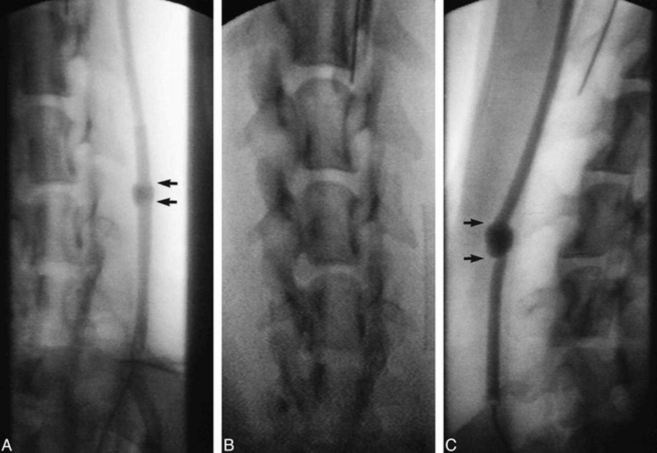
    Angiographic findings of a fusiform carotid artery aneurysm.

    A, Eight weeks after stent placement there is near-complete ablation of the aneurysmal lumen (arrows).

    B, The Wallstent within the carotid artery.

    C, The control fusiform aneurysm (arrows) and the carotid artery remain widely patent throughout the 8-week period of observation.

  • fig 3.
    • Download figure
    • Open in new tab
    • Download powerpoint
    fig 3.

    Gross pathologic examination.

    A, Gross end-block specimen of control aneurysm shows the walls remain widely dilated.

    B, The outer walls of the stented aneurysm have contracted along the length of the stent, producing a narrower profile of the carotid artery as compared with the profile of the control aneurysm.

    C, Control aneurysm, cross section. Section 3 shows the markedly dilated lumen of the fusiform aneurysm.

    D, Stented aneurysm. Section 4 is through the center of the fusiform aneurysm. Note the contraction of the outer walls of the aneurysm along the stent, again producing a narrower profile of the carotid artery and aneurysm as compared with the control aneurysm. The lumen between the stent and outer wall of the aneurysm is filled with white, organized thrombus.

    E, Longitudinal section. All stents were embedded in a glistening, translucent neointima. The stented aneurysms appear solid except for small channels, which connect the lumens of the carotid arteries with those of the aneurysms (arrows).

  • fig 4.
    • Download figure
    • Open in new tab
    • Download powerpoint
    fig 4.

    Histopathologic findings.

    A, Mason's trichrome stain on stented aneurysm shows the aneurysmal lumen is nearly completely filled in with organized fibrous connective tissue (curved arrows). The stent wires are surrounded by neointima (straight arrows).

    B, Mason's trichrome stain on control aneurysm in cross section shows markedly distended thin wall of the aneurysm.

    C and D, Trichrome stain on stented aneurysm in longitudinal section shows a small, patent endothelial-lined channel at one margin, connecting the carotid lumen with the aneurysmal lumen (arrow). These small channels were seen in all aneurysmal specimens. The remainder of the aneurysmal lumens were filled in by fibrous connective tissue (organized thrombus). Within the neointima surrounding the stent wires there was mild inflammatory cell infiltration (macrophages). Many macrophages contained blood pigment (resolving thrombus).

    E, Magnified cross-sectional view of aneurysmal lumen with Mason's trichrome stain. Fibrin and collagen stain blue. Dark blue represents more mature fibrin and collagen. Mature fibrous elements and collagen (dark blue) were noted near the periphery of the aneurysmal wall (thin arrows); less mature fibrous elements (light blue) lie near the central lumen of the aneurysm (thick arrows), suggesting that thrombus organization begins along the outer wall and progresses in a centripetal fashion toward the central lumen of the aneurysm.

    F, Verhoeff-van Gieson elastic stain shows heavily vascularized organized thrombus (arrows).

  • fig 5.
    • Download figure
    • Open in new tab
    • Download powerpoint
    fig 5.

    Diagram of flow patterns immediately after stent deployment (A) and 8 weeks later (B). Complex streams of flow are seen within the residual aneurysmal lumen between the outer aneurysmal wall and the stent wires (A). Eight weeks after stent deployment, the aneurysmal lumen is occupied with organized fibrous connective tissue (B)

PreviousNext
Back to top

In this issue

American Journal of Neuroradiology
Vol. 21, Issue 4
1 Apr 2000
  • Table of Contents
  • Index by author
Advertisement
Print
Download PDF
Email Article

Thank you for your interest in spreading the word on American Journal of Neuroradiology.

NOTE: We only request your email address so that the person you are recommending the page to knows that you wanted them to see it, and that it is not junk mail. We do not capture any email address.

Enter multiple addresses on separate lines or separate them with commas.
Occlusion of Experimentally Created Fusiform Aneurysms with Porous Metallic Stents
(Your Name) has sent you a message from American Journal of Neuroradiology
(Your Name) thought you would like to see the American Journal of Neuroradiology web site.
CAPTCHA
This question is for testing whether or not you are a human visitor and to prevent automated spam submissions.
Cite this article
Glen Geremia, Todd Brack, Luke Brennecke, Michael Haklin, Richard Falter
Occlusion of Experimentally Created Fusiform Aneurysms with Porous Metallic Stents
American Journal of Neuroradiology Apr 2000, 21 (4) 739-745;

Citation Manager Formats

  • BibTeX
  • Bookends
  • EasyBib
  • EndNote (tagged)
  • EndNote 8 (xml)
  • Medlars
  • Mendeley
  • Papers
  • RefWorks Tagged
  • Ref Manager
  • RIS
  • Zotero
0 Responses
Respond to this article
Share
Bookmark this article
Occlusion of Experimentally Created Fusiform Aneurysms with Porous Metallic Stents
Glen Geremia, Todd Brack, Luke Brennecke, Michael Haklin, Richard Falter
American Journal of Neuroradiology Apr 2000, 21 (4) 739-745;
del.icio.us logo Twitter logo Facebook logo Mendeley logo
  • Tweet Widget
  • Facebook Like
  • Google Plus One

Jump to section

  • Article
    • Abstract
    • Methods
    • Results
    • Discussion
    • Conclusion
    • Footnotes
    • References
  • Figures & Data
  • Info & Metrics
  • Responses
  • References
  • PDF

Related Articles

  • No related articles found.
  • PubMed
  • Google Scholar

Cited By...

  • Stent alone treatment for dissections and dissecting aneurysms involving the basilar artery
  • Safety and Efficacy of Neuroform for Treatment of Intracranial Aneurysms: A Prospective, Consecutive, French Multicentric Study
  • Reconstructive Endovascular Treatment of Intracranial Fusiform Aneurysms: A 1-Stage Procedure with Stent and Balloon
  • Clinical and Angiographic Follow-Up of Stent-Only Therapy for Acute Intracranial Vertebrobasilar Dissecting Aneurysms
  • Crossref
  • Google Scholar

This article has not yet been cited by articles in journals that are participating in Crossref Cited-by Linking.

More in this TOC Section

  • A Retrospective Study in Tentorial DAVFs
  • Proximal Protection Devices for Carotid Stenting
  • Contour Neurovascular System: Five Year Follow Up
Show more NEUROINTERVENTION

Similar Articles

Advertisement

Indexed Content

  • Current Issue
  • Accepted Manuscripts
  • Article Preview
  • Past Issues
  • Editorials
  • Editor's Choice
  • Fellows' Journal Club
  • Letters to the Editor
  • Video Articles

Cases

  • Case Collection
  • Archive - Case of the Week
  • Archive - Case of the Month
  • Archive - Classic Case

More from AJNR

  • Trainee Corner
  • Imaging Protocols
  • MRI Safety Corner
  • Book Reviews

Multimedia

  • AJNR Podcasts
  • AJNR Scantastics

Resources

  • Turnaround Time
  • Submit a Manuscript
  • Submit a Video Article
  • Submit an eLetter to the Editor/Response
  • Manuscript Submission Guidelines
  • Statistical Tips
  • Fast Publishing of Accepted Manuscripts
  • Graphical Abstract Preparation
  • Imaging Protocol Submission
  • Evidence-Based Medicine Level Guide
  • Publishing Checklists
  • Author Policies
  • Become a Reviewer/Academy of Reviewers
  • News and Updates

About Us

  • About AJNR
  • Editorial Board
  • Editorial Board Alumni
  • Alerts
  • Permissions
  • Not an AJNR Subscriber? Join Now
  • Advertise with Us
  • Librarian Resources
  • Feedback
  • Terms and Conditions
  • AJNR Editorial Board Alumni

American Society of Neuroradiology

  • Not an ASNR Member? Join Now

© 2025 by the American Society of Neuroradiology All rights, including for text and data mining, AI training, and similar technologies, are reserved.
Print ISSN: 0195-6108 Online ISSN: 1936-959X

Powered by HighWire