Advertisement

Main menu

  • Home
  • Content
    • Current Issue
    • Accepted Manuscripts
    • Article Preview
    • Past Issue Archive
    • Video Articles
    • AJNR Case Collection
    • Case of the Week Archive
    • Case of the Month Archive
    • Classic Case Archive
  • Special Collections
    • AJNR Awards
    • Low-Field MRI
    • Alzheimer Disease
    • ASNR Foundation Special Collection
    • Photon-Counting CT
    • View All
  • Multimedia
    • AJNR Podcasts
    • AJNR SCANtastic
    • Trainee Corner
    • MRI Safety Corner
    • Imaging Protocols
  • For Authors
    • Submit a Manuscript
    • Submit a Video Article
    • Submit an eLetter to the Editor/Response
    • Manuscript Submission Guidelines
    • Statistical Tips
    • Fast Publishing of Accepted Manuscripts
    • Graphical Abstract Preparation
    • Imaging Protocol Submission
    • Author Policies
  • About Us
    • About AJNR
    • Editorial Board
    • Editorial Board Alumni
  • More
    • Become a Reviewer/Academy of Reviewers
    • Subscribers
    • Permissions
    • Alerts
    • Feedback
    • Advertisers
    • ASNR Home

User menu

  • Alerts
  • Log in

Search

  • Advanced search
American Journal of Neuroradiology

American Journal of Neuroradiology

ASHNR American Society of Functional Neuroradiology ASHNR American Society of Pediatric Neuroradiology ASSR
  • Alerts
  • Log in

Advanced Search

  • Home
  • Content
    • Current Issue
    • Accepted Manuscripts
    • Article Preview
    • Past Issue Archive
    • Video Articles
    • AJNR Case Collection
    • Case of the Week Archive
    • Case of the Month Archive
    • Classic Case Archive
  • Special Collections
    • AJNR Awards
    • Low-Field MRI
    • Alzheimer Disease
    • ASNR Foundation Special Collection
    • Photon-Counting CT
    • View All
  • Multimedia
    • AJNR Podcasts
    • AJNR SCANtastic
    • Trainee Corner
    • MRI Safety Corner
    • Imaging Protocols
  • For Authors
    • Submit a Manuscript
    • Submit a Video Article
    • Submit an eLetter to the Editor/Response
    • Manuscript Submission Guidelines
    • Statistical Tips
    • Fast Publishing of Accepted Manuscripts
    • Graphical Abstract Preparation
    • Imaging Protocol Submission
    • Author Policies
  • About Us
    • About AJNR
    • Editorial Board
    • Editorial Board Alumni
  • More
    • Become a Reviewer/Academy of Reviewers
    • Subscribers
    • Permissions
    • Alerts
    • Feedback
    • Advertisers
    • ASNR Home
  • Follow AJNR on Twitter
  • Visit AJNR on Facebook
  • Follow AJNR on Instagram
  • Join AJNR on LinkedIn
  • RSS Feeds

AJNR Awards, New Junior Editors, and more. Read the latest AJNR updates

Research ArticlePediatric Neuroimaging

Proton MR Spectroscopy in Children with Bipolar Affective Disorder: Preliminary Observations

Mauricio Castillo, Lester Kwock, Helen Courvoisie and Stephen R. Hooper
American Journal of Neuroradiology May 2000, 21 (5) 832-838;
Mauricio Castillo
  • Find this author on Google Scholar
  • Find this author on PubMed
  • Search for this author on this site
Lester Kwock
  • Find this author on Google Scholar
  • Find this author on PubMed
  • Search for this author on this site
Helen Courvoisie
  • Find this author on Google Scholar
  • Find this author on PubMed
  • Search for this author on this site
Stephen R. Hooper
  • Find this author on Google Scholar
  • Find this author on PubMed
  • Search for this author on this site
  • Article
  • Figures & Data
  • Info & Metrics
  • Responses
  • References
  • PDF
Loading

Article Figures & Data

Figures

  • Tables
  • fig 1.
    • Download figure
    • Open in new tab
    • Download powerpoint
    fig 1.

    A and B, Axial T1-weighted images show placement of voxel in the left frontal lobe (A) and in the left temporal lobe (B)
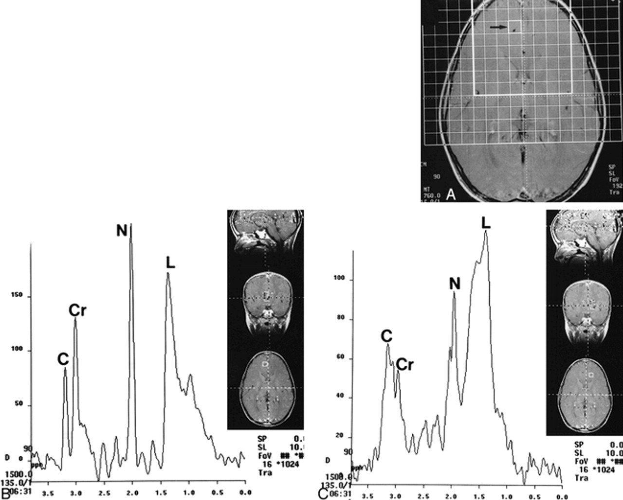
  • fig 2.
    • Download figure
    • Open in new tab
    • Download powerpoint
    fig 2.

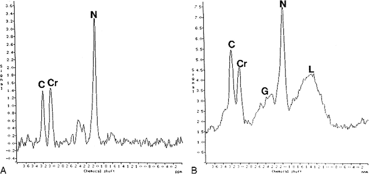
    MR spectra from the frontal lobes.

    A, Proton spectra from the left frontal lobe in a control subject show normal metabolites. C, choline; Cr, creatine; N, NAA.

    B, Proton spectra from the left frontal lobe in a patient with BPAD show prominent resonances between 2.1 and 2.5 ppm, corresponding to Glu/Gln (G). The resonances between 0.8 and 1.8 ppm correspond to lipids (L). C, choline; Cr, creatine; N, NAA.

  • fig 3.
    • Download figure
    • Open in new tab
    • Download powerpoint
    fig 3.

    MR spectra from the temporal lobes.

    A, Proton spectra from the left temporal lobe in a control subject show normal metabolites.

    B, Proton spectra from the left temporal lobe in a patient with BPAD show prominent Glu/Gln (G) but no lipids. C, choline; Cr, creatine; N, NAA.

  • fig 4.
    • Download figure
    • Open in new tab
    • Download powerpoint
    fig 4.

    MR spectra from the frontal lobes in a patient with BPAD obtained with smaller voxels.

    A, Axial T1-weighted localizer image with grid shows dense. White square outlines the area of analysis. Study was obtained with 2D PRESS technique (TE = 135). Note location of individual voxel (arrow), corresponding to spectra in B.

    B, Proton spectra from voxel (arrow, A; 1 × 1 × 1.5 cm) show marked elevation of lipids (L). Note that voxel is far away from any fat-containing structure, precluding extraneous contamination. C, choline; Cr, creatine; N, NAA.

    C, Proton spectra of individual voxel placed contralateral and in nearly a mirror position to B still show marked elevation of lipids (L). Again, voxel is placed away from fat-containing structures that could conceivably contaminate it. C, choline; Cr, creatine; N, NAA.

Tables

  • Figures
  • TABLE 1:
    • View popup
    • Download powerpoint
    TABLE 1:

    Means and SDs on the NEPSY test for children with bipolar affective disorder

  • TABLE 2:
    • View popup
    • Download powerpoint
    TABLE 2:

    Means and SDs for the Woodcock-Johnson Psychoeducational Battery in children with bipolar affective disorder

  • TABLE 3:
    • View popup
    • Download powerpoint
    TABLE 3:

    Metabolite ratios in the frontal lobes of children with bipolar affective disorder (BPAD) (n = 10) and unaffected control subjects (n = 10)

  • TABLE 4:
    • View popup
    • Download powerpoint
    TABLE 4:

    Metabolite ratios in the basal ganglia for children with bipolar affective disorder (BPAD) (n = 10) and unaffected control subjects (n = 10)

PreviousNext
Back to top

In this issue

American Journal of Neuroradiology
Vol. 21, Issue 5
1 May 2000
  • Table of Contents
  • Index by author
Advertisement
Print
Download PDF
Email Article

Thank you for your interest in spreading the word on American Journal of Neuroradiology.

NOTE: We only request your email address so that the person you are recommending the page to knows that you wanted them to see it, and that it is not junk mail. We do not capture any email address.

Enter multiple addresses on separate lines or separate them with commas.
Proton MR Spectroscopy in Children with Bipolar Affective Disorder: Preliminary Observations
(Your Name) has sent you a message from American Journal of Neuroradiology
(Your Name) thought you would like to see the American Journal of Neuroradiology web site.
CAPTCHA
This question is for testing whether or not you are a human visitor and to prevent automated spam submissions.
Cite this article
Mauricio Castillo, Lester Kwock, Helen Courvoisie, Stephen R. Hooper
Proton MR Spectroscopy in Children with Bipolar Affective Disorder: Preliminary Observations
American Journal of Neuroradiology May 2000, 21 (5) 832-838;

Citation Manager Formats

  • BibTeX
  • Bookends
  • EasyBib
  • EndNote (tagged)
  • EndNote 8 (xml)
  • Medlars
  • Mendeley
  • Papers
  • RefWorks Tagged
  • Ref Manager
  • RIS
  • Zotero
0 Responses
Respond to this article
Share
Bookmark this article
Proton MR Spectroscopy in Children with Bipolar Affective Disorder: Preliminary Observations
Mauricio Castillo, Lester Kwock, Helen Courvoisie, Stephen R. Hooper
American Journal of Neuroradiology May 2000, 21 (5) 832-838;
del.icio.us logo Twitter logo Facebook logo Mendeley logo
  • Tweet Widget
  • Facebook Like
  • Google Plus One

Jump to section

  • Article
    • Abstract
    • Methods
    • Results
    • Discussion
    • Conclusion
    • Footnotes
    • References
  • Figures & Data
  • Info & Metrics
  • Responses
  • References
  • PDF

Related Articles

  • No related articles found.
  • PubMed
  • Google Scholar

Cited By...

  • Steatosis in the Amygdala and Frontal Cortex: Potential Magnetic Resonance Imaging Biomarkers for Alzheimers Disease
  • Aiding and Abetting Anhedonia: Impact of Inflammation on the Brain and Pharmacological Implications
  • A Review of MR Spectroscopy Studies of Pediatric Bipolar Disorder
  • Proton MR Spectroscopy Correlates of Frontal Lobe Function in Healthy Children
  • A comparison of affected and unaffected relatives of patients with bipolar disorder using proton magnetic resonance spectroscopy
  • Are the Brains of Children with Bipolar Disorder Different?
  • Crossref
  • Google Scholar

This article has not yet been cited by articles in journals that are participating in Crossref Cited-by Linking.

More in this TOC Section

  • FRACTURE MR in Congenital Vertebral Anomalies
  • Comparing MRI Perfusion in Pediatric Brain Tumors
  • Sodium MRI in Pediatric Brain Tumors
Show more Pediatric Neuroimaging

Similar Articles

Advertisement

Indexed Content

  • Current Issue
  • Accepted Manuscripts
  • Article Preview
  • Past Issues
  • Editorials
  • Editor's Choice
  • Fellows' Journal Club
  • Letters to the Editor
  • Video Articles

Cases

  • Case Collection
  • Archive - Case of the Week
  • Archive - Case of the Month
  • Archive - Classic Case

More from AJNR

  • Trainee Corner
  • Imaging Protocols
  • MRI Safety Corner
  • Book Reviews

Multimedia

  • AJNR Podcasts
  • AJNR Scantastics

Resources

  • Turnaround Time
  • Submit a Manuscript
  • Submit a Video Article
  • Submit an eLetter to the Editor/Response
  • Manuscript Submission Guidelines
  • Statistical Tips
  • Fast Publishing of Accepted Manuscripts
  • Graphical Abstract Preparation
  • Imaging Protocol Submission
  • Evidence-Based Medicine Level Guide
  • Publishing Checklists
  • Author Policies
  • Become a Reviewer/Academy of Reviewers
  • News and Updates

About Us

  • About AJNR
  • Editorial Board
  • Editorial Board Alumni
  • Alerts
  • Permissions
  • Not an AJNR Subscriber? Join Now
  • Advertise with Us
  • Librarian Resources
  • Feedback
  • Terms and Conditions
  • AJNR Editorial Board Alumni

American Society of Neuroradiology

  • Not an ASNR Member? Join Now

© 2025 by the American Society of Neuroradiology All rights, including for text and data mining, AI training, and similar technologies, are reserved.
Print ISSN: 0195-6108 Online ISSN: 1936-959X

Powered by HighWire