
Abstract
BACKGROUND AND PURPOSE: Although atrophy of structures in the medial temporal lobe has been considered an indication of Alzheimer's disease (AD), atrophic changes on MR images have also been associated with other dementing diseases and are not specific to AD. This study was undertaken to determine whether characteristic alterations in the hippocampus of patients with AD are detectable with magnetization transfer (MT) imaging.
METHODS: Coronal MT imaging was performed in 35 patients with probable AD, in 14 patients with vascular dementia, in 13 patients with other types of dementia, and in 23 control subjects to measure MT ratios of the hippocampus. Medial temporal lobe atrophy was graded subjectively on a five-point scale.
RESULTS: Scores of medial temporal lobe atrophy in all dementia groups were significantly higher than those in control subjects, but no differences were found among the dementia groups. MT ratios in the hippocampus were significantly lower in patients with AD than in those with non-AD dementia and in the control subjects; however, no differences were found between the non-AD dementia patients and the control subjects. MT ratio measurements were better than visual analysis of atrophy for differentiating AD patients from those with non-AD dementia (an overall discrimination rate of 77% versus 65%). MT ratios significantly correlated with scores on the Mini-Mental State Examination and with medial temporal lobe atrophy in AD patients but not in patients with non-AD dementia.
CONCLUSION: MT measurements may be more specific than visual analysis in detecting structural damage of the hippocampus in AD patients and might be useful in discriminating AD from vascular dementia and other types of dementia.
The hippocampus and entorhinal cortex are involved early and most severely by the pathologic process of Alzheimer's disease (AD) (1). Several studies with MR imaging have shown significant atrophy (2–11) and high signal intensity (12) in the medial temporal lobe, including the hippocampal formation, in patients with AD. Although volumetric measurements of medial temporal lobe structures have been proposed to aid in the diagnosis of AD, atrophic changes also occur in other dementing diseases (13–17) and may not be a specific marker for AD. Kirsch et al (18) reported that hippocampal T2 relaxation time is markedly prolonged in AD, but other studies have shown it to be only mildly elongated (19, 20). A recent study by Campeau et al (21) found no significant difference in T2 relaxation time between patients with AD and control subjects. These authors concluded that measurements of MR relaxation time in the hippocampus are not useful for the detection of AD. In a preliminary study, we also found no significant prolongation of hippocampal T2 relaxation time in AD patients as compared with control subjects (unpublished data). MR imaging can detect in vivo abnormalities of the brain, but conventional techniques fail to show characteristic features in the brain of AD patients.
Magnetization transfer (MT) imaging is a technique based on interactions between immobile protons (on macromolecular proteins, probably contained in cell walls) and free protons of tissue (22, 23). Measurements of the MT ratio may differ depending on the macromolecular concentration and exchange rates characteristic of the different underlying histologic structures of the brain. In a recent preliminary study of three patients with temporal lobe sclerosis, Tofts et al (24) reported a decreased MT ratio in the hippocampus on the affected side, probably reflecting the presence of gliosis and neuronal loss, which are characteristic of hippocampal sclerosis. However, to the best of our knowledge, the MT technique has not been used to study the hippocampus of patients with AD. Therefore, we decided to explore the possibility that characteristic tissue alterations in AD patients could be detected with MT imaging. To this end, we measured the MT ratios of the hippocampus in patients with AD and compared them with MT ratios in patients with vascular dementia and other types of dementia and with those in control subjects to determine whether MT measurement is more specific than visual analysis in the diagnosis of AD.
Methods
Subjects
We studied 35 patients with AD (15 men and 20 women; mean age, 77 years), 14 patients with vascular dementia (six men and eight women; mean age, 78 years), 13 patients with other types of dementia (five men and eight women; mean age, 79 years), and 23 healthy control subjects (10 men and 13 women; mean age, 78 years). All patients with AD met the criteria for probable AD formulated by the NINCDS-ADRDA Work Group (25) and had scores of less than 4 on the ischemic scale developed by Hachinski et al (26). Patients with evidence of stroke, as determined either by history or by imaging findings, were excluded. Patients with AD were in the mild to moderate stages of the disease, with a mean Mini-Mental State Examination (MMSE) (27) score of 18.1 ± 4.8 (range, 9–25). T2-weighted MR images showed no or mild periventricular hyperintensity lesions (mild caps or rim in the periventricular white matter) and a few high-intensity spots in the subcortical white matter. Moreover, characteristic findings on single-photon emission CT scans were added to the clinical criteria for diagnosis of AD. All patients with AD showed definite hypoperfusion in the parietotemporal lobe, which was thought to be characteristic of AD (28).
Fourteen patients were considered to have probable vascular dementia according to the criteria of the NINDS-AIREN International Workshop (29). In addition to periventricular and deep white matter lesions, all patients in whom vascular dementia was diagnosed had multiple lacunar strokes of the basal gray matter and thalamus, with scores of more than 7 on the ischemic scale developed by Hachinski et al (26). Patients in whom the hippocampus was affected were excluded. Those with other types of dementia, identified on the basis of clinical criteria, included two patients with idiopathic Parkinson's disease, who were demented; two patients with probable dementia with Lewy bodies, according to the Consortium on DLB International Workshop criteria (30); four patients with idiopathic normal pressure hydrocephalus; three patients with probable progressive supranuclear palsy, according to the criteria of the NINDS-SPSP International Workshop (31); one patient with frontotemporal dementia, according to the criteria of the Lund and Manchester Groups (32); and one patient with Korsakoff's syndrome. The mean MMSE score of patients with vascular dementia and other dementias was 17.5 ± 4.7 (range, 6–23) and 18.5 ± 5.1 (range, 8–25), respectively.
Control subjects were cognitively normal and free of any neurologic or psychiatric illness and had minimal white matter changes. None of the subjects—including those with AD, vascular dementia, other types of dementia, and control subjects—had abnormal signal intensity lesions in the hippocampus on T2-weighted images. Informed consent for MR studies was obtained from all control subjects. For the patients, consent for participation was given by them or their closest relatives.
MR Studies
MR studies were performed using a 1.5-T whole-body imager with a transmit-receive head coil. To detect brain lesions associated with dementia, axial T2-weighted images were obtained with a fast spin-echo sequence with parameters of 4000/110/2 (TR/TE/excitations), a section thickness of 8 mm with an intersection gap of 2 mm, a matrix size of 256 × 238, and a field of view of 25 cm. Coronal oblique MT images were obtained with a 2D gradient-echo sequence with parameters of 390/12/2 and a flip angle of 60°. The tilting angle was oriented perpendicular to the longitudinal axis of the hippocampus. The saturation pulse on MT images had the following parameters: off-resonance gaussian pulse centered 1.0 kHz below water frequency with a duration of 8.192 milliseconds, a bandwidth of 121.5 Hz, and an irradiation pulse amplitude of 13 μ T. The amount of MT was quantified by calculation of the MT ratio defined as (Mo − Ms) × 100/Mo, where Mo and Ms represent the signal intensity of an area without and with the saturation pulse, respectively. The section thickness was 5 mm, with an intersection gap of 1 mm, and the matrix size and field of view were the same as those used for the axial T2-weighted studies. Six coronal slices, including the hippocampus, were obtained.
Medial temporal lobe atrophy was visually assessed on coronal T1-weighted images obtained with a gradient-echo sequence of 390/12/2 (ie, Mo images). The quantification method was not used in this study because the 5-mm-slice image is not recommended for reliable and valid volumetry (33) and volumetric measurement is time-consuming and not yet readily applicable to clinical settings. We therefore used a subjective five-point rating scale (0 = absent, 1 = minimal, 2 = mild, 3 = moderate, and 4 = severe) (Fig 1). The medial temporal lobe atrophy score was defined by the height of the hippocampal formation (including the hippocampus proper, the subiculum, and the parahippocampal gyrus and dentate gyrus) and by enlargement of the surrounding CSF spaces, including the choroid fissure and temporal horn, according to the visual rating score of medial temporal lobe atrophy proposed by Scheltens et al (34). All imaging studies were rated blindly by consensus between two experienced observers. When rating of medial temporal lobe atrophy was asymmetrical, right and left scores were averaged.

Sample images and the subjective rating scale of medial temporal lobe atrophy: 0 = absent, 1 = minimal, 2 = mild, 3 = moderate, 4 = severe
The MT ratio of the hippocampus was calculated from circular regions of interest (ROIs) with 30 to 50 pixels (32–53 mm2) placed in the right and left hippocampus at the level of the hippocampal body. ROIs for the hippocampus, including the right and left sides, were measured by one observer blinded to clinical data, from which averaged values were calculated. Care was taken to avoid the partial volume effect of CSF when defining the ROIs. Observer reliability was evaluated in 25 subjects with or without neurologic diseases. High intraobserver and interobserver reliability was found for the measurements of MT ratios (r = .95 and r = .92, respectively).
Statistical Analysis
Values were expressed as mean ± SD. Differences among groups on continuous variables and MT ratios were analyzed by one-way analysis of variance with a post hoc Scheffé F test. Comparisons of scores of medial temporal lobe atrophy among groups were performed with the Kruskall-Wallis analysis of variance and the Mann-Whitney U test. Logistic regression was used between the AD group and the non-AD group (patients with vascular dementia and other dementias) to determine the ability to differentiate among dementia groups using MR imaging studies. Correlations between the MT ratio and the MMSE score, between the MT ratio and the medial temporal lobe atrophy score, and between the medial temporal lobe atrophy score and the MMSE score were calculated using Pearson's correlation test and Spearman's nonparametric rank correlation test, respectively. A P value of less than .05 was considered to indicate statistical significance.
Results
The clinical data of the study groups are presented in the Table. There were no significant differences in age and sex among the four subject groups. Duration of dementia did not differ among the dementia groups. As expected, the MMSE scores of the patients in the dementia groups were significantly lower than those of the control subjects; however, severity of dementia did not differ significantly among the dementia groups.
Clinical characteristics of patients and control subjects
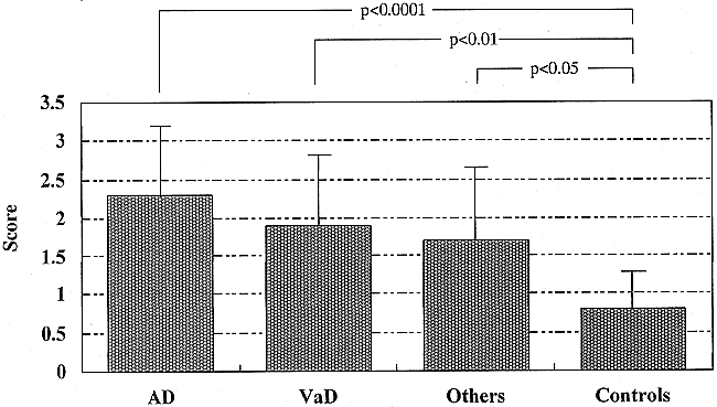
Mean scores of medial temporal lobe atrophy were 2.3 ± 0.9 in patients with AD, 1.9 ± 0.9 in patients with vascular dementia, 1.7 ± 1.0 in patients with other dementias, and 0.8 ± 0.5 in control subjects, as shown in Figure 2. Atrophy scores were significantly different (P < .0001) for the four subject groups. A post hoc Mann-Whitney U test showed significantly higher values in patients with AD, vascular dementia, and other dementias than in control subjects (P < .0001, P < .01, and P < .05, respectively). However, there were no significant differences in atrophy scores among the dementia groups.

Mean scores of medial temporal lobe atrophy in patients with AD, vascular dementia (VaD), other dementias, and control subjects. Post hoc Mann-Whitney U test showed significant differences between all dementia groups and control subjects
Mean MT ratios of the hippocampus were 36.9 ± 2.1 in patients with AD, 39.2 ± 1.9 in patients with vascular dementia, 40.1 ± 1.7 in patients with other dementias, and 40.6 ± 1.8 in control subjects, as shown in Figure 3. Analysis of variance showed significant group differences in MT ratios (F = 20.06, P < .0001). Post hoc Scheffé test showed that MT ratios in patients with AD were significantly lower than in patients with vascular dementia and other dementias, and in control subjects (P < .01, P < .0001, and P < .0001, respectively). However, no significant differences were found in MT ratios between non-AD groups and control subjects. Figure 4 shows Mo, Ms, and calculated MT ratio images from a representative patient with AD.

Mean MT ratios in patients with AD, vascular dementia, other dementias, and in control subjects. Post hoc Scheffé test showed significant differences between AD and vascular dementia (VaD) (P < .01), between AD and other dementias (P < .0001), and between AD and control subjects (P < .0001)

76-year-old woman with AD.
A, Gradient-echo Mo image (390/12/2), acquired without a radio frequency saturation pulse, shows moderate atrophy of the medial temporal lobe, graded as 3 using the visual rating scale.
B, Ms image, acquired with a radio frequency saturation pulse, shows slightly high signal intensity in areas of hippocampal formation (arrowheads).
C, Calculated MT ratio image shows slightly low signal intensity in areas of the hippocampus (arrowheads) (MT ratio = 34.2%).
Logistic regression was used to estimate the ability to differentiate among dementia groups by means of MT measurements and visual assessment. The visual rating scale for atrophy correctly identified 29 (83%) of 35 patients with AD and 11 (41%) of 27 patients with non-AD dementia. Measurement of the MT ratio correctly identified 28 (80%) of 35 patients with AD and 20 (74%) of 27 patients with non-AD dementia. Thus, the overall discrimination rate with MT imaging was 77% compared with 65% for visual analysis of atrophy.
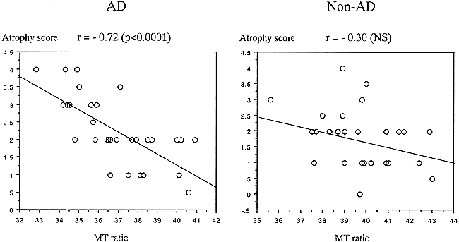
The MT ratio significantly correlated with the MMSE score in patients with AD (r = .70, P < .0001) but not in patients with non-AD dementia (r = .18, not significant) (Fig 5). The MT ratio also significantly correlated with the score of medial temporal lobe atrophy in patients with AD (r = −.72, P < .0001) but not in patients with non-AD dementia (r = −.30, not significant) (Fig 6). The score of medial temporal lobe atrophy significantly correlated with the MMSE score in patients with AD (r = −.73, P < .0001) but not in patients with non-AD dementia (r = −.30, not significant).

Correlations between MMSE scores and MT ratios in AD and non-AD dementia (Pearson's correlation coefficients). The MT ratio significantly correlated with the MMSE score in patients with AD but not in patients with non-AD dementia. Non-AD dementia includes vascular dementia and other dementias (NS indicates not significant)

Correlations between MT ratios and scores of medial temporal lobe atrophy in AD and non-AD dementia (Spearman's rank correlation coefficients). The MT ratio significantly correlated with the score of medial temporal lobe atrophy in patients with AD but not in patients with non-AD dementia. Non-AD dementia includes vascular dementia and other dementias (NS indicates not significant)
Discussion
Findings of several MR studies have established that volumetry of medial temporal lobe structures is useful in assisting in the clinical diagnosis of AD (2–11). However, atrophy has been shown in vascular dementia (13, 14), Parkinson's disease (13, 17), dementia with Lewy bodies (15, 17), and frontotemporal dementia (16). Although volumetric measurement of specific medial temporal lobe structures may provide a more detailed analysis of atrophy, it is impractical because of the time required to analyze each scan. Since there is good agreement between volumetric measurements and visual ratings (35), we used a standardized visual rating scale. Our findings are consistent with such volumetric studies and with a recent study by Barber et al (36), in which atrophy was assessed by visual analysis. On the basis of these studies and our own findings, medial temporal lobe atrophy may be a common phenomenon in some dementias and not restricted to AD.
MT imaging can provide pathophysiological information about the microscopic structure of the brain, reflecting underlying histopathologic changes. Recent reports have documented that significant decreases in the MT ratio occur in demyelinating plaques of multiple sclerosis (37, 38), in edema (37, 38), in ischemic white matter lesions (38–40), in infarcts (40–42), in wallerian degeneration (43), and in the deep gray matter, including the basal ganglia and thalamus, in liver cirrhosis (44). The precise mechanism for reduction of the MT ratio in the hippocampus of AD patients is not yet clear. In AD, histopathologic findings in the hippocampus show loss of pyramidal cells accompanied by an increase in the number of astrocytes, microglia, and oligodendrocytes, as well as by an accumulation of plaques and neurofibrillary tangles (1). Although it is unclear whether plaques and tangles decrease the MT ratio, pathologic changes, including loss of neurons and gliosis, are likely to result in a decrease in MT ratio due to a decrease in bound water and/or an increase in free water. AD is also characterized by the degeneration of neurons that interconnect the hippocampal formation with the associated cortices and other brain structures (1). Microscopically, the stratum lacunosum-radiatum of the hippocampus has shown both loss of myelin and fibrillary gliosis in patients with AD, indicating degeneration of intrahippocampal nerve fibers, including the perforant pathway (45). Therefore, demyelination and axonal loss may be other causative factors of a decreased MT ratio.
On the other hand, it is more difficult to explain the medial temporal lobe atrophy without alteration in the MT ratio seen in the patients in the non-AD group. In vascular dementia, structural abnormalities of the medial temporal lobe seem to differ depending on the location and extent of cerebrovascular lesions. We restricted our investigations to patients with subcortical or Binswanger's type vascular dementia. Although patients had no strategic infarcts in the hippocampus, it is conceivable that a cerebral volume deficit, including the medial temporal lobe, may be caused mainly or solely by cerebral substance loss induced by multiple subcortical infarcts. In an experimental study with aged rats, chronic vascular insufficiency without infarctions caused mimicked AD-type changes in the hippocampal regions, including CA1 neuron (the Sommer sector) damage and gliosis (46). Since neurons of the hippocampal formation, especially neurons of CA1 and CA3 to CA4 (the end folium), are particularly sensitive to ischemia and anoxia, acute hypoxic episodes are likely to result in nerve cell damage and subsequent gliosis. Recent postmortem findings have shown that hippocampal damage, including neuronal loss and reactive astrocytosis, is also present in patients with dementia of purely vascular origin (47, 48). Therefore, vascular factors appear to be of pathogenetic significance for the appearance of hippocampal damage in some but not all patients with vascular dementia. The patterns of medial temporal lobe damage probably represent the results of different vascular lesions or different causative factors.
The group with other dementias had a variety of dementia syndromes, and medial temporal lobe involvement varied among patients. The typical dementia in Parkinson's disease is a subcortical type, resulting from dopaminergic insufficiency. It can be hypothesized that loss of dopaminergic neurons projecting to the frontal lobe results in subsequent degeneration of medial temporal lobe structures (17). Some patients with Parkinson's disease and dementia have concomitant AD disorders (49), and hippocampal damage may occur as a result of neuropathologic changes caused by the coexistence of AD. Like AD, dementia with Lewy bodies affects the medial temporal lobe structures preferentially. However, as neuronal counts in the hippocampus of patients with dementia with Lewy bodies are higher than they are in patients with AD, hippocampal involvement in dementia with Lewy bodies is relatively preserved (50). Medial temporal lobe damage also occurs in frontotemporal dementia, but its severity is less than that in AD. Medial temporal lobe atrophy might be a deafferentation phenomenon associated with frontal and temporal damage (16). The pathologic heterogeneity in other types of dementia could be related to medial temporal lobe atrophy without altering the MT ratio.
Medial temporal lobe structures include not only the dentate gyrus, hippocampus proper, and subicular complex but also the parahippocampal gyrus, including the entorhinal cortex and surrounding white matter. Medial temporal lobe atrophy in AD might result primarily from neuronal degeneration in the hippocampal formation, as neurons in the CA1 region are severely reduced, whereas in non-AD dementia, atrophy is unlikely to be invariably caused by direct neuronal damage of the hippocampus proper. At the least, secondary degeneration in extrahippocampal lesions after primary brain damage might be a cause of medial temporal lobe atrophy in non-AD dementia. Therefore, our results are probably related to the difference in the pathologic processes underlying the medial temporal lobe damage, including the hippocampus, in patients with AD and non-AD dementia. In the future, a correlative study of histopathologic findings in the hippocampus with MT imaging is needed to substantiate our findings.
No pathologic specimens were available for the patients in this study. Although the diagnosis of dementia was based on internationally accepted criteria, some of our patients may well have been misdiagnosed. The presence of both AD and vascular dementia, so-called mixed dementia, is far more common than previously acknowledged. The AD group may have included patients whose disorder was that of another degenerative dementia. However, all patients were followed clinically for at least 2 years. Although this does not provide certainty of diagnosis, it should at least help to reduce diagnostic error.
Since CSF has a lower MT ratio than brain tissue, partial volume averaging of CSF may have occurred when measuring the atrophied hippocampus. This is unlikely, however, as patients with non-AD dementia showed no significant decrease in the MT ratio, despite their atrophy. Although the equipment we used is limited to a slice thickness of 5 mm, imaging with thinner slices would have had little influence on the data.
Conclusion
MT measurements are more specific than visual analysis in detecting structural damage of the hippocampus in patients with AD and might be useful in discriminating AD from non-AD dementia with medial temporal lobe atrophy. Although the mechanism of decrease in the hippocampal MT ratio of patients with AD is unclear, the difference in the pathologic process underlying medial temporal lobe damage, including the hippocampus, in AD and non-AD dementia is probably related to a decrease in the MT ratio. The MT technique is practical for routine clinical use because of the short examination time (about 5 minutes), and should be added to neuroimaging studies of patients with AD.
Acknowledgments
We thank K. Shimizu and other engineers in the Department of Medical Applications of the Shimadzu Corporation for their support and technical assistance. We are also grateful to J. Patrick Barron of the International Medical Communications Center of Tokyo Medical University for his review of the manuscript.
Footnotes
↵1 Address reprint requests to Haruo Hanyu, MD, Department of Geriatric Medicine, Tokyo Medical University, 6–7–1 Nishishinjuku, Shinjuku-ku, Tokyo 160–0023, Japan.
References
- Received September 7, 1999.
- Accepted after revision January 27, 2000.
- Copyright © American Society of Neuroradiology
In this issue


Jump to section
Related Articles
Cited By...
- Microstructural Tissue Changes in Alzheimer Disease Brains: Insights from Magnetization Transfer Imaging
- Longitudinal Magnetization Transfer Imaging in Mild to Severe Alzheimer Disease
- Magnetization Transfer Ratio in Alzheimer Disease: Comparison with Volumetric Measurements
- A review of structural magnetic resonance neuroimaging